. 2008 Mar;9(1):33-43.
doi: 10.1007/s10162-008-0110-6. Epub 2008 Jan 30.

Cooperative function of Tbx1 and Brn4 in the periotic mesenchyme is necessary for cochlea formation

Affiliations

Cooperative function of Tbx1 and Brn4 in the periotic mesenchyme is necessary for cochlea formation

Evan M Braunstein et al. J Assoc Res Otolaryngol. 2008 Mar.

Abstract

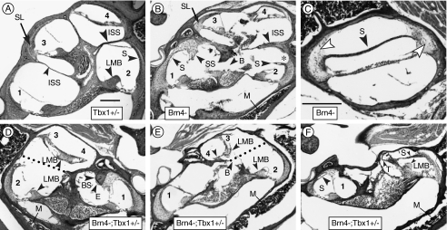
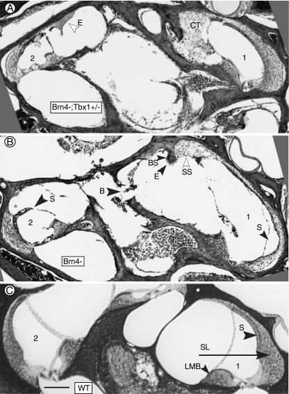
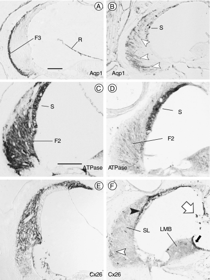
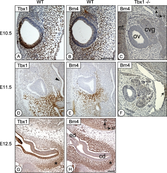
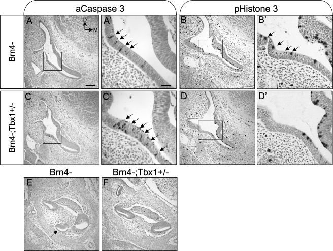
The T-box transcription factor TBX1 has been identified as the major gene responsible for the etiology of velocardiofacial syndrome/DiGeorge syndrome (VCFS/DGS). Conductive hearing loss occurs in a majority of patients with this syndrome, while sensorineural deafness has also been reported in some cases. Mutations in POU3F4/BRN4, a POU domain transcription factor, cause DFN3, an X-linked nonsyndromic form of deafness characterized by mixed conductive and sensorineural hearing loss. Inactivation of the murine orthologues of these genes causes similar defects to those seen in humans and has provided excellent models for the study of inner ear development. Tbx1 and Brn4 are expressed in the mesenchymal cells surrounding the otic vesicle and have been shown to play roles in cochlear outgrowth. Furthermore, expression of Brn4 is reduced in Tbx1 null mutants, suggesting a possible genetic interaction between these genes. To test whether Tbx1 and Brn4 function in a common pathway, mice mutant for both genes were generated and analyzed for inner ear defects. Brn4-;Tbx1+/- mutants displayed a significant reduction in the number of turns of the cochlea compared to Brn4- or Tbx1+/- mice. In addition, Brn4-;Tbx1+/- mice displayed structural defects in the apical cochlea indicative of Mondini dysplasia found in patients with either VCFS/DGS or DFN3. These data establish a genetic interaction between Tbx1 and Brn4 relevant to human disease and indicate a function of these genes in signaling from the periotic mesenchyme to the otic vesicle to direct proper coiling of the cochlear duct.


Figures

FIG. 1
FIG. 1
Defects in mesenchymal structures of mutant mice. A Adult Tbx1+/− mice display the normal four to five half turns of the cochlea in a modiolar section. The organ of Corti of the initial four half turns are lower basal (1), upper basal (2), lower second (3), and upper second (4). B Left ear of a Brn4− mouse displaying four half turns of the cochlea. The interscalar septum (ISS) is abnormally thick. In contrast to the spiral ligament in the basal turn, in the second turn it is normal (SL). Increased bone marrow in the medial otic capsule is apparent (M). C A more tangential section of the same ear shown in B. The large void above the stria vascularis (S) is present because it is not adhered to the spiral ligament. The spiral ligament has far fewer cells within it than normal (arrowheads). D Right ear of a Brn4−;Tbx1+/− mouse displaying an inverted second turn of the cochlea with no spiral ligament present. The apical surface of the spiral limbus (LMB) is oriented towards the cochlear base in this partially formed half turn. The dotted line indicates the approximate position of the missing interscalar septum. The cochlear duct terminated just beyond the organ of Corti labeled (4). E Left ear of a Brn4−;Tbx1+/− mouse exhibiting an organ of Corti in the second turn oriented at right angles to its normal position. At (3), the basilar membrane is attached to the lateral wall but there is no spiral ligament present. The cochlear duct is shortened, with its apical-most extremity noted by the arrowhead (4). F Left ear of a Brn4−;Tbx1+/− mouse that developed only a basal turn of the cochlea. The stria vascularis in the lower basal turn (1) contains a cyst (arrowhead) and the suprastrial region is hypertrophied. The spiral ligament of the upper basal turn is poorly developed and is barely attached to the stria vascularis, which forms the apical wall of the scala media. The apical cochlear duct is tightly coiled and terminates near the arrowhead (T). B, missing bone; BS, bone spur; E, eosinophillic substance attached to bone spur; ISS, interscalar septum; LMB, spiral limbus; M, marrow; SL, spiral ligament; S, stria vascularis; SS, suprastrial region; and T, termination of cochlear duct. An asterisk marks detachment of the stria from the spiral ligament. Scale bar inA is 100 μm and applies to all panels except C. The scale bar inC indicates 100 μm.
FIG. 2
FIG. 2
Defects in the basal turn of mutant mice. A Basal turn of a Brn4−;Tbx1+/− mouse. Excessive connective tissue (CT) has filled the space normally occupied by perilymph in the scala vestibuli. Abnormal eosinophilic material (E) is present in the upper basal turn. B Basal turn of a Brn4− mouse. Hypertrophied suprastrial connective tissue (SS) is present in the lower basal turn, and the stria vascularis (S) is missing. Note the adjacent spiral ligament is much thinner than in the corresponding area in A and C. In the upper basal turn, the spiral ligament is missing (asterisk) where it would normally be attached to the stria vascularis, and there are voids in the modiolar bone (B). C Basal turn of wild type cochlea showing normal morphology. (1) and (2) indicate lower basal and upper basal half-turns, respectively. B, missing bone; BS, bone spur; CT, connective tissue; E, eosinophillic substance; LMB, spiral limbus; SL, spiral ligament; S, stria vascularis; and SS, suprastrial region. The bar inC indicates 100 μm.
FIG. 3
FIG. 3
Spiral ligament fibrocytes are abnormal in mutant mice. Immunostaining of wild type (A, C, and E) and Brn4−;Tbx1+/− (B, D, and F) inner ears. A Normal distribution of type III fibrocytes (F3) at the junction of the spiral ligament and the petrous bone immunostained for Aquaporin 1 (Aqp1). B Aqp1 immunostained cells in Brn4−;Tbx1+/− ears are situated broadly throughout the spiral ligament (arrowheads). C Type II fibrocytes (F2) in the spiral ligament express Na+, K+–ATPase in wild type inner ears. The stria vascularis (S) also exhibits positive immunostaining. D The type II fibrocytes in Brn4−;Tbx1+/− mice only weakly express Na+, K+–ATPase; however, the stria remains strongly positive. E Connexin 26 (Cx26) is expressed broadly in the spiral ligament of a wild type inner ear. F Cx26 immunostaining of the inner ear of a Brn4−;Tbx1+/− mouse shows only a few positive cells within the spiral ligament (open arrowhead), but strongly stains the strial basal and intermediate cells (closed arrowhead). The spiral limbus (LMB) expresses Cx26 in wild type mice (not shown); however, it fails to stain in Brn4−;Tbx1+/− mice save for a dark strip of supralimbal cells (closed arrow). Melanocytes stain nonspecifically (open arrow). F2, type II fibrocytes; F3, type III fibrocytes; R, Reissner’s membrane; S, stria vascularis; SL, spiral ligament; and LMB, spiral limbus. Scale bar is 100 μm.
FIG. 4
FIG. 4
Tbx1 and Brn4 are coexpressed in the periotic mesenchyme during development. Adjacent sections of the inner ear at E10.5 (A and B), E11.5 (D and E), and E12.5 (G and H) immunostained for either Tbx1 (A, D, and G) or Brn4 (B, E, and H) protein. C and F are stage matched sections of Tbx1−/− embryos stained for Brn4. AF are sagittal sections, while G and H are transverse. At E10.5, both Tbx1 (A) and Brn4 (B) are expressed in the ventral and posterior periotic mesenchyme surrounding the otic vesicle. Tbx1 is also expressed in the posterior-dorsal otic vesicle epithelium. At E11.5 in medial sections through the otic vesicle, Tbx1 (D) and Brn4 (E) are coexpressed in the posterior mesenchyme. Tbx1 expression can also be detected in the pharyngeal endoderm (arrowhead inD). Tbx1−/− embryos (which have an enlarged ganglion), show absent or strongly reduced Brn4 expression in the periotic mesenchyme at E10.5 (C) or E11.5 (F). Brn4 expression in the neural tube (nt) and the second pharyngeal arch mesenchyme remains (arrow inF). At E12.5, Tbx1 expression is confined to only the most medial region of the periotic mesenchyme (G), while Brn4 is expressed in a much broader cell population (H). An asterisk marks the area of coexpression in (G) and (H). cd, cochlear duct; cvg, cochleovestibular ganglion; ed, endolymphatic duct; nt, neural tube; and ov, otic vesicle. Orientation shown in C applies to AF, while orientation in H applies to G and H. Scale bar inH is 200 μm and applies to CH. Scale bar inB is 200 μm and applies to A and B.
FIG. 5
FIG. 5
Cell survival is abnormal in the basal cochlea of Brn4−;Tbx1+/ embryos. Transverse sections through the otic vesicle of E12.5 Brn4− (A and B) and Brn4−;Tbx1+/ (C and D) embryos stained for the apoptosis marker activated Caspase 3 (aC3; A, A′, C, and C′) or the mitotic marker phospho-Histone 3 (pH3; B, B′, D, and D′). A and A′ At E12.5, apoptotic cells are present in the lateral wall of Brn4− embryos (arrows). A magnified view of the boxed region is shown in A′. B and B′ Proliferating cells are detected throughout cochlear duct in Brn4− embryos. C and C′Brn4−;Tbx1+/ embryos display increased apoptosis in the lateral wall of the cochlea, and a reduced proliferation in this same region (D and D′). E and F While some dying cells were detected, no significant differences in apoptosis were seen in the cochlear duct or periotic mesenchyme between Brn4− and Brn4−;Tbx1+/ embryos; however, the apical cochlea (arrow inE) is missing in Brn4−;Tbx+/ embryos. Scale bar indicates 200 μm in AF and 50 μm in A′D′.

References

    1. {'text': '', 'ref_index': 1, 'ids': [{'type': 'PubMed', 'value': '10979118', 'is_inner': True, 'url': 'https://pubmed.ncbi.nlm.nih.gov/10979118/'}]}
    2. Arellano B, Ramirez Camacho R, Garcia Berrocal JR, Villamar M, del Castillo I, Moreno F. Sensorineural hearing loss and Mondini dysplasia caused by a deletion at locus DFN3. Arch. Otolaryngol. Head Neck Surg. 126:1065–1069, 2000. - PubMed
    1. {'text': '', 'ref_index': 1, 'ids': [{'type': 'DOI', 'value': '10.1093/hmg/ddl084', 'is_inner': False, 'url': 'https://doi.org/10.1093/hmg/ddl084'}, {'type': 'PMC', 'value': 'PMC2563157', 'is_inner': False, 'url': 'https://pmc.ncbi.nlm.nih.gov/articles/PMC2563157/'}, {'type': 'PubMed', 'value': '16600992', 'is_inner': True, 'url': 'https://pubmed.ncbi.nlm.nih.gov/16600992/'}]}
    2. Arnold JS, Braunstein EM, Ohyama T, Groves AK, Adams JC, Brown MC, Morrow BE. Tissue-specific roles of Tbx1 in the development of the outer, middle and inner ear, defective in 22q11DS patients. Hum. Mol. Genet. 15:1629–1639, 2006a. - PMC - PubMed
    1. {'text': '', 'ref_index': 1, 'ids': [{'type': 'DOI', 'value': '10.1242/dev.02264', 'is_inner': False, 'url': 'https://doi.org/10.1242/dev.02264'}, {'type': 'PubMed', 'value': '16452092', 'is_inner': True, 'url': 'https://pubmed.ncbi.nlm.nih.gov/16452092/'}]}
    2. Arnold JS, Werling U, Braunstein EM, Liao J, Nowotschin S, Edelmann W, Hebert JM, Morrow BE. Inactivation of Tbx1 in the pharyngeal endoderm results in 22q11DS malformations. Development 133:977–987, 2006b. - PubMed
    1. {'text': '', 'ref_index': 1, 'ids': [{'type': 'DOI', 'value': '10.1242/dev.000760', 'is_inner': False, 'url': 'https://doi.org/10.1242/dev.000760'}, {'type': 'PubMed', 'value': '17395647', 'is_inner': True, 'url': 'https://pubmed.ncbi.nlm.nih.gov/17395647/'}]}
    2. Bok J, Dolson DK, Hill P, Ruther U, Epstein DJ, Wu DK. Opposing gradients of Gli repressor and activators mediate Shh signaling along the dorsoventral axis of the inner ear. Development 134:1713–1722, 2007. - PubMed
    1. {'text': '', 'ref_index': 1, 'ids': [{'type': 'DOI', 'value': '10.1006/dbio.2002.0822', 'is_inner': False, 'url': 'https://doi.org/10.1006/dbio.2002.0822'}, {'type': 'PubMed', 'value': '12435365', 'is_inner': True, 'url': 'https://pubmed.ncbi.nlm.nih.gov/12435365/'}]}
    2. Chang W, ten Dijke P, Wu DK. BMP pathways are involved in otic capsule formation and epithelial–mesenchymal signaling in the developing chicken inner ear. Dev. Biol. 251:380–394, 2002. - PubMed

Publication types