

Determinants of natural immunity against tuberculosis in an endemic setting: factors operating at the level of macrophage-Mycobacterium tuberculosis interaction
- PMID: 18234054
- PMCID: PMC2276965
- DOI: 10.1111/j.1365-2249.2007.03585.x
Determinants of natural immunity against tuberculosis in an endemic setting: factors operating at the level of macrophage-Mycobacterium tuberculosis interaction
Abstract
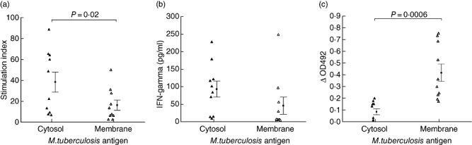
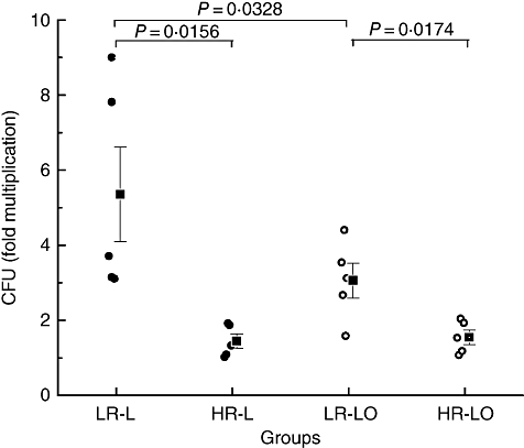
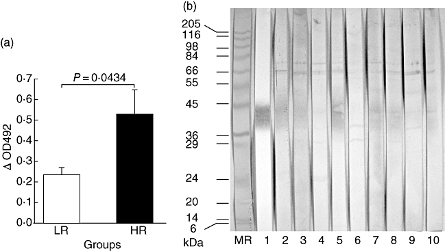
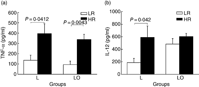
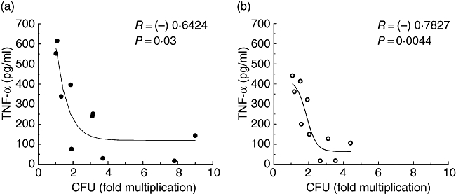
We aimed to delineate factors operating at the interface of macrophage-mycobacterium interaction which could determine the fate of a 'subclinical' infection in healthy people of a tuberculosis-endemic region. Ten study subjects (blood donors) were classified as 'high' or 'low' responders based on the ability of their monocyte-derived macrophages to restrict or promote an infection with Mycobacterium tuberculosis. Bacterial multiplication between days 4 and 8 in high responder macrophages was significantly lower (P < 0.02) than low responders. All donor sera were positive for antibodies against cell-membrane antigens of M. tuberculosis and bacilli opsonized with heat-inactivated sera were coated with IgG. In low responder macrophages, multiplication of opsonized bacilli was significantly less (P < 0.04) than that of unopsonized bacilli. The levels of tumour necrosis factor (TNF)-alpha and interleukin (IL)-12 produced by infected high responder macrophages was significantly higher (P < 0.05) than low responders. However, infection with opsonized bacilli enhanced the production of IL-12 in low responders to its level in high responders. The antibody level against membrane antigens was also significantly higher (P < 0.05) in high responders, although the antigens recognized by two categories of sera were not remarkably different. Production of certain other cytokines (IL-1beta, IL-4, IL-6 and IL-10) or reactive oxygen species (H2O2 and NO) by macrophages of high and low responders did not differ significantly. The study highlights the heterogeneity of Indian subjects with respect to their capability in handling subclinical infection with M. tuberculosis and the prominent role that TNF-alpha, opsonizing antibodies and, to a certain extent, IL-12 may play in containing it.
Figures
Similar articles
-
Naturally produced opsonizing antibodies restrict the survival of Mycobacterium tuberculosis in human macrophages by augmenting phagosome maturation.Open Biol. 2015 Dec;5(12):150171. doi: 10.1098/rsob.150171. Open Biol. 2015. PMID: 26674415 Free PMC article.
-
Role of cellular activation and tumor necrosis factor-alpha in the early expression of Mycobacterium tuberculosis 85B mRNA in human alveolar macrophages.J Infect Dis. 2004 Jul 15;190(2):341-51. doi: 10.1086/421522. Epub 2004 Jun 18. J Infect Dis. 2004. PMID: 15216471
-
Differences in IgG responses against infection phase related Mycobacterium tuberculosis (Mtb) specific antigens in individuals exposed or not to Mtb correlate with control of TB infection and progression.Tuberculosis (Edinb). 2017 Sep;106:25-32. doi: 10.1016/j.tube.2017.06.001. Epub 2017 Jun 8. Tuberculosis (Edinb). 2017. PMID: 28802401
-
Tuberculosis and the art of macrophage manipulation.Pathog Dis. 2018 Jun 1;76(4):fty037. doi: 10.1093/femspd/fty037. Pathog Dis. 2018. PMID: 29762680 Free PMC article. Review.
-
Role of cytokines in tuberculosis.Immunobiology. 1993 Nov;189(3-4):316-39. doi: 10.1016/S0171-2985(11)80364-5. Immunobiology. 1993. PMID: 8125515 Review.
Cited by
-
Biomarker changes associated with Tuberculin Skin Test (TST) conversion: a two-year longitudinal follow-up study in exposed household contacts.PLoS One. 2009 Oct 14;4(10):e7444. doi: 10.1371/journal.pone.0007444. PLoS One. 2009. PMID: 19826490 Free PMC article.
-
Elevated serum levels of CCL17 correlate with increased peripheral blood platelet count in patients with active tuberculosis in China.Clin Vaccine Immunol. 2011 Apr;18(4):629-32. doi: 10.1128/CVI.00493-10. Epub 2011 Jan 26. Clin Vaccine Immunol. 2011. PMID: 21270281 Free PMC article.
-
Distinct and shared B cell responses of tuberculosis patients and their household contacts.PLoS One. 2022 Oct 25;17(10):e0276610. doi: 10.1371/journal.pone.0276610. eCollection 2022. PLoS One. 2022. PMID: 36282846 Free PMC article.
-
Immune responses to Mycobacterium tuberculosis membrane-associated antigens including alpha crystallin can potentially discriminate between latent infection and active tuberculosis disease.PLoS One. 2020 Jan 31;15(1):e0228359. doi: 10.1371/journal.pone.0228359. eCollection 2020. PLoS One. 2020. PMID: 32004357 Free PMC article.
-
Differential expression of immunogenic proteins on virulent Mycobacterium tuberculosis clinical isolates.Biomed Res Int. 2014;2014:741309. doi: 10.1155/2014/741309. Epub 2014 Jul 7. Biomed Res Int. 2014. PMID: 25105140 Free PMC article.
References
-
- Dye C. Global epidemiology of tuberculosis. Lancet. 2006;367:938–40. - PubMed
-
- Bloom BR, Murray CJ. Tuberculosis: commentary on a reemergent killer. Science. 1992;257:1055–64. - PubMed
-
- Olobo JO, Geletu M, Demissie A, et al. Circulating TNF-alpha, TGF-beta, and IL-10 in tuberculosis patients and healthy contacts. Scand J Immunol. 2001;53:85–91. - PubMed
-
- Denis M, Gregg EO, Ghandirian E. Cytokine modulation of Mycobacterium tuberculosis growth in human macrophages. Int J Immunopharmacol. 1990;12:721–7. - PubMed
Publication types
MeSH terms
Substances
LinkOut - more resources
Full Text Sources
Medical
