. 2008 Jan 30:9:16.
doi: 10.1186/1471-2202-9-16.

The pedunculopontine tegmental nucleus and the nucleus basalis magnocellularis: do both have a role in sustained attention?

Affiliations

The pedunculopontine tegmental nucleus and the nucleus basalis magnocellularis: do both have a role in sustained attention?

Claire L Rostron et al. BMC Neurosci. .

Abstract

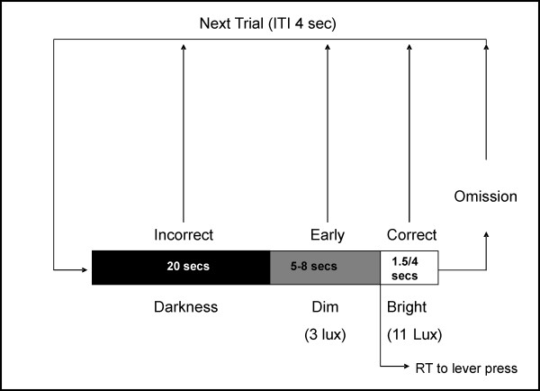
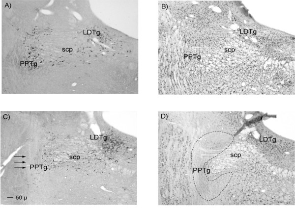
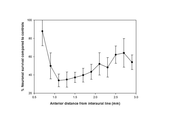
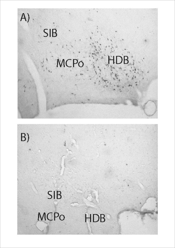
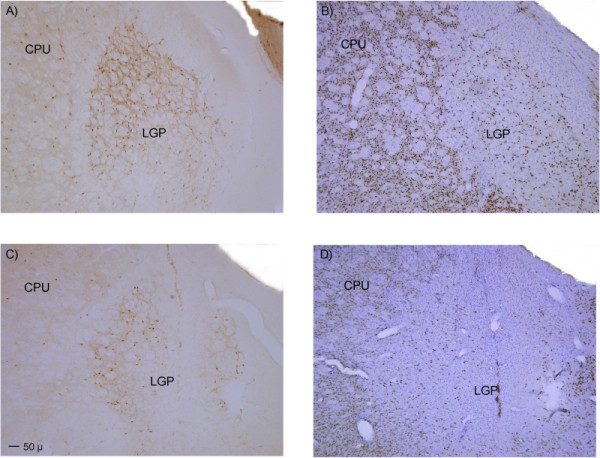
Background: It is well established that nucleus basalis magnocellularis (NbM) lesions impair performance on tests of sustained attention. Previous work from this laboratory has also demonstrated that pedunculopontine tegmental nucleus (PPTg) lesioned rats make more omissions on a test of sustained attention, suggesting that it might also play a role in mediating this function. However, the results of the PPTg study were open to alternative interpretation. We aimed to resolve this by conducting a detailed analysis of the effects of damage to each brain region in the same sustained attention task used in our previous work. Rats were trained in the task before surgery and post-surgical testing examined performance in response to unpredictable light signals of 1500 ms and 4000 ms duration. Data for PPTg lesioned rats were compared to control rats, and rats with 192 IgG saporin infusions centred on the NbM. In addition to operant data, video data of rats' performance during the task were also analysed.

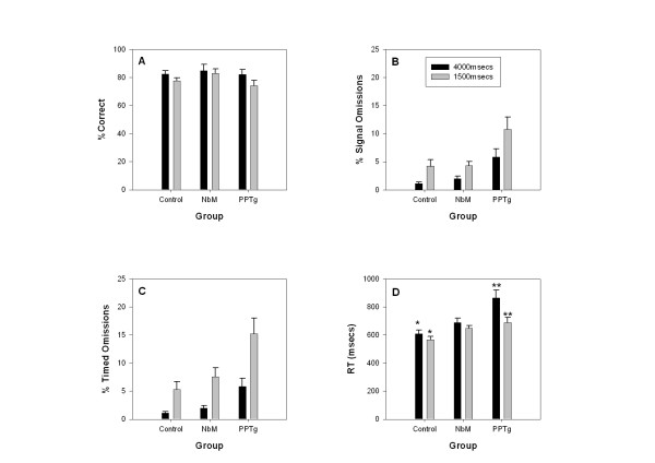
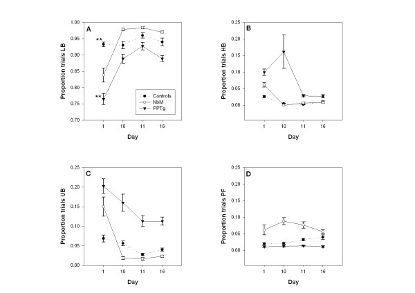
Results: Both lesion groups omitted trials relative to controls but the effect was milder and transient in NbM rats. The number of omitted trials decreased in all groups when tested using the 4000 ms signal compared to the 1500 ms signal. This confirmed previous findings for PPTg lesioned rats. Detailed analysis revealed that the increase in omissions in PPTg rats was not a consequence of motor impairment. The video data (taken on selected days) showed reduced lever orientation in PPTg lesioned rats, coupled with an increase in unconditioned behaviours such as rearing and sniffing. In contrast NbM rats showed evidence of inadequate lever pressing.

Conclusion: The question addressed here is whether the PPTg and NbM both have a role in sustained attention. Rats bearing lesions of either structure showed deficits in the test used. However, we conclude that the most parsimonious explanation for the deficit observed in PPTg rats is inadequate response organization, rather than impairment in sustained attention. Furthermore the impairment observed in NbM lesioned rats included lever pressing difficulties in addition to impaired sustained attention. Unfortunately we could not link these deficits directly to cholinergic neuronal loss.


Figures

Figure 1
Figure 1
A schematic representation of the sustained attention task.
Figure 2
Figure 2
Mean (+/- SE) performance of lesion and control animals in the sustained attention task over post-surgical days 1–10. The 4 panels show: A – Percent correct responses (response to bright light), B – Percent early responses (response to dim light), C – Percent signal omissions, D – Latency to lever press on a correct response. Full statistical analysis appears in the text. There were significant effects of day on signal omissions and correct responses suggesting an element of relearning following lesion surgery. PPTg lesioned animals made significantly more signal omissions (p < 0.001) and had significantly longer latencies (p < 0.005) compared to controls. NbM lesioned animals showed reduced correct responses (p < 0.05) and longer latencies (p < 0.05) compared to controls. Significant group × day interactions were found only in relation to early responses and are depicted in panel B: on day 1 there were significant differences between the PPTg group and controls (* p < 0.05) and between PPTg and NBM groups (***p < 0.001); on day 2 the PPTg group were significantly different to the NBM group (*p < 0.05).
Figure 3
Figure 3
Mean (+/- SE) performance of lesion and control animals under different signal length conditions. Data for the 1500 ms signal represent combined data from days 11–15 while data for the 4000 ms signal represent combined data from days 16–20. The 4 panels show: A – Percent correct responses (response to bright light), B – Percent signal omissions (failure to respond during the bright signal), C – Percent timed omissions (failure to respond within 4000 ms of the onset of the bright signal), D – Latency to lever press on a correct response. Full statistical analysis appears in the text. Main effects of signal length (without interaction) were found for timed omissions (p < 0.001), signal omissions (p < 0.001) and percent correct responses (p < 0.001). Significant signal length effects were only present in the PPTg group (p < 0.005) and the controls (p < 0.05) for the latency measure. Significant task × group interactions were found only in relation to the reaction time data depicted in panel B: there were significant differences between the 1500 and 4000 msec conditions in the control group (* p < 0.01) and in the PPTg group (**p < 0.009).
Figure 4
Figure 4
Mean (+/- SE) proportion of trials on which coded behaviours were observed in the video footage. Data are taken from days 1, 10, 11, 16. The 4 panels show: A – Bright light lever orientation (LB), B – Bright light houselight orientation (HB), C – Expression of unconditioned behaviours during the bright light (UB), D – Lever pressing failures (PF). Full statistical analysis appears in the text. PPTg lesioned animals had reduced bright lever orientation on day 1 compared to controls (p < 0.01) and showed significantly increased houselight orientation with increased unconditioned behaviours. NbM lesioned animals showed only increased lever pressing failures compared to PPTg lesioned animals (p < 0.05). Only the data for lever orientation to the bright light (panel A) showed a significant group × day interaction. On the first day, there was a significant difference between the PPTg lesioned group and the control group (** p < 0.009).
Figure 5
Figure 5
A representation of ibotenate lesions of the PPTg included in the data analyses. The largest lesion is shown in black and the smallest in grey. The location of the PPTg is indicated by a dashed black outline on the right of the diagrammatic sections. Distance is given from the interaural line in mm.
Figure 6
Figure 6
Photomicrographs illustrating the extent of survival of presumed cholinergic neurons following ibotenate lesions of the PPTg. The four panels show: A – NADPH diaphorase staining in control tissue, B – NeuN/cresyl violet staining in control tissue, C – NADPH diaphorase staining in lesioned tissue with arrows to indicate the expected location of NADPH positive neurons, D – NeuN/cresyl violet staining in lesioned tissue with the borders of the PPTg indicated by black dashed lines. Abbreviations: PPTg = pedunculopontine tegmental nucleus, LDTg = laterodorsal tegmental nucleus, SCP = superior cerebellar peduncle.
Figure 7
Figure 7
Graph depicting the degree of survival of NADPH diaphorase positive neurons throughout the rostral caudal extent of the PPTg. Data are expressed as the mean (+/- SE) percentage neuronal count of control animals.
Figure 8
Figure 8
Photomicrographs illustrating the extent of ChAT positive neuronal loss in the region of the nucleus basalis magnocellularis. The two panels show: A – ChAT positive neurons in a control animal (infusion of Dulbeccos saline), B – Extensive loss of ChAT positive neurons in the same location in an animal who received bilateral infusions of 192 IgG Saporin. Abbreviations: HDB = Horizontal diagonal band of Broca, MCPo = Magnocellular preoptic nucleus, SIB = Substantia innominata basal part.
Figure 9
Figure 9
Photomicrographs illustrating the extent of neuronal damage to the lateral globus pallidus. The two panels show: A – Loss of parvalbumin immunopositive neurons, B – Loss of NeuN/cresyl violet reactivity in the same area. Abbreviations: LGP = lateral globus pallidus, CPU = caudate putamen.

References

    1. Dellu F, Mayo W, Cherkaoui J, Le Moal M, Simon H. Learning disturbances following excitotoxic lesion of cholinergic pedunculo-pontine nucleus in the rat. Br Res. 1991;544:126–132. doi: 10.1016/0006-8993(91)90893-Z. - DOI - PubMed
    1. Sturm W, de Simone A, Krause BJ, Specht K, Hesselman V, Radermacher I, Herzog H, Tellman L, Muller-Gartner H-W, Wilmes K. Functional anatomy of intrinsic alertness: evidence for a fronto-parietal-thalamic-brainstem network in the right hemisphere. Neuropsychologia. 1999;37:797–805. doi: 10.1016/S0028-3932(98)00141-9. - DOI - PubMed
    1. Kozak R, Bowman EM, Latimer MP, Rostron CL, Winn P. Excitotoxic lesions of the pedunculopontine tegmental nucleus in rats impair performance on a test of sustained attention. Exp Br Res. 2005;162:257–264. doi: 10.1007/s00221-004-2143-3. - DOI - PubMed
    1. Winn P. Frontal syndrome as a consequence of lesions in the pedunculopontine tegmental nucleus: a short theoretical review. Br Res Bull. 1998;47:551–563. doi: 10.1016/S0361-9230(98)00136-1. - DOI - PubMed
    1. Winn P. How best to consider the structure and function of the pedunculopontine tegmental nucleus: Evidence from animal studies. J Neurol Sci. 2006;248:234–250. doi: 10.1016/j.jns.2006.05.036. - DOI - PubMed

Publication types

MeSH terms

LinkOut - more resources