. 2008 Jan 31:8:6.
doi: 10.1186/1472-6807-8-6.

Crystal structures of a purple acid phosphatase, representing different steps of this enzyme's catalytic cycle

Affiliations

Crystal structures of a purple acid phosphatase, representing different steps of this enzyme's catalytic cycle

Gerhard Schenk et al. BMC Struct Biol. .

Abstract

Background: Purple acid phosphatases belong to the family of binuclear metallohydrolases and are involved in a multitude of biological functions, ranging from bacterial killing and bone metabolism in animals to phosphate uptake in plants. Due to its role in bone resorption purple acid phosphatase has evolved into a promising target for the development of anti-osteoporotic chemotherapeutics. The design of specific and potent inhibitors for this enzyme is aided by detailed knowledge of its reaction mechanism. However, despite considerable effort in the last 10 years various aspects of the basic molecular mechanism of action are still not fully understood.

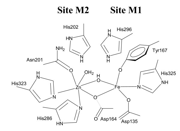
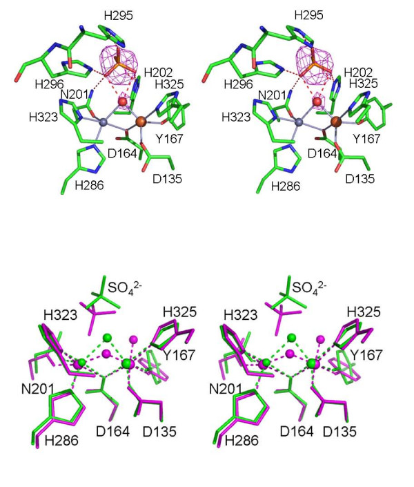
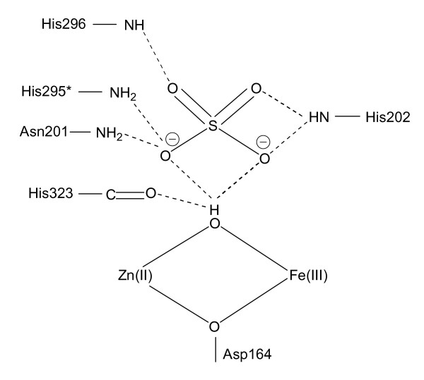
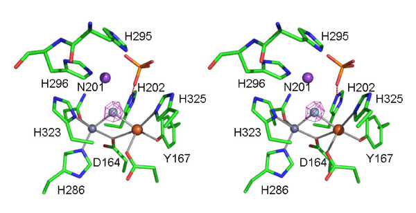
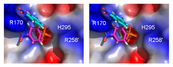
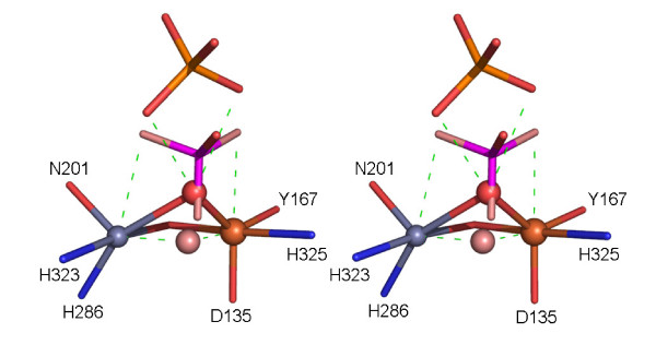
Results: Red kidney bean purple acid phosphatase is a heterovalent enzyme with an Fe(III)Zn(II) center in the active site. Two new structures with bound sulfate (2.4 A) and fluoride (2.2 A) provide insight into the pre-catalytic phase of its reaction cycle and phosphorolysis. The sulfate-bound structure illustrates the significance of an extensive hydrogen bonding network in the second coordination sphere in initial substrate binding and orientation prior to hydrolysis. Importantly, both metal ions are five-coordinate in this structure, with only one nucleophilic mu-hydroxide present in the metal-bridging position. The fluoride-bound structure provides visual support for an activation mechanism for this mu-hydroxide whereby substrate binding induces a shift of this bridging ligand towards the divalent metal ion, thus increasing its nucleophilicity.

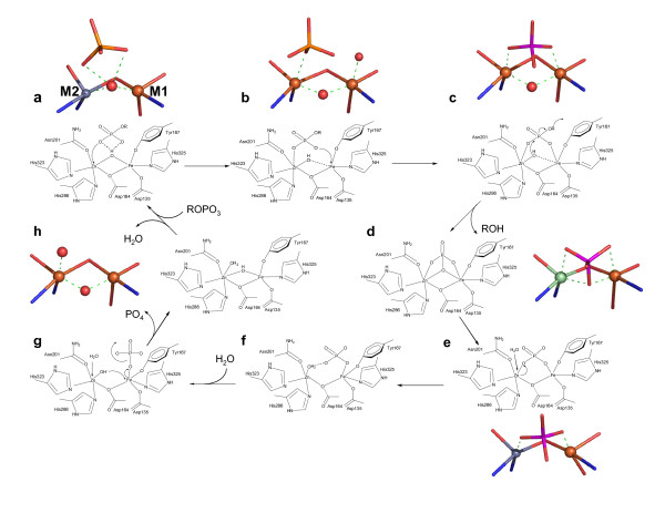
Conclusion: In combination with kinetic, crystallographic and spectroscopic data these structures of red kidney bean purple acid phosphatase facilitate the proposal of a comprehensive eight-step model for the catalytic mechanism of purple acid phosphatases in general.


Figures

Figure 1
Figure 1
Schematic illustration of the active site of red kidney bean purple acid phosphatase (rkbPAP), a representative binuclear metallohydrolase. In most (if not all) binuclear metallohydrolases the binding affinities of the two metal centers vary, with M1 representing the tight binding site and M2 the lower affinity site [8]. In rkbPAP M1 and M2 are occupied by Fe(III) and Zn(II), respectively. Combined crystallographic and spectroscopic data for PAPs indicate the presence of a bridging (hydr)oxo group and one terminal water ligand (see text). The presence of a terminal Fe(III)-bound hydroxide is currently debated with spectroscopic data suggesting its absence [8], but the crystal structure of rat PAP supporting its presence [8].
Figure 2
Figure 2
(a) Stereodiagram of the active site of the rkbPAP-sulfate complex. Fo-Fc electron density for the sulfate group is overlayed. The sulfate group is bound in the second coordination sphere via extensive hydrogen bonding interactions. General legend: Fe(III) is in tan, Zn(II) in grey, carbon in green, oxygen in red, nitrogen in blue and sulphur in orange. Hydrogen bonds and other contacts are shown as dashed lines. (b) Stereodiagram of the superimposition of the active site of the rkbPAP-sulfate complex (green) with the active site of the rat PAP-sulfate complex [8] (magenta). In the rkbPAP-sulfate complex the bridging hydroxide adopts an elevated position, precluding the binding of a terminal water to Fe(III). Both metals in the rkbPAP-sulfate complex structure are five coordinate.
Figure 3
Figure 3
Hydrogen bond network in the active site of rkbPAP. The hydrogen bond pattern demonstrates how the initial, pre-catalytic enzyme-substrate complex may be stabilized. The protonation state of sulfate at the pH of crystallization (pH 4.0) is SO42-. Residue His295 (indicated by a "*") is the only amino acid that is not invariant among PAPs from different sources.
Figure 4
Figure 4
Stereodiagram of the active site of the rkbPAP-fluoride complex. Fo-Fc electron density for the bridge is overlayed. The fluoride replaces the hydroxide in the bridging position. General legend: Fe(III) is in tan, Zn(II) in grey, sodium in purple, fluoride in cyan, carbon in green, oxygen in red, nitrogen in blue and sulphur in orange. Hydrogen bonds and other contacts are shown as dashed lines.
Figure 5
Figure 5
Model of substrate (para-nitrophenyl phosphate) binding to rkbPAP based on the sulfate complex (this study; cyan carbons) and on the structure of the phosphate complex (magenta carbons). The ' symbol represents a residue from the neighboring subunit.
Figure 6
Figure 6
Proposed mechanism of binuclear metallohydrolase-catalyzed esterolysis. Following the binding of the substrate in a pre-catalytic complex, structural rearrangements lead to "quasi-monodentate" and bidentate coordination of the μ-hydroxide and phosphate groups, respectively (a-c). Nucleophilic attack by the μ-hydroxide is followed by the release of the leaving group, and the active site is returned to its resting state by the exchange of the bound phosphate group by two water molecules (d-h). A terminal M1-bound hydroxide is observed in the structure of rat PAP (b), which appears to be an artefact of crystallization [46] and is not supported by solution studies on resting PAP [17]. Where available crystallographic pictures of relevant active site structures are included. (a) rkbPAP-sulfate complex; (b) rat PAP-sulfate complex [43]; (c) pig PAP-phosphate complex [42]; (d) sweet potato PAP-phosphate complex [18] ; (e) rkbPAP-phosphate complex [41]; (h) rkbPAP [40] (the bridging and terminal M2-bound water ligands were modelled based on ENDOR studies [17]).
Figure 7
Figure 7
Overlay of the rkbPAP-sulfate complex (with the bridging oxygen in red), the bridging hydroxide (pink) as observed in pig PAP [42] and the phosphate as observed in sweet potato PAP [18]. The image demonstrates a plausible trajectory for the substrate with the bridging hydroxide as the nucleophile in catalysis. Inversion of configuration around the phosphorous atom is observed. General legend: Fe(III) is in tan, Zn(II) in grey, carbon in green, oxygen in red, nitrogen in blue, sulphur in orange, hydrogen in white and phosphorous in magenta.

References

    1. Lippard SJ, Berg JM. Principles of Bioinorganic Chemistry, University Science Books, Mill Valley, CA; 1994.
    1. Wilcox DE. Binuclear metallohydrolases. Chem Rev. 1996;96:2435–2458. doi: 10.1021/cr950043b. - DOI - PubMed
    1. Dismukes GC. Manganese enzymes with binuclear active sites. Chem Rev. 1996;96:2909–2926. doi: 10.1021/cr950053c. - DOI - PubMed
    1. Barford D, Das AK, Egloff MP. The structure and mechanism of protein phosphatases: Insights into catalysis and regulation. Annu Rev Biophys Biomol Struct. 1998;27:133–164. doi: 10.1146/annurev.biophys.27.1.133. - DOI - PubMed
    1. Jackson MD, Denu JM. Molecular reactions of protein phosphatases – Insights from structure and chemistry. Chem Rev. 2001;101:2313–2340. doi: 10.1021/cr000247e. - DOI - PubMed

Publication types