. 2008 Apr 25;374(1):33-49.
doi: 10.1016/j.virol.2007.12.028. Epub 2008 Jan 30.

Nucleotidylylation of the VPg protein of a human norovirus by its proteinase-polymerase precursor protein

Affiliations

Nucleotidylylation of the VPg protein of a human norovirus by its proteinase-polymerase precursor protein

Gaël Belliot et al. Virology. .

Abstract

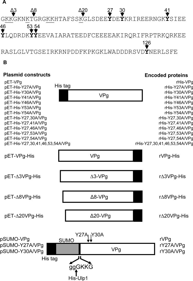
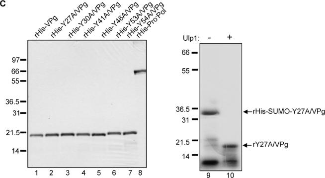
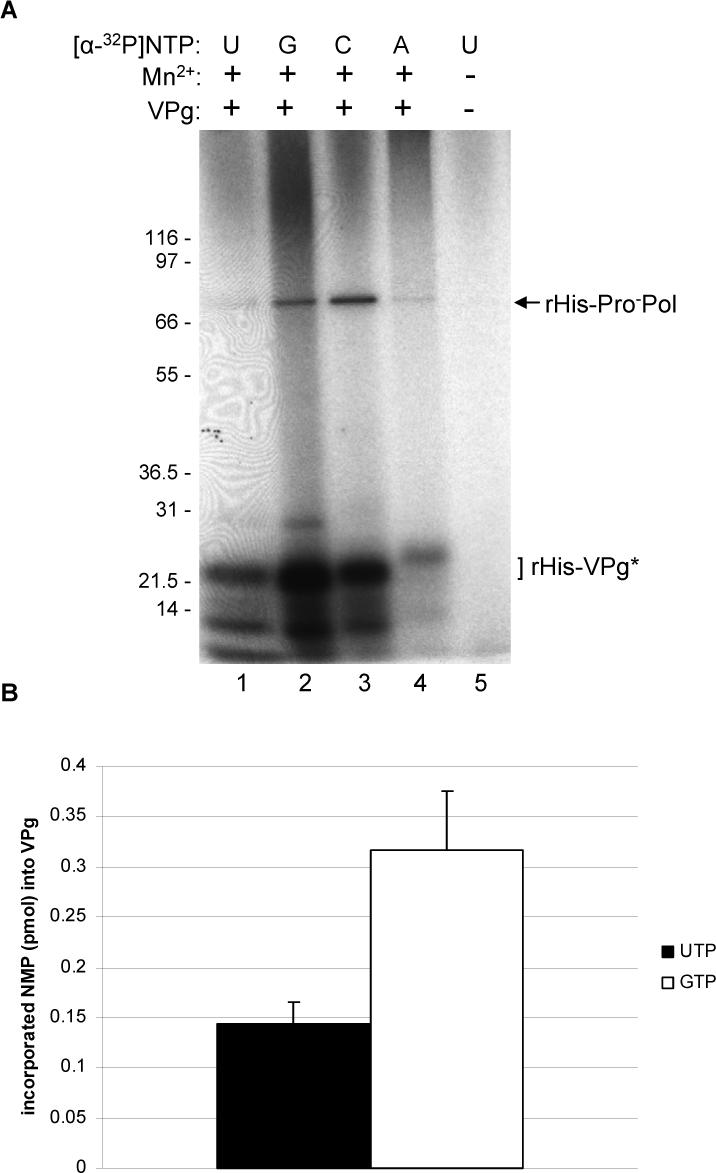
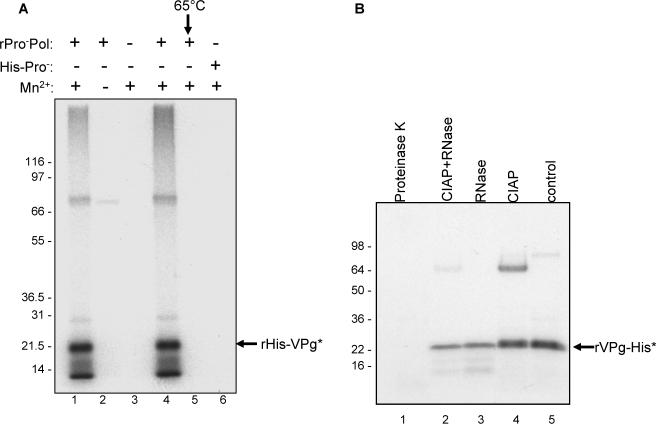
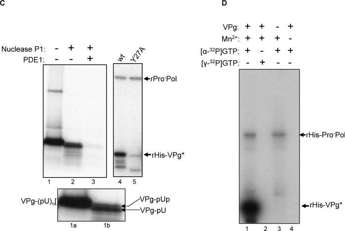
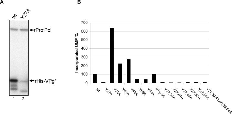
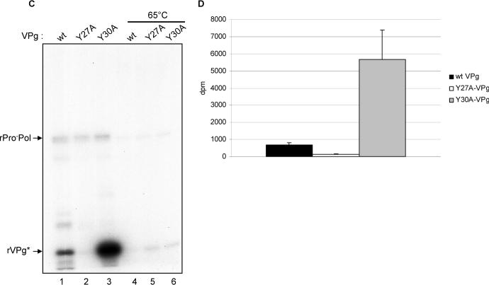
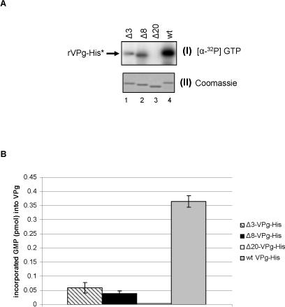
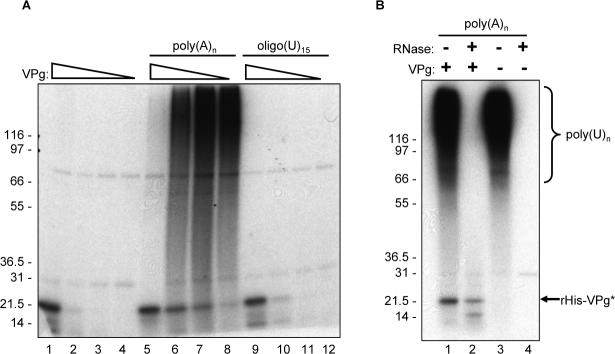
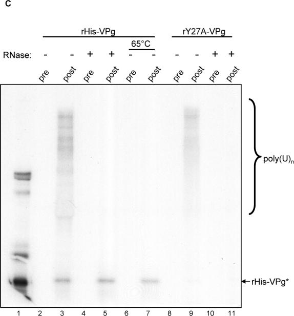
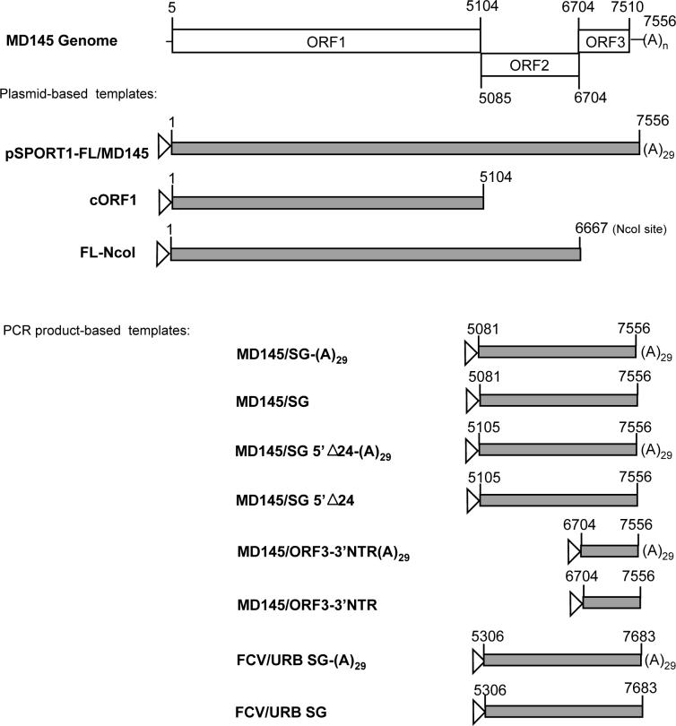
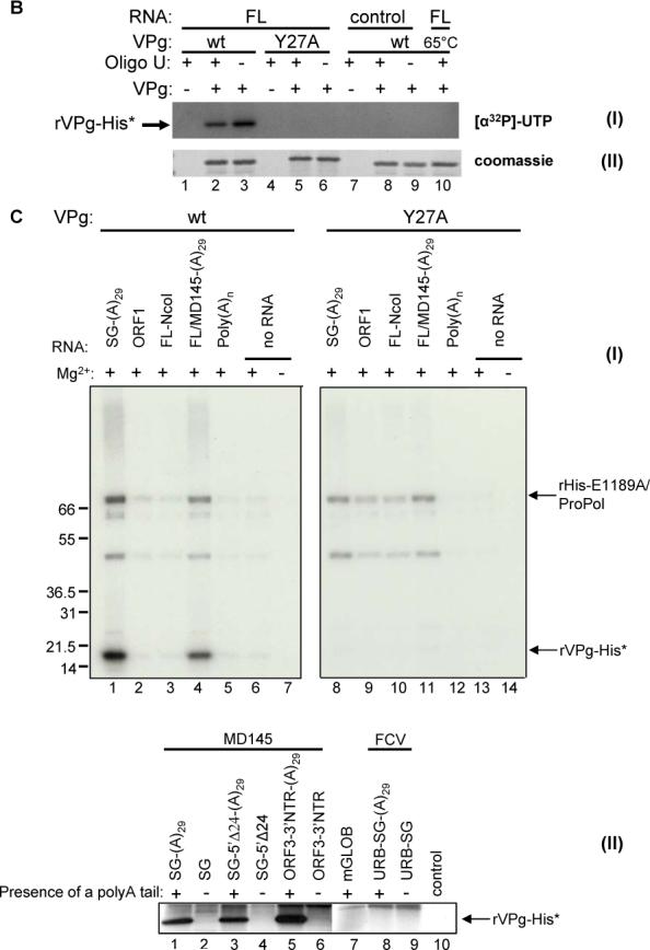
Caliciviruses have a positive strand RNA genome covalently-linked at the 5'-end to a small protein, VPg. This study examined the biochemical modification of VPg by the ProPol form of the polymerase of human norovirus strain MD145 (GII.4). Recombinant norovirus VPg was shown to be nucleotidylylated in the presence of Mn2+ by MD145 ProPol. Phosphodiesterase I treatment of the nucleotidylylated VPg released the incorporated UMP, which was consistent with linkage of RNA to VPg via a phosphodiester bond. Mutagenesis analysis of VPg identified Tyrosine 27 as the target amino acid for this linkage, and suggested that VPg conformation was important for the reaction. Nucleotidylylation was inefficient in the presence of Mg2+; however the addition of full- and subgenomic-length MD145 RNA transcripts led to a marked enhancement of the nucleotidylylation efficiency in the presence of this divalent cation. Furthermore, evidence was found for the presence of an RNA element near the 3'-end of the polyadenylated genome that enhanced the efficiency of nucleotidylylation in the presence of Mg2+.


Figures

Figure 1
Figure 1
MD145 VPg DNA constructs generated for study. (A) The primary deduced amino acid sequence of the MD145 VPg is shown. The tyrosine residues at positions 27, 30, 41, 46, 53, 54, and 126 are shown in bold type, and the lysine residues in the N-terminal region are underlined. The N-terminus of the VPg encoded in truncated VPg constructs Δ3-, Δ8- and Δ20-VPg-His is indicated by arrows. (B) cDNA clones engineered for expression of the MD145 VPg and its mutated forms in bacteria. His-tag is represented by the black box in the diagram, and is located at the N- or C-terminus. The VPg constructs were designated according to the mutagenized tyrosine (e.g., pET-His-Y27A/VPg). Double tyrosine mutated VPg constructs were designated similarly. VPg proteins with sequential deletions from the N-terminus were designated according to the first amino acid in the truncated VPg (e.g., pET-Δ3VPg-His). For the SUMO-VPg constructs, the SUMO peptide is indicated by a grey box. For the Ulp1 cleavage site, SUMO peptide and VPg amino acids are indicated by lower and lower cases, respectively. The resulting construct was named as indicated above. (C) SDS-PAGE analysis of 1μg bacterially-expressed recombinant VPg proteins (lanes 1−7) and ProPol (lane 8) that were engineered with an N-terminal His tag. The mutated rY27A/VPg (Figure 1C, lanes 9 and 10) was expressed without a His-tag (see Materials and Methods). The rHis-SUMO-Y27A/VPg fusion protein (lane 9) and tag-free rY27A/VPg (lane 10) were resolved by SDS-PAGE. Ulp1 proteinase treatment is indicated above the gel. The migration of the molecular weight marker (Mark XII, Invitrogen) is indicated in kDa here and in the following figures.
Figure 1
Figure 1
MD145 VPg DNA constructs generated for study. (A) The primary deduced amino acid sequence of the MD145 VPg is shown. The tyrosine residues at positions 27, 30, 41, 46, 53, 54, and 126 are shown in bold type, and the lysine residues in the N-terminal region are underlined. The N-terminus of the VPg encoded in truncated VPg constructs Δ3-, Δ8- and Δ20-VPg-His is indicated by arrows. (B) cDNA clones engineered for expression of the MD145 VPg and its mutated forms in bacteria. His-tag is represented by the black box in the diagram, and is located at the N- or C-terminus. The VPg constructs were designated according to the mutagenized tyrosine (e.g., pET-His-Y27A/VPg). Double tyrosine mutated VPg constructs were designated similarly. VPg proteins with sequential deletions from the N-terminus were designated according to the first amino acid in the truncated VPg (e.g., pET-Δ3VPg-His). For the SUMO-VPg constructs, the SUMO peptide is indicated by a grey box. For the Ulp1 cleavage site, SUMO peptide and VPg amino acids are indicated by lower and lower cases, respectively. The resulting construct was named as indicated above. (C) SDS-PAGE analysis of 1μg bacterially-expressed recombinant VPg proteins (lanes 1−7) and ProPol (lane 8) that were engineered with an N-terminal His tag. The mutated rY27A/VPg (Figure 1C, lanes 9 and 10) was expressed without a His-tag (see Materials and Methods). The rHis-SUMO-Y27A/VPg fusion protein (lane 9) and tag-free rY27A/VPg (lane 10) were resolved by SDS-PAGE. Ulp1 proteinase treatment is indicated above the gel. The migration of the molecular weight marker (Mark XII, Invitrogen) is indicated in kDa here and in the following figures.
Figure 2
Figure 2
Nucleotidylylation of rHis-VPg by the rHis-ProPol polymerase. (A) Two μg of rHis-VPg were assayed in the presence of [α32P]-NTP (either UTP, lanes 1 and 5; GTP, lane 2; CTP, lane 3 and ATP, lane 4). Lane 5 contains a negative control without VPg and Mn2+. The NTP used in each assay, and the presence or absence of VPg and Mn2+ (0.5 mM final) is indicated above the gel. The identities of the radiolabeled proteins are indicated on the right, and the modified rVPg protein is indicated by an asterisk here and the following figures. (B) In a separate experiment, guanylylation and uridylylation reactions of rHis-VPg were conducted in triplicate. Five μl of each assay was analyzed by SDS-PAGE. Labeled VPg separated by SDS-PAGE, was excised from the gel and the amount of UMP (black column) and GMP (white column) incorporated into VPg was determined. The graph represents the mean value of the triplicate assay for each NTP. Incorporated NMP is given in pmol/μg of rHis-VPg (left side of the graph). Standard deviations are indicated by vertical bars.
Figure 3
Figure 3
Characterization of the VPg modification by ProPol. (A) Autoradiography of the VPg-uridylylation assay. Two μg of rHis-VPg was incubated in the presence of [α32P]-UTP and several combinations of rHis-ProPol (1 μg), rHis-Pro (1 μg) and Mn2+ as indicated above the gel. rHis-ProPol was also heated at 65°C for 10 min prior to addition to the assay (lane 5). (B) Autoradiography of the UTP-labeled VPg after enzymatic treatment. Uridylylation assay and enzymatic treatments are described in the Materials and Methods. The 15 μl reaction was Proteinase K- or RNase-treated (lanes 1 and 3). The 15 μl reaction was also CIAP-treated (Calf Intestinal Alkaline Phosphatase, lane 4) or CIAP- and RNase-treated (lane 2). Enzymatic treatments are indicated above the gel. For the control (lane 5), 5 μl of the uridylylation assay were directly resolved by SDS-PAGE. (C) Nuclease P1 and phosphodiesterase 1 (PDE1) treatment of the uridylylated VPg. For the control (lane 1), 5 μl of the assay was directly resolved by SDS-PAGE. Enzymatic treatments are indicated above the gel. The insert contains a magnified view of the labeled VPg from lane 1 (lane 1a) and 2 (lane 2a). The putative forms of labeled VPg are indicated by arrows (VPg-pUpU and VPg-pU) or bracket (VPg-p(U)n). (D) Two μg of rHis-VPg were assayed for nucleotidylylation in the presence of [α32P]-GTP (lanes 1, 3 and 4) or [γ32P]-GTP (lane 2). The combinations of VPg, Mn2+ (1 mM final), [α32P]-GTP and [γ32P]-GTP are indicated above the gel. rHis-ProPol and rHis-VPg are indicated by arrows.
Figure 3
Figure 3
Characterization of the VPg modification by ProPol. (A) Autoradiography of the VPg-uridylylation assay. Two μg of rHis-VPg was incubated in the presence of [α32P]-UTP and several combinations of rHis-ProPol (1 μg), rHis-Pro (1 μg) and Mn2+ as indicated above the gel. rHis-ProPol was also heated at 65°C for 10 min prior to addition to the assay (lane 5). (B) Autoradiography of the UTP-labeled VPg after enzymatic treatment. Uridylylation assay and enzymatic treatments are described in the Materials and Methods. The 15 μl reaction was Proteinase K- or RNase-treated (lanes 1 and 3). The 15 μl reaction was also CIAP-treated (Calf Intestinal Alkaline Phosphatase, lane 4) or CIAP- and RNase-treated (lane 2). Enzymatic treatments are indicated above the gel. For the control (lane 5), 5 μl of the uridylylation assay were directly resolved by SDS-PAGE. (C) Nuclease P1 and phosphodiesterase 1 (PDE1) treatment of the uridylylated VPg. For the control (lane 1), 5 μl of the assay was directly resolved by SDS-PAGE. Enzymatic treatments are indicated above the gel. The insert contains a magnified view of the labeled VPg from lane 1 (lane 1a) and 2 (lane 2a). The putative forms of labeled VPg are indicated by arrows (VPg-pUpU and VPg-pU) or bracket (VPg-p(U)n). (D) Two μg of rHis-VPg were assayed for nucleotidylylation in the presence of [α32P]-GTP (lanes 1, 3 and 4) or [γ32P]-GTP (lane 2). The combinations of VPg, Mn2+ (1 mM final), [α32P]-GTP and [γ32P]-GTP are indicated above the gel. rHis-ProPol and rHis-VPg are indicated by arrows.
Figure 4
Figure 4
Role of the tyrosine residues in VPg uridylylation. (A) The expression and purification of the His tag-free rProPol has been described previously (Belliot et al., 2005). Two μg of rHis-VPg (lane 1) or rHis-Y27A/VPg (lane 2) were incubated in the presence of rProPol, 1 mM MnCl2 and [α32P]-UTP. Labeled proteins are indicated by arrows on the right side of the gel. (B) Effect of the tyrosine residues at positions 27, 30, 41, 46, 53 and 54 on the uridylylation of the VPg proteins. The VPg mutated proteins are indicated below the graph. rVPg protein (1.5 μg) was incubated with 1 μg of SUMO expressed rProPol in the presence of 5 μCi [α32P]-UTP and 0.5 mM MnCl2. Five μl of each reaction were analyzed by SDS-PAGE. The relative amount of incorporated UMP was determined by using a Phosphorimager. Incorporation values are given in percentage of the level of incorporation for the wt VPg protein (100%). (C) SUMO-expressed wild type (wt, lanes 1 and 4) VPg, rY27A/VPg (lanes 2 and 5) and rY30A/VPg (lanes 3 and 6) were assayed for uridylylation in the presence of tag-free rProPol, 1 mM MnCl2, and [α32P]-UTP. Wild type and mutated VPgs were also heated at 65°C for 10 min prior to incubation with rPro-Pol (lanes 4 to 6). Uridylylation assays were then conducted the same way. The proteins of interest are indicated by arrows. (D) Role of the tyrosine residues at positions 27 and 30 in uridylylation. Tag-free- wt rVPg (black column), rY27A/VPg (white column) and rY30A/VPg (grey column) were assayed for uridylylation in the presence of tag-free rProPol. The uridylylation assays were conducted in triplicate and 5 μl of each assays were analyzed by SDS-PAGE. The amount of incorporated UMP is given in disintegration per min (dpm) per μg of VPg. Columns represent mean values and standard deviations are represented by vertical bars.
Figure 4
Figure 4
Role of the tyrosine residues in VPg uridylylation. (A) The expression and purification of the His tag-free rProPol has been described previously (Belliot et al., 2005). Two μg of rHis-VPg (lane 1) or rHis-Y27A/VPg (lane 2) were incubated in the presence of rProPol, 1 mM MnCl2 and [α32P]-UTP. Labeled proteins are indicated by arrows on the right side of the gel. (B) Effect of the tyrosine residues at positions 27, 30, 41, 46, 53 and 54 on the uridylylation of the VPg proteins. The VPg mutated proteins are indicated below the graph. rVPg protein (1.5 μg) was incubated with 1 μg of SUMO expressed rProPol in the presence of 5 μCi [α32P]-UTP and 0.5 mM MnCl2. Five μl of each reaction were analyzed by SDS-PAGE. The relative amount of incorporated UMP was determined by using a Phosphorimager. Incorporation values are given in percentage of the level of incorporation for the wt VPg protein (100%). (C) SUMO-expressed wild type (wt, lanes 1 and 4) VPg, rY27A/VPg (lanes 2 and 5) and rY30A/VPg (lanes 3 and 6) were assayed for uridylylation in the presence of tag-free rProPol, 1 mM MnCl2, and [α32P]-UTP. Wild type and mutated VPgs were also heated at 65°C for 10 min prior to incubation with rPro-Pol (lanes 4 to 6). Uridylylation assays were then conducted the same way. The proteins of interest are indicated by arrows. (D) Role of the tyrosine residues at positions 27 and 30 in uridylylation. Tag-free- wt rVPg (black column), rY27A/VPg (white column) and rY30A/VPg (grey column) were assayed for uridylylation in the presence of tag-free rProPol. The uridylylation assays were conducted in triplicate and 5 μl of each assays were analyzed by SDS-PAGE. The amount of incorporated UMP is given in disintegration per min (dpm) per μg of VPg. Columns represent mean values and standard deviations are represented by vertical bars.
Figure 5
Figure 5
Deletional mutagenesis of the N-terminus of the norovirus VPg. (A) Three μg of wild type rVPg-His (wt, lane 4), rΔ3-VPg-His (Δ3, lane 1), rΔ8-VPg-His (Δ8, lane lane 2) and rΔ20-VPg-His (Δ20, lane 3) were assayed for guanylylation in the presence of 1 μg of rE1189A/ProPol-His, [α32P]-GTP and 1 mM MnCl2. Wild type and truncated VPg proteins are indicated above the gel. The autoradiography and Coomassie blue staining of the modified VPg proteins are shown on panels I, and II, respectively. Panels I and II represent the same gel. (B) Amount of incorporated GMP into wt and truncated VPgs. Three μg of wild type rVPg-His (grey column), rΔ3-VPg-His (Δ3, hatched column), rΔ8-VPg-His (Δ8, black column) and rΔ20-VPg-His (Δ20, white column) were assayed for guanylylation as described above. The assays were conducted in triplicate. The amount of incorporated UMP was calculated as described in Materials and Methods and reported here as pmol per μg of VPg. Columns represent the mean value of three experiments and the standard deviation is indicated by vertical bars.
Figure 6
Figure 6
VPg uridylylation in the presence of homopolymeric poly(A)n templates. (A) Three μg (lanes 1, 5 and 9), 1.5 μg (lanes 2, 6 and 10), 0.75 μg (lanes 3, 7 and 11) and 0.375 μg (lanes 4, 8 and 12) of rHis-VPg (corresponding to 180, 90, 45, and 22.5 pmol per assay, respectively) were assayed for uridylylation in the presence of rHis-ProPol (13 pmol), [α32P]-UTP and 1 mM MnCl2. Uridylylation assays were also performed in the presence of 5 μg of homopolymeric poly(A)n templates (Roche) (lanes 5 to 8) or 10 μM oligo(rU)15 primer (Invitrogen) (lanes 9 to 12). The decreasing amounts of rHis-VPg are represented by an open triangle above the gel. The presence of poly(A)n and oligo(U)15 in each reaction is indicated above the gel. (B) One μg of rHis-ProPol was incubated with 5 μg of poly(A)n, 5 μCi [α32P]-UTP, and 1 mM MnCl2 in the presence (lanes 1 and 2) or absence (lanes 3 and 4) of 1.5 μg rHis-VPg). Five μl of each reaction were resolved by SDS-PAGE without treatment (lanes 1 and 3) or following treatment with RNase prior to SDS-PAGE (lanes 2 and 4). The combinations of RNase and VPg are indicated above the gel. Labeled poly(U)n and rHis-VPg are indicated by bracket and arrow, respectively on the right side of the gel. (C) Immunoprecipitation assay of the uridylylation reaction mixture with an antiserum raised against bacterially-expressed rHis-VPg (Belliot et al., 2003). For each immunoprecipitation assay, a uridylylation assay was first performed with rHis-Pro-Pol, rHis-VPg (lanes 2 to 7), and rHis-Y27A/VPg (lanes 8 to 11). The reaction mixture was then assayed by immunoprecipitation without treatment (lanes 2, 3, 8 and 9) or following treatment with RNase (lanes 4, 5, 10 and 11) or heating at 65°C for 10 min (lanes 6 and 7). Labeled materials were resuspended with electrophoresis lysis buffer, released by boiling and directly resolved onto a 12% Tris-glycine polyacrylamide denaturing gel, or used for RIPA. The serum (VPg pre or post-immunization) used in each reaction is indicated. The positive control in lane 1 was 5μl of a VPg uridylylation assay as described above. Labeled poly(U)n and rHis-VPg are indicated on the right with a bracket and arrow, respectively.
Figure 6
Figure 6
VPg uridylylation in the presence of homopolymeric poly(A)n templates. (A) Three μg (lanes 1, 5 and 9), 1.5 μg (lanes 2, 6 and 10), 0.75 μg (lanes 3, 7 and 11) and 0.375 μg (lanes 4, 8 and 12) of rHis-VPg (corresponding to 180, 90, 45, and 22.5 pmol per assay, respectively) were assayed for uridylylation in the presence of rHis-ProPol (13 pmol), [α32P]-UTP and 1 mM MnCl2. Uridylylation assays were also performed in the presence of 5 μg of homopolymeric poly(A)n templates (Roche) (lanes 5 to 8) or 10 μM oligo(rU)15 primer (Invitrogen) (lanes 9 to 12). The decreasing amounts of rHis-VPg are represented by an open triangle above the gel. The presence of poly(A)n and oligo(U)15 in each reaction is indicated above the gel. (B) One μg of rHis-ProPol was incubated with 5 μg of poly(A)n, 5 μCi [α32P]-UTP, and 1 mM MnCl2 in the presence (lanes 1 and 2) or absence (lanes 3 and 4) of 1.5 μg rHis-VPg). Five μl of each reaction were resolved by SDS-PAGE without treatment (lanes 1 and 3) or following treatment with RNase prior to SDS-PAGE (lanes 2 and 4). The combinations of RNase and VPg are indicated above the gel. Labeled poly(U)n and rHis-VPg are indicated by bracket and arrow, respectively on the right side of the gel. (C) Immunoprecipitation assay of the uridylylation reaction mixture with an antiserum raised against bacterially-expressed rHis-VPg (Belliot et al., 2003). For each immunoprecipitation assay, a uridylylation assay was first performed with rHis-Pro-Pol, rHis-VPg (lanes 2 to 7), and rHis-Y27A/VPg (lanes 8 to 11). The reaction mixture was then assayed by immunoprecipitation without treatment (lanes 2, 3, 8 and 9) or following treatment with RNase (lanes 4, 5, 10 and 11) or heating at 65°C for 10 min (lanes 6 and 7). Labeled materials were resuspended with electrophoresis lysis buffer, released by boiling and directly resolved onto a 12% Tris-glycine polyacrylamide denaturing gel, or used for RIPA. The serum (VPg pre or post-immunization) used in each reaction is indicated. The positive control in lane 1 was 5μl of a VPg uridylylation assay as described above. Labeled poly(U)n and rHis-VPg are indicated on the right with a bracket and arrow, respectively.
Figure 7
Figure 7
Effect of MD145 genomic and subgenomic RNAs on VPg uridylylation in the presence of Mg2+. (A) Diagram of the constructs used in the experiment. The genomic organization of the MD145 RNA genome is based upon the sequence available in Genbank (AY032605). Positions of the first and last nucleotides for each ORF are indicated. pSPORT1-FL/MD145 and pSPORT1-SG/MD145 were linearized by AatII for RNA production. Both constructs were engineered with a T7 promoter upstream of the 5’-end (open triangle) and a 29 nucleotide length polyA tail at the 3’-end (poly(A)29). To generate genomic RNA lacking ORF3 (named FL-NcoI), the pSPORT1-FL/MD145 plasmid construct was linearized at the NcoI restriction site naturally present at position 6667 of the MD145 genome. Linearized constructs were then used to produce RNA. The cORF1 construct was described previously (linearized by NotI for RNA production) (Belliot et al., 2003). For the generation of PCR DNA templates, a T7 promoter sequence was engineered into the forward primer, and the reverse primer was engineered either with or without a poly(A)29 tail. The pSPORT1-FL/MD145 (above) and pQ14 (Sosnovtsev and Green, 1995) plasmids were used as the templates for generation of the MD145 and Urbana (URB) feline calicivirus PCR products, respectively. (B) VPg uridylylation in the presence of genomic RNA and Mg2+. Five μl of each reaction were directly analyzed by SDS-PAGE (panels I and II). The gel was Coomassie-stained (panel I), dried and subjected to autoradiography (panel II). rVPg-His (lanes 1 to 3 and 7 to 10) and rHis-Y27A/VPg (lanes 4 to 6) used for the assays are indicated above panel I. The uridylylation assays were conducted in the presence of FL genomic RNA (lanes 1 to 6 and 10) or a control RNA without polyA tail (Promega) (lanes 7 to 9). Several combination of VPg and oligo(U) were tested, and they are indicated above panel I. For the negative control, rVPg-His was heated at 65°C for 10 min (lane 10) prior to the uridylylation assay in the presence of FL genomic RNA. Immunoprecipitated rVPg-His is indicated by arrow. (C) Panel I. Wild type rVPg-His (lanes 1 to 7) and rHis-Y27A/VPg (lanes 8 to 14) were assayed for uridylylation in the presence of 1 to 5 μg of subgenomic RNA (SG, lanes 1 and 8), ORF1 RNA (lanes 2 and 9), genomic RNA lacking ORF3 (FLNcoI, lanes 3 and 10), genomic RNA (FL, lanes 4 and 11), poly(A)n template (lane 5 and 12) or in the absence of any RNA (lanes 6, 7, 13 and 14). For the negative control, VPg was assayed in the absence of Mg2+ (lanes 7 and 14). Five μl of each reaction was analyzed by SDS-PAGE. rVPg-His and rHis-E1189A/ProPol are indicated by arrows. Panel II. Wild type rVPg-His was assayed for uridylylation in the presence of Mg2+ and RNA transcripts representing subgenomic regions of either norovirus MD145 (lanes 1−6) or feline calicivirus (FCV) Urbana (lanes 8 and 9) as indicated above the lane and diagrammed in 7A above. A polyadenylated globin messenger RNA (Life Technologies, Gaithersburg, Maryland) was included as a nonviral RNA control (lane 7). The negative control (Lane 10) excluded ProPol and template RNA from the uridylylation reaction mixture.
Figure 7
Figure 7
Effect of MD145 genomic and subgenomic RNAs on VPg uridylylation in the presence of Mg2+. (A) Diagram of the constructs used in the experiment. The genomic organization of the MD145 RNA genome is based upon the sequence available in Genbank (AY032605). Positions of the first and last nucleotides for each ORF are indicated. pSPORT1-FL/MD145 and pSPORT1-SG/MD145 were linearized by AatII for RNA production. Both constructs were engineered with a T7 promoter upstream of the 5’-end (open triangle) and a 29 nucleotide length polyA tail at the 3’-end (poly(A)29). To generate genomic RNA lacking ORF3 (named FL-NcoI), the pSPORT1-FL/MD145 plasmid construct was linearized at the NcoI restriction site naturally present at position 6667 of the MD145 genome. Linearized constructs were then used to produce RNA. The cORF1 construct was described previously (linearized by NotI for RNA production) (Belliot et al., 2003). For the generation of PCR DNA templates, a T7 promoter sequence was engineered into the forward primer, and the reverse primer was engineered either with or without a poly(A)29 tail. The pSPORT1-FL/MD145 (above) and pQ14 (Sosnovtsev and Green, 1995) plasmids were used as the templates for generation of the MD145 and Urbana (URB) feline calicivirus PCR products, respectively. (B) VPg uridylylation in the presence of genomic RNA and Mg2+. Five μl of each reaction were directly analyzed by SDS-PAGE (panels I and II). The gel was Coomassie-stained (panel I), dried and subjected to autoradiography (panel II). rVPg-His (lanes 1 to 3 and 7 to 10) and rHis-Y27A/VPg (lanes 4 to 6) used for the assays are indicated above panel I. The uridylylation assays were conducted in the presence of FL genomic RNA (lanes 1 to 6 and 10) or a control RNA without polyA tail (Promega) (lanes 7 to 9). Several combination of VPg and oligo(U) were tested, and they are indicated above panel I. For the negative control, rVPg-His was heated at 65°C for 10 min (lane 10) prior to the uridylylation assay in the presence of FL genomic RNA. Immunoprecipitated rVPg-His is indicated by arrow. (C) Panel I. Wild type rVPg-His (lanes 1 to 7) and rHis-Y27A/VPg (lanes 8 to 14) were assayed for uridylylation in the presence of 1 to 5 μg of subgenomic RNA (SG, lanes 1 and 8), ORF1 RNA (lanes 2 and 9), genomic RNA lacking ORF3 (FLNcoI, lanes 3 and 10), genomic RNA (FL, lanes 4 and 11), poly(A)n template (lane 5 and 12) or in the absence of any RNA (lanes 6, 7, 13 and 14). For the negative control, VPg was assayed in the absence of Mg2+ (lanes 7 and 14). Five μl of each reaction was analyzed by SDS-PAGE. rVPg-His and rHis-E1189A/ProPol are indicated by arrows. Panel II. Wild type rVPg-His was assayed for uridylylation in the presence of Mg2+ and RNA transcripts representing subgenomic regions of either norovirus MD145 (lanes 1−6) or feline calicivirus (FCV) Urbana (lanes 8 and 9) as indicated above the lane and diagrammed in 7A above. A polyadenylated globin messenger RNA (Life Technologies, Gaithersburg, Maryland) was included as a nonviral RNA control (lane 7). The negative control (Lane 10) excluded ProPol and template RNA from the uridylylation reaction mixture.

References

    1. Ambros V, Baltimore D. Protein is linked to the 5′ end of poliovirus RNA by a phosphodiester linkage to tyrosine. J Biol Chem. 1978;253(15):5263–6. - PubMed
    1. Belliot G, Noel JS, Li JF, Seto Y, Humphrey CD, Ando T, Glass RI, Monroe SS. Characterization of capsid genes, expressed in the baculovirus system, of three new genetically distinct strains of “Norwalk-like viruses”. J Clin Microbiol. 2001;39(12):4288–95. - PMC - PubMed
    1. Belliot G, Sosnovtsev SV, Chang KO, Babu V, Uche U, Arnold JJ, Cameron CE, Green KY. Norovirus proteinase-polymerase and polymerase are both active forms of RNA-dependent RNA polymerase. J Virol. 2005;79(4):2393–403. - PMC - PubMed
    1. Belliot G, Sosnovtsev SV, Mitra T, Hammer C, Garfield M, Green KY. In vitro proteolytic processing of the MD145 norovirus ORF1 nonstructural polyprotein yields stable precursors and products similar to those detected in calicivirus-infected cells. J Virol. 2003;77(20):10957–74. - PMC - PubMed
    1. Chang KO, Sosnovtsev SV, Belliot G, King AD, Green KY. Stable expression of a Norwalk virus RNA replicon in a human hepatoma cell line. Virology. 2006;353(2):463–73. - PubMed

Publication types

MeSH terms