. 2008 Apr 25;374(1):11-22.
doi: 10.1016/j.virol.2007.12.034. Epub 2008 Jan 29.

Nbs1-dependent binding of Mre11 to adenovirus E4 mutant viral DNA is important for inhibiting DNA replication

Affiliations

Nbs1-dependent binding of Mre11 to adenovirus E4 mutant viral DNA is important for inhibiting DNA replication

Shomita S Mathew et al. Virology. .

Abstract

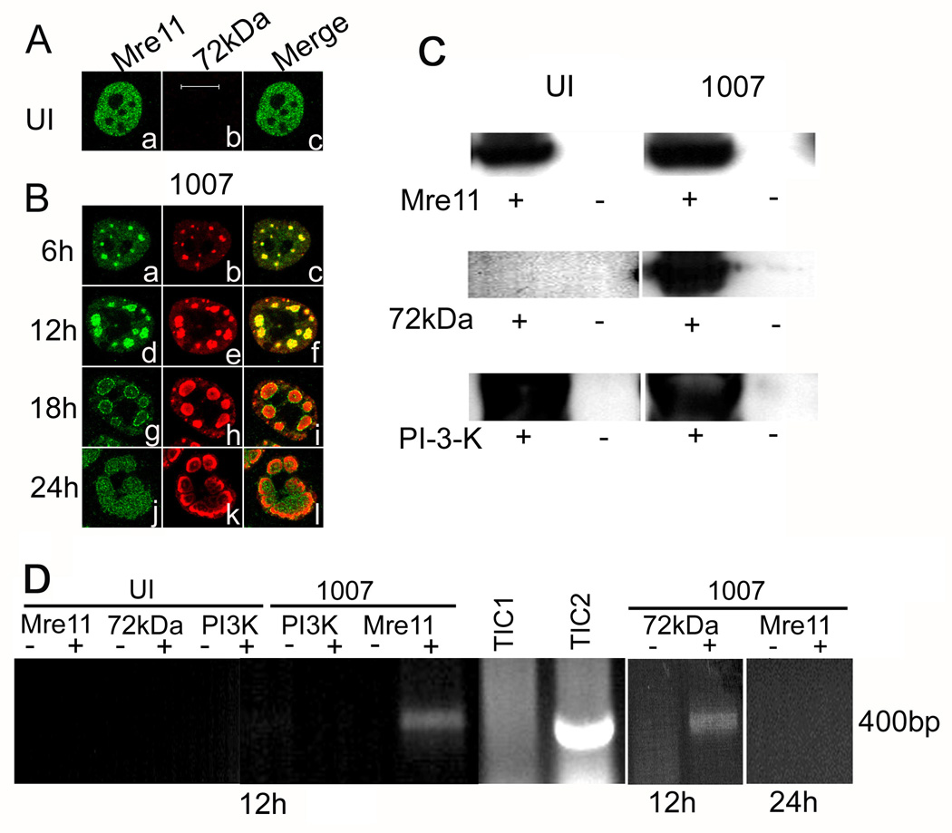
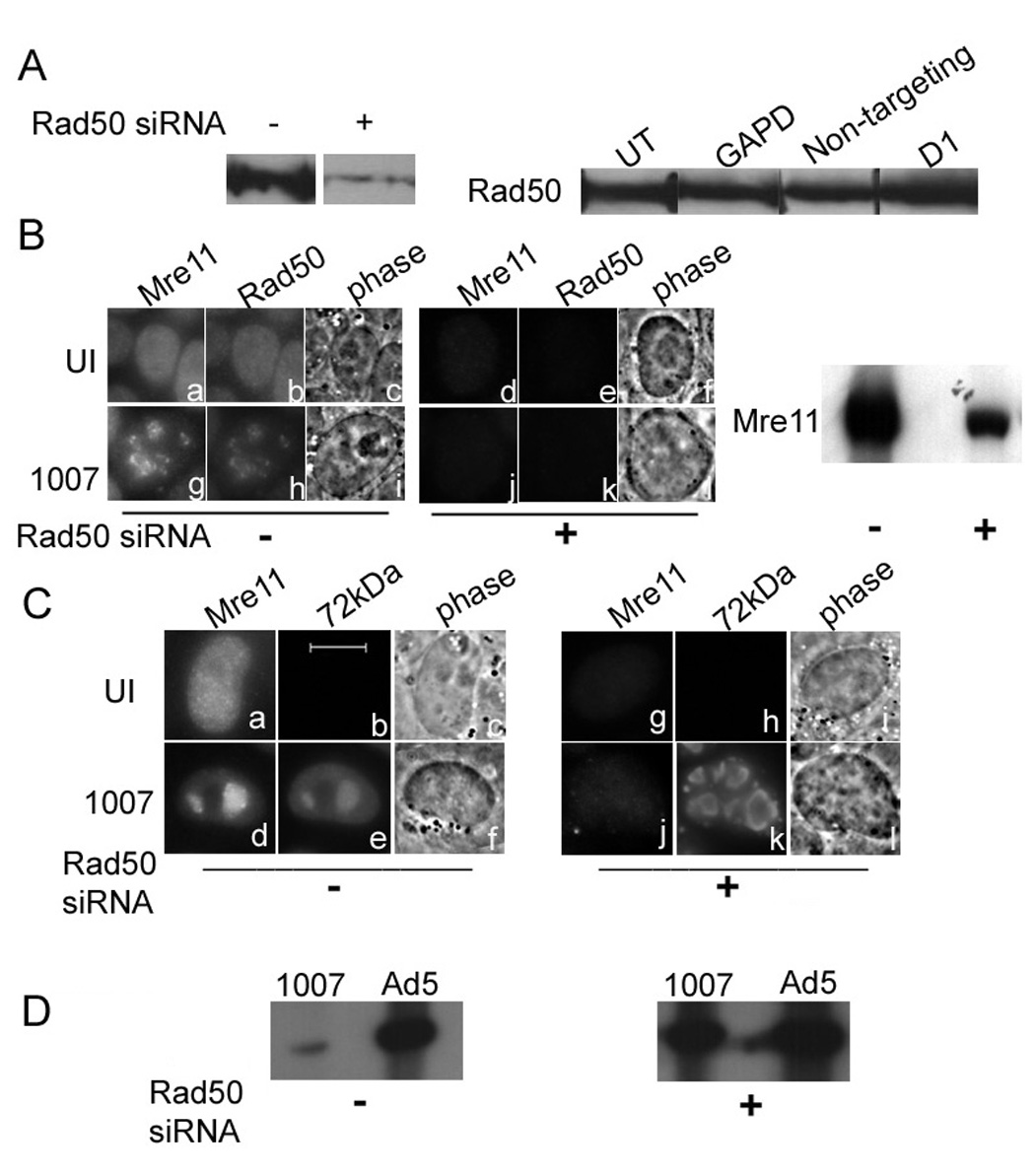
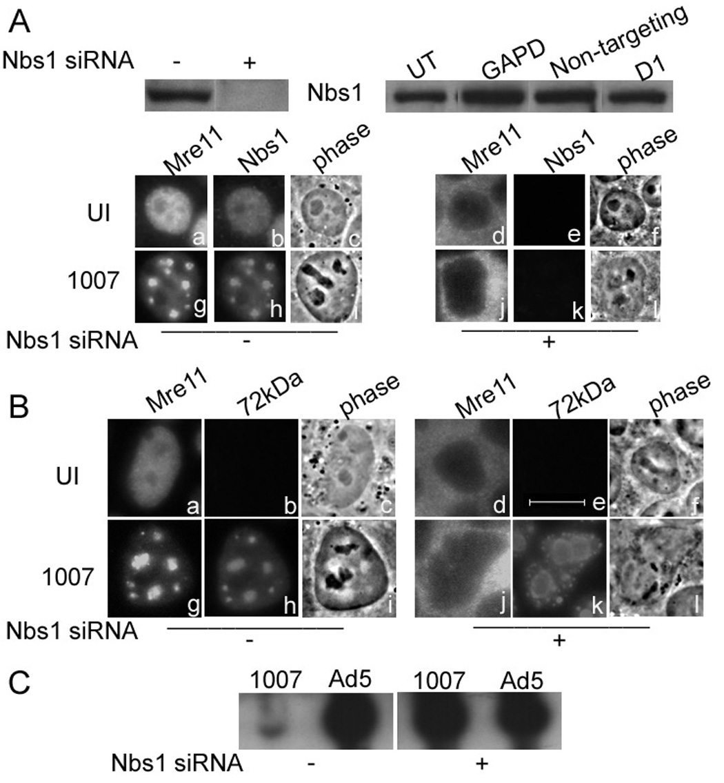
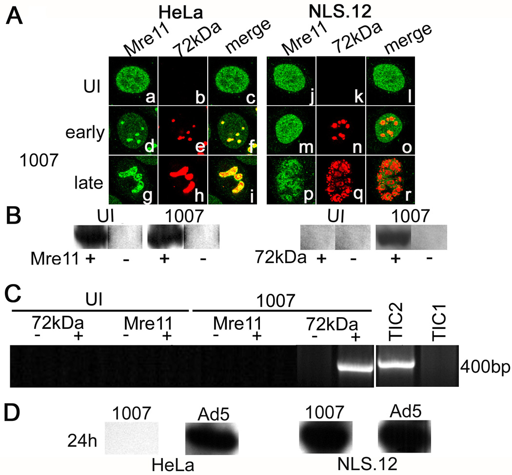
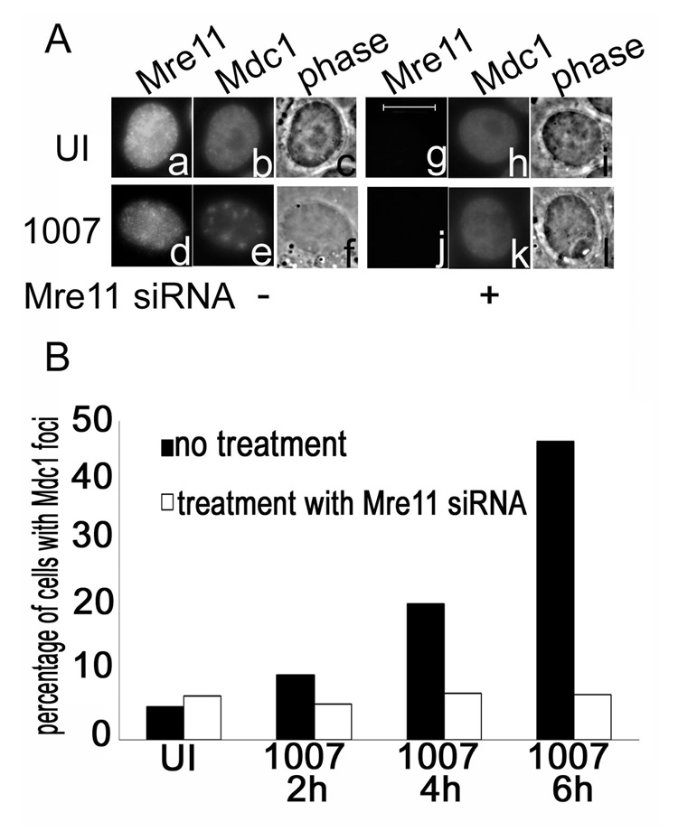
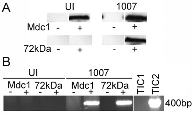
Adenovirus (Ad) infections stimulate the activation of cellular DNA damage response and repair pathways. Ad early regulatory proteins prevent activation of DNA damage responses by targeting the MRN complex, composed of the Mre11, Rad50 and Nbs1 proteins, for relocalization and degradation. In the absence of these viral proteins, Mre11 colocalizes with viral DNA replication foci. Mre11 foci formation at DNA damage induced by ionizing radiation depends on the Nbs1 component of the MRN complex and is stabilized by the mediator of DNA damage checkpoint protein 1 (Mdc1). We find that Nbs1 is required for Mre11 localization at DNA replication foci in Ad E4 mutant infections. Mre11 is important for Mdc1 foci formation in infected cells, consistent with its role as a sensor of DNA damage. Chromatin immunoprecipitation assays indicate that both Mre11 and Mdc1 are physically bound to viral DNA, which could account for their localization in viral DNA containing foci. Efficient binding of Mre11 to E4 mutant DNA depends on the presence of Nbs1, and is correlated with a significant E4 mutant DNA replication defect. Our results are consistent with a model in which physical interaction of Mre11 with viral DNA is mediated by Nbs1, and interferes with viral DNA replication.


Figures

Figure 1
Figure 1. Mre11 localizes to E2-72kDa containing viral replication foci in E4 mutant infection and it is bound to E4 mutant DNA
HeLa cells were uninfected (UI) or infected with H5dl1007 (1007) at 30 FFU/cell for the times indicated. (A) and (B) Immunofluorescence staining was performed with antibodies specific for Mre11 (1A, panel a; 1B, panels a, d, g, and j) and the viral E2-72kDa protein (1A, panel b; 1B, panels b, e, h, and k). Bar 10µm. Uninfected cells and cells infected for 12 or 24 h with H5dl1007 were treated with formaldehyde and used for chIP experiments as described in materials and methods. (C) Western blotting was performed to confirm immunoprecipitation of Mre11, E2-72kDa and PI3K with their respective antibodies; representative blots are shown. Lanes from samples that were immunoprecipitated with specific Ab or mock immunoprecipitated are labeled (+) and (−), respectively. (D) PCR amplification using primers specific to the E1b region was performed on chromatin samples prepared from uninfected and H5dl1007 infected cells that were immunoprecipitated (+) with Mre11, PI3K, or E2-72kDa Abs as indicated, or mock immunoprecipitated in parallel without the addition of Ab (−). PCR reactions were fractionated on a 1% agarose gel and stained with ethidium bromide to visualize the 400bp expected PCR product. Total input chromatin (TIC) samples prepared from UI (TIC1) and H5dl1007 infected cells (TIC2) were included to indicate input DNA levels. ChIP experiments with the Mre11 and E2-72kDa antibodies were performed 3 times with similar results. ChIP experiments with Mre11 and PI3K antibodies were performed twice with similar results.
Figure 2
Figure 2. Rad50 is important for Mre11 stability and the inhibition of E4 mutant DNA replication
HeLa cells were transfected with control siRNA or Rad50 siRNA prior to infection with Ad5 or H5dl1007 at 3 FFU/cell for 24h. (A) Rad50 knockdown was monitored by western blotting of 75µg of total protein prepared 96 hours after mock (−) or Rad50 specific siRNA (+) transfection, using rabbit polyclonal Ab against Rad50. Additional controls demonstrating the specificity of Rad50 expression knockdown included no treatment (UT), non-targeting siRNA (non targeting), siRNA against GAPD, and treatment with the transfection reagent alone (D1). (B) Mre11 and Rad50 distribution in untransfected uninfected (panels a to c) and cells infected with H5dl1007 (panels g to i) for 24 hpi are shown. The distribution of Mre11 and Rad50 in siRNA transfected HeLa cells that were either uninfected or infected for 24 h with H5dl1007 are shown in panels d to f and j to l respectively. A western blot depicting levels of Mre11 in Rad50 siRNA treated cells is also shown (right panel). (C) Panels a to f show the distribution of host Mre11 and viral E2-72kDa in uninfected and H5dl1007 infected cells in the absence of siRNA transfection. Panels g to l show the distribution of Mre11 and E2-72kDa in cells transfected with Rad50 siRNA that were subsequently uninfected or infected with H5dl1007. Bar 10µm. (D) Levels of viral DNA synthesis in Ad5 and H5dl1007 infected HeLa with (+) and without (−) Rad50 siRNA transfection were quantified by Southern analysis of 10µg of Eco RI digested total DNA prepared at 24 hpi. The Eco R1 C fragment from the DNA digestion was used for comparison between Ad5 and H5dl1007.
Figure 3
Figure 3. Nbs1 is important for Mre11 localization and the inhibition of E4 mutant DNA replication
HeLa cells were transfected with control siRNA or Nbs1 siRNA prior to infection with Ad5 or H5dl1007 at 3 FFU/cell for 24h. (A) Nbs1 knockdown was monitored by western blotting using 75µg of total protein prepared 96 hours after mock (−) or Nbs1 specific siRNA (+) transfection, using goat polyclonal Ab against Mre11. Additional controls demonstrating the specificity of Nbs1 expression knockdown included no treatment (UT), non-targeting siRNA (non targeting), siRNA against GAPD, and treatment with the transfection reagent alone (D1). Mre11 and Nbs1 distribution in untransfected, uninfected cells (panels a to c) and cells infected with H5dl1007 (Panels g to i) for 24 h are shown. The distribution of Mre11 and Nbs1 in siRNA transfected HeLa cells that were either uninfected or infected for 24 h with H5dl1007 are shown in panels d to f and j to l respectively. (B) Mre11 and viral E2-72kDa protein distribution in untransfected cells that were either uninfected (panels a–c) or infected (panels g to I) are shown. The distribution of Mre11 and E2-72kDa distribution in Nbs1 siRNA transfected cells that were either uninfected or infected with H5dl1007 is shown in panels d to f and j to l, respectively. Bar 10µm. (C) Levels of viral DNA synthesis in Ad5 and H5dl1007 infected HeLa with (+) and without (−) Nbs1 siRNA transfection were quantified by Southern analysis of 10µg of Eco RI digested total DNA prepared at 24 hpi. The C fragment from the DNA digestion was used to compare Ad5 and H5dl1007 DNA levels.
Figure 4
Figure 4. Nbs1 is important for Mre11 localization in E4 mutant replication centers and Mre11 binding to viral DNA
(A) HeLa and NLS.12 cells were infected with H5dl1007 for 10 h (early) and 24 h (late) at 3 FFU/cell. Confocal microscopy was used to analyze the distribution of host Mre11 (panels a,d,g,j,m,p) and viral E2-72kDa (panels b,e,h,k,n,q) proteins in uninfected and infected cells. Merged images of these staining patterns are shown in panels c, f, i, and l, o, r. ChIP experiments were performed as described in Fig. 1 using samples prepared from NLS.12 cells that were uninfected or infected with H5dl1007 for 12h at 30FFU/cell. (B) Western blotting was performed to confirm immunoprecipitation of Mre11 and E2-72kDa from chromatin prepared from H5dl1007 infected and uninfected NLS.12 cells, in preparation for chIP analysis. Lanes from samples that were immunoprecipitated with specific Ab or mock immunoprecipitated are labeled (+) and (−), respectively. (C) PCR amplification using primers specific to the E1b region was performed on UI and H5dl1007 infected samples prepared at 12 hpi and immunoprecipitated with Mre11 or E2-72kDa. PCR products were analyzed as described in Fig. 1. Lanes from samples that were immunoprecipitated with specific Ab or mock immunoprecipitated in parallel are labeled (+) and (−), respectively. Total input chromatin samples from uninfected (TIC1) and H5dl1007-infected (TIC2) cells were included to indicate input DNA levels. The chIP experiments were performed twice with similar results. (D) Levels of viral DNA synthesis in HeLa and NLS.12 cells infected with Ad5 and H5dl1007 at 3FFU/cellwere quantified by Southern analysis of 10µg of Eco RI digested total DNA prepared at 24 hpi. The C fragment from the DNA digestion was used for comparison between Ad5 and H5dl1007.
Figure 5
Figure 5. Mre11 is required for the formation of Mdc1 foci in response to E4 mutant infections
HeLa cells were transfected with control siRNA or Mre11 siRNA prior to infection with H5dl1007 at 3 FFU/cell for 24h. (A) Immunofluorescence staining was performed to analyze the distribution of Mre11 and Mdc1. Panels a to f show Mre11 and Mdc1 distribution in untransfected HeLa cells that were either uninfected (a–c) or infected with H5dl1007 (d–f). Panels g to l show Mre11 and Mdc1 distribution in Mre11 siRNA transfected cells that were either uninfected (g–i) or infected with H5dl1007 (j–l). Bar 10µm. Immunostained cells from the time course were scored blind for Mdc1 foci formation in the presence and absence of Mre11. The graph presented in (B) shows the percentage of cells with Mdc1 foci in uninfected and infected cells prepared at the times indicated, that were untransfected (black bars) or transfected (white bars) with Mre11 siRNA.
Figure 6
Figure 6. Mdc1 binds to E4 mutant viral DNA
HeLa cells were infected with H5dl1007 at 30 FFU/cell for 6h and processed for chIP as described in Fig. 1. (A) Western blotting was performed to confirm immunoprecipitation of Mdc1 and E2-72kDa and representative blots are shown. Lanes from samples that were immunoprecipitated with specific Ab or mock immunoprecipitated are labeled (+) and (−), respectively. (B) PCR amplification using primers specific to the E1b region was performed on uninfected (UI) and H5dl1007 (1007) infected samples immunoprecipitated with Mdc1 or E2-72kDa Abs (+) or mock immunoprecipitated in parallel (−). PCR products were analyzed as described in Fig. 1. Total input chromatin (TIC) samples were included to indicate input DNA levels in uninfected (TIC1) and H5dl1007 (TIC2) infected cells. The Mdc1 chIP experiment was performed 3 times with similar results.

References

    1. Aspegren A, Rabino C, Bridge E. Organization of splicing factors in adenovirus-infected cells reflects changes in gene expression during the early to late phase transition. Exp. Cell Res. 1998;245:203–213. - PubMed
    1. Assenmacher N, Hopfner KP. MRE11/RAD50/NBS1: complex activities. Chromosoma. 2004;63:631–638. - PubMed
    1. Bridge E, Ketner G. Redundant control of adenovirus late gene expression by early region 4. J. Virol. 1989;63:631–638. - PMC - PubMed
    1. Carney JP, Maser RS, Olivares H, Davis EM, Le Beau M, Yates JR, 3rd, Hays L, Morgan WF, Petrini JH. The hMre11/hRad50 protein complex and Nijmegen breakage syndrome: linkage of double strand break repair to the cellular DNA damage response. Cell. 1998;93:477–486. - PubMed
    1. Carson CT, Schwartz RA, Stracker TH, Lilley CE, Lee DV, Weitzman MD. The Mre11 complex is required for ATM activation and the G2/M checkpoint. EMBO J. 2003;22:6610–6620. - PMC - PubMed

Publication types