. 2008 Apr;82(7):3574-83.
doi: 10.1128/JVI.02038-07. Epub 2008 Jan 30.

A dormant internal ribosome entry site controls translation of feline immunodeficiency virus

Affiliations

A dormant internal ribosome entry site controls translation of feline immunodeficiency virus

Valentina Camerini et al. J Virol. 2008 Apr.

Abstract

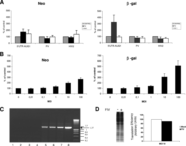
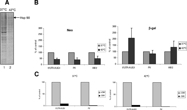
The characterization of internal ribosome entry sites (IRESs) in virtually all lentiviruses prompted us to investigate the mechanism used by the feline immunodeficiency virus (FIV) to produce viral proteins. Various in vitro translation assays with mono- and bicistronic constructs revealed that translation of the FIV genomic RNA occurred both by a cap-dependent mechanism and by weak internal entry of the ribosomes. This weak IRES activity was confirmed in feline cells expressing bicistronic RNAs containing the FIV 5' untranslated region (UTR). Surprisingly, infection of feline cells with FIV, but not human immunodeficiency virus type 1, resulted in a great increase in FIV translation. Moreover, a change in the cellular physiological condition provoked by heat stress resulted in the specific stimulation of expression driven by the FIV 5' UTR while cap-dependent initiation was severely repressed. These results reveal the presence of a "dormant" IRES that becomes activated by viral infection and cellular stress.


Figures

FIG. 1.
FIG. 1.
FIV translation is cap dependent in the RRL. Capped and uncapped FIV-Gag and FIV-LacZ transcripts (schematically represented on the upper part of each panel) were translated in the RRL at various RNA concentrations, as indicated. After 45 min of incubation at 30°C in the RRL, the samples were analyzed by 12% SDS-PAGE and the dried gel was submitted to autoradiography. The shorter Gag isoforms (a, b, and c) and the molecular weight markers are indicated. Data are representative of at least three experiments. MW, molecular mass.
FIG. 2.
FIG. 2.
The FMDV L protease inhibits translation driven by the FIV 5′ UTR. (A) The RRL was preincubated for 10 min without (lanes 1, 4, 7, and 10) or with 0.4 μl (lanes 2, 5, 8, and 11) or 0.6 μl (lanes 3, 6, 9, and 12) of in vitro-expressed FMDV L protease. Different capped RNA transcripts, namely, FIV-Gag (100 ng), FIV-LacZ (100 ng), globin-lacZ (10 ng), and EMCV-LacZ (100 ng), were translated. After 45 min of incubation at 30°C in the RRL, the samples were analyzed by 12% SDS-PAGE and the dried gel was submitted to autoradiography. The relative intensities of the bands were quantified, and the results, expressed as percentages of the control (no protease added), are presented in the histogram at the bottom of each panel. The Gag isoforms are indicated by the arrows (a, b, and c). Data are representative of at least three experiments. (B) At the end of the 10-min preincubation period, samples (1 μl) from the experiment described above were subjected to 10% SDS-PAGE and the proteins were transferred to polyvinylidene difluoride membrane and incubated with antibodies specific to the C-terminal part of eIF4GI. The positions of the intact molecule and the cleavage products (Cp) are indicated on the right. MW, molecular mass.
FIG. 3.
FIG. 3.
In vitro translation driven by the FIV 5′ UTR is inefficient in a bicistronic vector. (A) Schematic diagram of the constructs used. (B) Capped and uncapped monocistronic transcripts together with uncapped bicistronic transcripts containing the EMCV IRES (left panel), the FIV 5′ UTR alone (lanes 7 to 10, 15, and 16), or the FIV 5′ UTR followed by the gag coding region (lanes 11 to 14, 17, and 18) were translated in the RRL at two different concentrations (23 and 29 nM) as indicated. After 45 min of incubation at 30°C, the samples were processed by 12% SDS-PAGE and submitted to autoradiography. Data are representative of at least three experiments. MW, molecular mass.
FIG. 4.
FIG. 4.
The FIV 5′ UTR has genuine IRES activity. (A) Increasing concentrations (10 and 25 μM, denoted by the triangle) of 2′-O-methyloligoribonucleotides that are complementary to the region downstream of AUG1 or of the PBS region were annealed to 0.3 pmol of capped FIV-Gag RNA or (B) uncapped bicistronic transcript containing the FIV 5′ UTR followed by a segment of the gag coding region. The position of annealing of the 2′-O-methyloligoribonucleotides on the two transcripts is schematically represented at the top of each panel. After 45 min of incubation at 30°C in the RRL, the samples were processed by 12% SDS-PAGE and submitted to autoradiography. (C) The resulting oligonucleotide-mRNA duplex that is formed upon hybridization of the 2′-O-methyloligoribonucleotides in panel A was visualized on a nondenaturing agarose gel. MW, molecular mass.
FIG. 5.
FIG. 5.
The FIV 5′ UTR is inefficient at supporting internal initiation in CrFK cells. (A) Comparative analysis of β-gal activity expressed from the CMV and ΔCMV pBi-5′UTR-AUG1 and pBi-5′UTR-AUG4 plasmids that were transiently transfected into CrFK cells. Results are expressed as percentages of the β-gal activity from each parental plasmid (containing the CMV promoter and set at 100%). (B) Agarose nondenaturing electrophoretic analysis of 5 μg of total cytoplasmic RNA extracted from CrFK cells stably expressing Bi-AUG1 and Bi-AUG4 after RT and PCR (lanes 4 and 7) or PCR without prior RT (lanes 3 and 6) or RT-PCR without RNAs (lane 1). PCR products from the parental plasmids pBi-5′UTR-AUG1 and pBi-5′UTR-AUG4 (lanes 2 and 5) were run in parallel as size markers. The region amplified by PCR is schematically depicted at the top of the panel. (C) Translational activities of the bicistronic constructs pBi-5′UTR-AUG1, pBi-5′UTR-AUG4, pBi-PV, and pBi-HIV2 that were stably transfected into CrFK cells. The results are expressed as the ratio of β-gal (second gene) to neomycin (first gene) activities and compared to the activity of the positive control, pBi-PV, which was arbitrary set to 100%.
FIG. 6.
FIG. 6.
FIV, but not HIV-1, infection enhances translational activity from its cognate IRES. (A) CrFK cells expressing the bicistronic construct pBi-5′UTR-AUG1, pBi-PV, or pBi-HIV2 were infected with equal amounts of VSV-G-pseudotyped FIV and HIV-1 at an MOI of 10 (see Materials and Methods). At 48 h postinfection, neomycin (left panel) and β-gal (right panel) activities in mock-infected (gray), FIV-infected (black), or HIV-1-infected (white) cells were measured. The Neo and β-gal activities in mock-infected cells were arbitrarily set to 100%. (B) CrFK cells expressing the pBi-5′UTR-AUG1 construct were infected with VSV-G-pseudotyped FIV virions at MOIs ranging from 0.01 to 100, as indicated. At 48 h postinfection, Neo and β-gal activities were determined and plotted as percentages of the control (mock-infected cells, set at 100%). (C) Agarose nondenaturing electrophoretic analysis of 5 μg of total cytoplasmic RNA extracted from CrFK cells stably expressing Bi-AUG1 and infected with FIV at MOIs of 0 (lanes 2 and 5), 10 (lanes 3 and 6), and 100 (lanes 4 and 7). RNA were analyzed after RT and PCR (lanes 5, 6, and 7) or PCR without prior RT (lanes 2, 3, and 4) or RT-PCR without RNAs (lane 1). PCR products from the parental plasmid pBi-5′UTR-AUG1 (lane 8) were run in parallel as size markers. Molecular size markers were run at the right side of the gel, and the sizes of the bands are indicated in kilobases. (D) CrFK cells stably transfected with bicistronic plasmid pBi-5′UTR-AUG1 were infected with pseudotyped FIV particles (MOI = 10). The effect on the overall cellular translation was analyzed by incubating the cells with [35S]methionine for 1 h. Cell extracts were processed by SDS-PAGE followed by autoradiography. The intensities of the bands, corresponding to global protein synthesis, were quantified, and the results, expressed as percentages of the noninfected cells, are presented in the histogram on the right of the autoradiogram.
FIG. 7.
FIG. 7.
Prolonged heat shock reveals FIV IRES activity. CrFK cells stably expressing bicistronic plasmids pBi-AUG1, pBi-PV, and pBi-HIV2 (see Fig. 5) were exposed to heat shock at 42°C for 15 h. (A) The effect of heat shock on cellular translation was quantified by labeling the cells with [35S]methionine for 1 h. Cell extracts were then processed by SDS-PAGE followed by autoradiography. The position of heat shock protein Hsp90 is indicated. (B) Analysis of the effects of heat shock on neomycin and β-gal activities (left and right panels, respectively) for each of the constructs described above. The results are expressed as percentages of the activity at 37°C. (C) Activity of β-gal expressed by bicistronic vectors pBi-AUG1 and pBi-PV with or without a CMV promoter at 37°C or 42°C. Results are expressed as percentages of β-gal activity.

References

    1. Balvay, L., M. L. Lastra, B. Sargueil, J.-L. Darlix, and T. Ohlmann. 2007. Translational control of retroviruses. Nat. Rev. Microbiol. 5128-140. - PMC - PubMed
    1. Belsham, G. J., and N. Sonenberg. 2000. Picornavirus RNA translation: roles for cellular proteins. Trends Microbiol. 8330-335. - PubMed
    1. Bendinelli, M., M. Pistello, S. Lombardi, A. Poli, C. Garzelli, D. Matteucci, L. Ceccherini-Nelli, G. Malvaldi, and F. Tozzini. 1995. Feline immunodeficiency virus: an interesting model for AIDS studies and an important cat pathogen. Clin. Microbiol. Rev. 887-112. - PMC - PubMed
    1. Berlioz, C., and J.-L. Darlix. 1995. An internal ribosomal entry mechanism promotes translation of murine leukemia virus gag polyprotein precursors. J. Virol. 692214-2222. - PMC - PubMed
    1. Brasey, A., M. Lopez-Lastra, T. Ohlmann, N. Beerens, B. Berkhout, J.-L. Darlix, and N. Sonenberg. 2003. The leader of human immunodeficiency virus type 1 genomic RNA harbors an internal ribosome entry segment that is active during the G2/M phase of the cell cycle. J. Virol. 773939-3949. - PMC - PubMed

LinkOut - more resources