Comparative Study
. 2008 Jan 30;28(5):1109-17.
doi: 10.1523/JNEUROSCI.4853-07.2008.

The proneural basic helix-loop-helix gene ascl1a is required for retina regeneration

Affiliations
Comparative Study

The proneural basic helix-loop-helix gene ascl1a is required for retina regeneration

Blake V Fausett et al. J Neurosci. .

Abstract

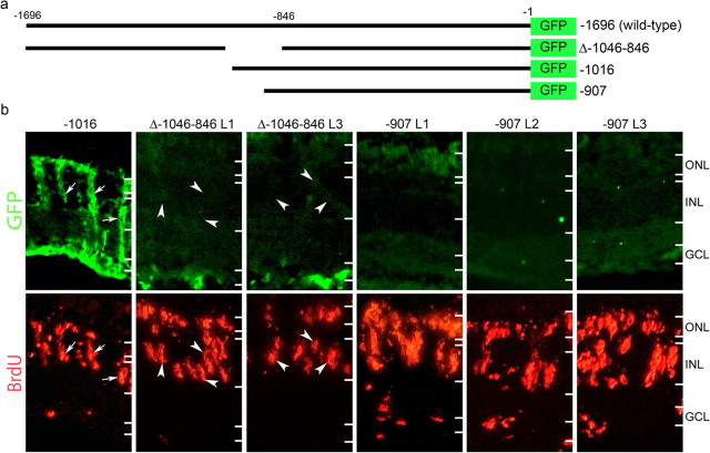
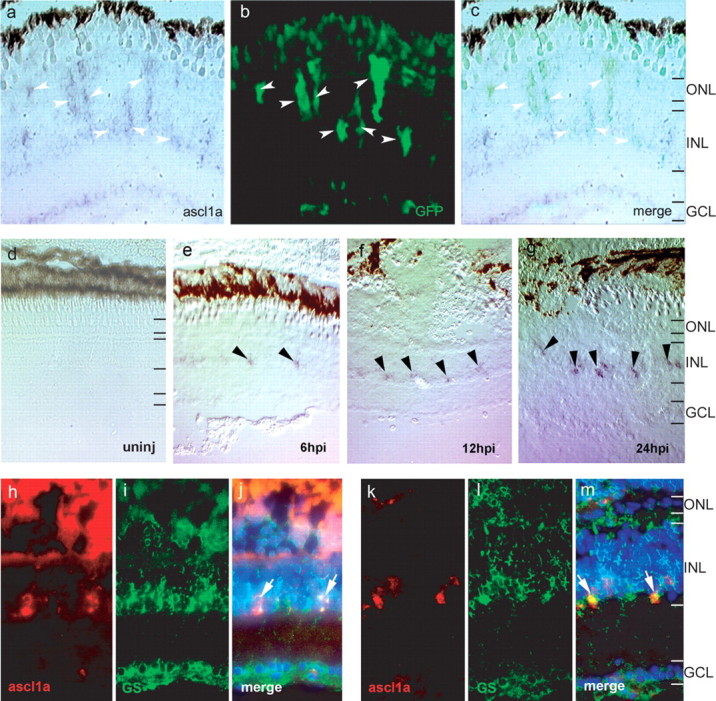
Unlike mammals, teleost fish can regenerate an injured retina, restoring lost visual function. Little is known of the molecular events that underlie retina regeneration. We previously found that in zebrafish, retinal injury stimulates Müller glia to generate multipotent alpha1-tubulin (alpha1T) and pax6-expressing progenitors for retinal repair. Here, we report the identification of a critical E-box in the alpha1T promoter that mediates transactivation by achaete-scute complex-like 1a (ascl1a) during retina regeneration. More importantly, we show that ascl1a is essential for retina regeneration. Within 4 h after retinal injury, ascl1a is induced in Müller glia. Knockdown of ascl1a blocks the induction of alpha1T and pax6 as well as Müller glial proliferation, consequently preventing the generation of retinal progenitors and their differentiated progeny. These data suggest ascl1a is required to convert quiescent Müller glia into actively dividing retinal progenitors, and that ascl1a is a key regulator in initiating retina regeneration.


Figures

Figure 1.
Figure 1.
A 109 bp region of the α1T promoter is required for transgene expression in dedifferentiating Müller glia. a, Schematic representation of α1T promoter constructs. The bars represent promoter sequence, and the numbers indicate relative position from the start codon. −1696 is the wild-type promoter described previously (Hieber et al., 1998; Goldman et al., 2001). Δ-1046–846 has been described previously (Goldman and Ding, 2000). The −1016 promoter directs transgene expression in Müller glia (Fausett and Goldman, 2006). The −907 promoter lacks 789 bp of upstream sequence. b, Transgenic fish received retinal injuries on day 0 and were given a 4 h pulse of BrdU at 4 dpi. Transgenic fish, which carry the required DNA element, express GFP in BrdU-labeled Müller glia (-1016, arrows), whereas transgenic fish lacking the element do not (Δ-1046–846 and −907). Two independent lines of Δ-1046–846 and three independent lines of −907 transgenic fish all display a lack of GFP expression in BrdU-labeled cells (arrowheads in Δ-1046–846). The images for −1016 and Δ-1046–846 are from the same sections. Because the −907 transgenic fish display very weak GFP expression in general, we used serial sections to obtain the −907 images. GCL, Ganglion cell layer.
Figure 2.
Figure 2.
An E-box within the 109 bp region that is required for transgene expression in Müller glia binds nuclear extracts from zebrafish and rat brain and zebrafish retina. a, Electrophoretic mobility shift assay using a probe from the 109 bp region binds nuclear extracts from zebrafish and rat brain. The arrow indicates specific binding. Cold indicates where 50-fold molar excess unlabeled probe was added as competition. Extract indicates whether zebrafish (zf) or rat brain extracts were added. b, Nucleotide sequence of the probes used for electrophoretic mobility shift assay. The E-box is outlined in probe 4 with a box. Mutations are indicated by italicized and underlined text. c, Mutations to the E-box (lanes 3 and 4) render the probe unable to bind nuclear extracts from zebrafish brain, whereas mutations to non-E-box nucleotides do not affect binding (lanes 1, 2, and 5). Probes correspond to those shown in b. Unlabeled mutant probes compete with wild-type probe binding when the E-box is intact (lanes 6, 7, and 10), but not when the E-box is mutated (lanes 8 and 9), even at 50-fold molar excess. d, Nuclear extracts from zebrafish retina bind specifically to the E-box. e, An E-box probe from a different region of the promoter (Eb) is unable to compete with the E-box from probe 4 (lane 3) and does not bind to zebrafish brain nuclear extracts (lane 4).
Figure 3.
Figure 3.
Mutation of −954 E-box in the wild-type −1696 α1T promoter prevents transgene induction in proliferating Müller glia after retina injury. a, Schematic representation of α1T promoter:GFP constructs. The wild-type −1696α1T:GFP transgene (-1696) and the 2 bp mutation created in the E-box at position −954 in the −1696 construct (TG-954CA) are indicated. b, Three independent lines of transgenic fish received retinal injuries on day 0 and were given a 4 h pulse of BrdU at 4 dpi. Note that none of the lines harboring the 2 bp mutation induced transgene GFP expression in BrdU-labeled Müller glia (arrowheads). See Figure 1 for comparison with the wild-type −1696α1T:GFP transgene response. The arrows in the lowest right GFP panel show transgene induction in retinal ganglion cells, the axons of which were presumably damaged when we injured the retina in this fish.
Figure 4.
Figure 4.
Ascl1a is induced in proliferating Müller glia after retinal injury. a–c, Ascl1a expression is detected by in situ hybridization (a) in GFP+ Müller glia (b) at 4 dpi (arrowheads). c shows the merge image of a and b. d–g, In situ hybridization for ascl1a from 6–24 hpi. Ascl1a is not expressed in the uninjured retina (d). Ascl1a is induced in cells of the INL at 6 hpi (e, arrowheads). Ascl1a expression gradually increases and is easily detected at 24 hpi (g, arrowheads). h–m, Ascl1a in situ hybridization (red, h, k) and glutamine synthetase immunostaining (green, i, l) show ascl1a is induced in Müller glia at 6 hpi (arrows in merge, j) and 24 hpi (arrows in merge, m). DAPI nuclear staining is shown in the merged panels. GCL, Ganglion cell layer.
Figure 5.
Figure 5.
Ascl1a regulates the α1T promoter through the −954 E-box. a, Luciferase reporter vectors with either a minimal β-globin promoter alone (light gray bars) or three copies of the −954 E-box (dark gray bars) or the mutant E-box from probe 4–4 (see Fig. 2b) (black bars) were transfected in combination with ascl1a into HEK293T cells. Ascl1a transactivates the reporter when a functional E-box is present but not when the E-box is mutated. b, Full-length α1T constructs harboring either the wild-type (-1696) or mutant E-box promoters were transfected into HEK293T cells with or without ascl1a. Ascl1a induces reporter expression when the E-box is intact, but not when the E-box is mutated. Error bars indicate SEM for three replicates.
Figure 6.
Figure 6.
Ascl1a is required for transgene expression in vivo. −1016 transgenic fish received retinal injuries and morpholino treatment on day 0. Eyes were harvested on day 4. a, Control morpholino treatment has no effect on transgene expression in Müller glia (arrows), and there are many GFP+ Müller glia (arrowheads). Morpholinos targeting ascl1a (ascl1a and ascl1a5′UTR panels) prevent transgene expression in treated cells (arrows) and cause reduced transgene expression in general, although Müller glia that did not receive ascl1a morpholinos are able to express GFP (arrowheads). GCL, Ganglion cell layer.
Figure 7.
Figure 7.
Ascl1a knockdown prevents induction of endogenous α1T and pax6. Retinas from −1016 transgenic fish were injured and electroporated with lissamine-labeled MOs on day 0 and harvested on day 4. a–f, α1T expression detected by ISH is shown as a dark brown deposit (a, d), native GFP expression is shown in green (b, e), and lissamine-labeled MO is shown in red (b, e). The injury site is marked by an asterisk. a–c, Control MO treatment does not affect endogenous or transgene α1T induction (arrows). d–f, Ascl1a knockdown prevents endogenous and transgene α1T induction. Note the lack of α1T and GFP expression between the asterisk and the arrows, where the retina is treated with MO. Where the retina did not receive MO, α1T and GFP are expressed (arrows). g–l, Pax6 expression detected by ISH is shown as a blue/purple deposit, native GFP expression is shown in green, and MO is shown in red. g–i, Control MO treatment does not affect pax6 induction at 4 dpi. Arrows indicate pax6+/MO+/GFP+ Müller glia. j–l, Ascl1a MO treatment prevents pax6 induction. Arrowheads indicate MO+/pax6 cells. Rare GFP+ cells are sometimes present but were not treated with MO (arrow).
Figure 8.
Figure 8.
Ascl1a is required for proliferation of Müller glia. Morpholinos were electroporated into injured −1016 retinas on day 0 and fish were housed in BrdU-treated water from 36 to 60 h after injury to label dividing cells. The fish recovered until day 10 when eyes were harvested. The control morpholino does not prevent treated cells from labeling with BrdU (arrowheads; top 3 control panels). Morpholinos targeting ascl1a (ascl1a and ascl1a5′UTR) prevent cells from labeling with BrdU (arrows; ascl1a and ascl1a5′UTR panels). Some cells are able to proliferate but were not treated with morpholino (arrowheads; ascl1a and ascl1a5′UTR panels). GCL, Ganglion cell layer.

References

    1. Akagi T, Inoue T, Miyoshi G, Bessho Y, Takahashi M, Lee JE, Guillemot F, Kageyama R. Requirement of multiple basic helix-loop-helix genes for retinal neuronal subtype specification. J Biol Chem. 2004;279:28492–28498. - PubMed
    1. Barthel LK, Raymond PA. In situ hybridization studies of retinal neurons. Methods Enzymol. 2000;316:579–590. - PubMed
    1. Bernardos RL, Barthel LK, Meyers JR, Raymond PA. Late-stage neuronal progenitors in the retina are radial Müller glia that function as retinal stem cells. J Neurosci. 2007;27:7028–7040. - PMC - PubMed
    1. Bray S. Notch signalling in Drosophila: three ways to use a pathway. Semin Cell Develop Biol. 1998;9:591–597. - PubMed
    1. Cameron DA, Gentile KL, Middleton FA, Yurco P. Gene expression profiles of intact and regenerating zebrafish retina. Mol Vis. 2005;11:775–791. - PubMed

Publication types

MeSH terms