. 2008 Apr;99(4):1581-9.
doi: 10.1152/jn.01396.2007. Epub 2008 Jan 30.

Cutaneous sensory neurons expressing the Mrgprd receptor sense extracellular ATP and are putative nociceptors

Affiliations

Cutaneous sensory neurons expressing the Mrgprd receptor sense extracellular ATP and are putative nociceptors

G Dussor et al. J Neurophysiol. 2008 Apr.

Abstract

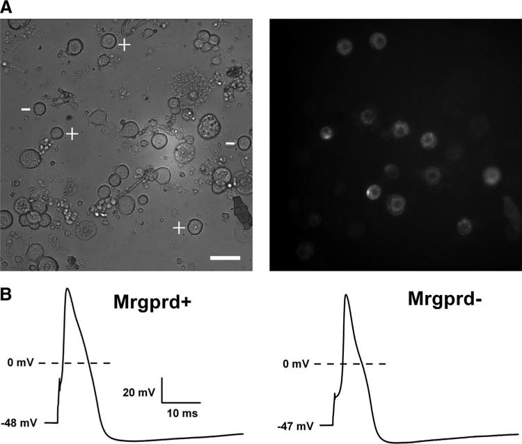
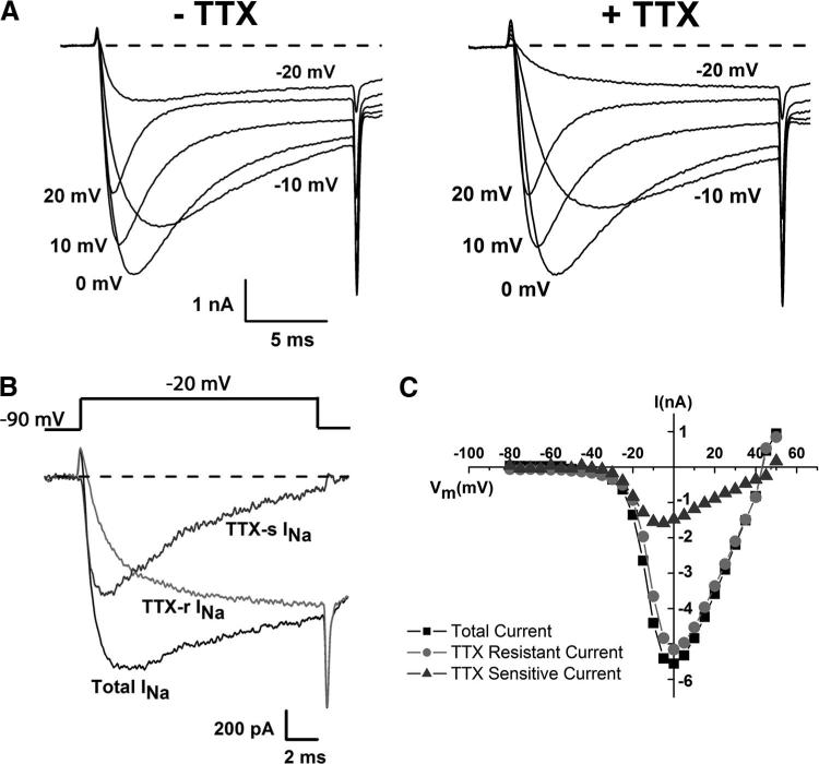
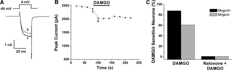
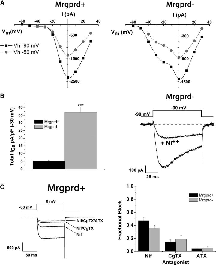
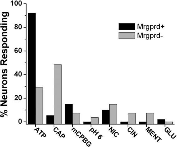
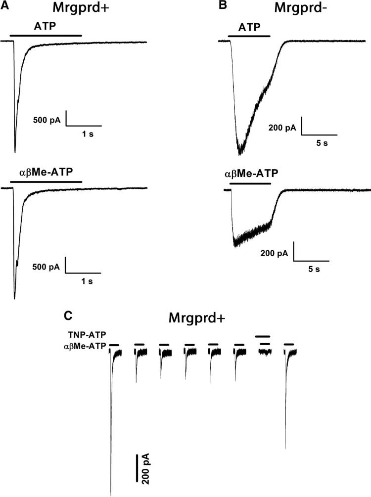
Sensory neurons expressing the Mrgprd receptor are known to innervate the outermost living layer of the epidermis, the stratum granulosum. The sensory modality that these neurons signal and the stimulus that they respond to are not established, although immunocytochemical data suggest they could be nonpeptidergic nociceptors. Using patch clamp of dissociated mouse dorsal root ganglion (DRG) neurons, the present study demonstrates that Mrgprd+ neurons have several properties typical of nociceptors: long-duration action potentials, TTX-resistant Na(+) current, and Ca(2+) currents that are inhibited by mu opioids. Remarkably, Mrgprd+ neurons respond almost exclusively to extracellular ATP with currents similar to homomeric P2X3 receptors. They show little or no sensitivity to other putative nociceptive agonists, including capsaicin, cinnamaldehyde, menthol, pH 6.0, or glutamate. These properties, together with selective innervation of the stratum granulosum, indicate that Mrgprd+ neurons are nociceptors in the outer epidermis and may respond indirectly to external stimuli by detecting ATP release in the skin.


Figures

FIG. 1
FIG. 1
Mrgprd+ dorsal root ganglion (DRG) neurons are small diameter and have long-duration action potentials. A: phase (left) and fluorescence (right) images show the size of isolated DRG neurons. Neurons were selected as positive (marked +) if clear expression of green fluorescent protein (GFP) was visualized after fluorescent excitation. Neurons were selected as negative (marked −) if no GFP could be observed after fluorescent excitation. Not all visible Mrgprd+ and Mrgprd− neurons are marked in the phase image on the left and Mrgprd− neurons the size of which was markedly larger or smaller than Mrgprd+ neurons were not used in this study. Scale bar, 40 μm. B: action potentials evoked in an Mrgprd+ neuron (left) and an Mrgprd− neuron (right)in response to a 2-nA current injection for 0.5 ms. Note the long duration (measured at 0 mV) and prominent shoulder on the falling phase in both Mrgprd+ and Mrgprd− neurons.
FIG. 2
FIG. 2
Mrgprd+ neurons display prominent TTX-resistant voltage-gated Na+ current. A: Na+ current evoked in a representative Mrgprd+ neuron by steps from a holding potential of −90 mV to the indicated test potentials before (left) and after (right) application of 1 μM TTX. Sweeps were selected from the protocol used to generate C and steps were given every 1 s. B: Na+ current components in the same Mrgprd+ neuron as in A evoked at −20 mV before (total INa+) and after (TTX-r INa+, TTX-resistant) application of 1 μM TTX. The TTX-s INa+ (TTX-sensitive) sweep was generated by digitally subtracting the TTX-r INa+ sweep from the total INa+ sweep. C: current-voltage relationship for the Mrgprd+ neuron detailed in A and B. Peak current amplitudes were taken before and after addition of TTX for Total INa+ (■) and TTX-r INa+ (●), respectively. The TTX-s INa+ (▲) was determined at each voltage using the same method as in B.
FIG. 3
FIG. 3
Mrgprd+ neurons display voltage-gated Ca2+ current that is negatively modulated by DAMGO. A: voltage-gated Ca2+ current in a representative Mrgprd+ neuron evoked at 0 mV from a holding potential of −80 mV before (1) and after (2) addition of 1 μM DAMGO. B: time course of peak Ca2+ current amplitude evoked every 20 s. DAMGO (1 μM) was applied for 60 s where indicated, and time points 1 and 2 correspond to the sweeps in A. C: a higher percentage of Mrgprd+ neurons (88%, n = 25) display sensitivity to DAMGO when compared with Mrgprd− neurons (61%, n = 18). No inhibition of Ca2+ current was observed in either population when 10 μM naloxone was given for 20 s before and during DAMGO application (n = 6).
FIG. 4
FIG. 4
Mrgprd+ neurons display several types of voltage-gated Ca2+ current but do not display low-threshold Ca2+ current. A: current-voltage relationships for Mrgprd+ (left) and Mrgprd− (right) neurons. Ca2+ current was evoked every 20 s from a holding potential of either −90 mV (■) or −50 mV (○). Note the larger Ca2+ current amplitude in Mrgprd− neurons at voltages negative to −20 mV. B: total ICa++ density (left) evoked from a holding potential of −90 mV to a test potential of −30 mV in Mrgprd+ (n = 13) and Mrgprd neurons (n = 12). The ICa++ at −30 mV in a representative Mrgprd− - neuron (right) is sensitive to block by 100 μM Ni2+. C: total ICa++ in a representative Mrgprd+ neuron (left) evoked at 0 mV from a holding potential of −60 mV before and after addition of 30 μM nifedipine, 1 μM ω-conotoxin GVIA, and 250 nM ω-agatoxin IVA. Antagonists were applied for ≥60 s. The fraction of ICa++ blocked by each antagonist (right) is shown for both Mrgprd+ (n = 10) and Mrgprd− (n = 10) neurons.
FIG. 5
FIG. 5
Mrgprd+ neurons respond predominantly to application of ATP. Concentrations of ligands applied were (in μM) 50 ATP, 2 CAP (capsaicin), 3 mCPBG (a 5-HT3 agonist), 500 NIC (nicotine), CIN (cinnamaldehyde) 100, MENT (menthol) 100, and GLU (glutamate) 1,000. Sample sizes were (listed as n = Mrgprd+, Mrgprd− for each ligand): ATP n = 40, 38; CAP n = 37, 39; mCPBG n = 21, 20; pH 6.0 n = 26, 23; NIC n = 22, 21; CIN n = 22, 25; MENT n = 24, 21; GLU n = 15, 15. An arbitrary cutoff of 50 pA was used to determine whether response was positive or negative.
FIG. 6
FIG. 6
Mrgprd+ neurons display current kinetics and pharmacology consistent with expression of the P2X3 receptor. A: representative traces from separate Mrgprd+ neurons responding to either 50 μM ATP (top) or 50 μM αβMe-ATP (bottom). B: representative traces from separate Mrgprd- neurons responding to either 50 μM ATP (top) or 50 μM αβMe-ATP (bottom). Note the change in the horizontal time scale in. C: the current in Mrgprd+ neurons is blocked by 50 nM TNP-ATP. An application of 50 μM αβMe-ATP was given for 1 s every 60 s (horizontal time between applications not to scale). TNP-ATP (50 nM) was applied for 10 s before and during the 7th application of αβMe-ATP and then washed off. All recordings were made at −70 mV.

References

    1. Bevan S, Winter J. Nerve growth factor (NGF) differentially regulates the chemosensitivity of adult rat cultured sensory neurons. J Neurosci. 1995;15:4918–4926. - PMC - PubMed
    1. Blair NT, Bean BP. Roles of tetrodotoxin (TTX)-sensitive Na+ current, TTX-resistant Na+ current, and Ca2+ current in the action potentials of nociceptive sensory neurons. J Neurosci. 2002;22:10277–10290. - PMC - PubMed
    1. Blair NT, Bean BP. Role of tetrodotoxin-resistant Na+ current slow inactivation in adaptation of action potential firing in small-diameter dorsal root ganglion neurons. J Neurosci. 2003;23:10338–10350. - PMC - PubMed
    1. Bourinet E, Soong TW, Stea A, Snutch TP. Determinants of the G protein-dependent opioid modulation of neuronal calcium channels. Proc Natl Acad Sci USA. 1996;93:1486–1491. - PMC - PubMed
    1. Chateau Y, Misery L. Connections between nerve endings and epidermal cells: are they synapses? Exp Dermatol. 2004;13:2–4. - PubMed

Publication types

MeSH terms