. 2008 Feb;49(2):743-50.
doi: 10.1167/iovs.07-1072.

Complement-associated deposits in the human retina

Affiliations

Complement-associated deposits in the human retina

Aditya Seth et al. Invest Ophthalmol Vis Sci. 2008 Feb.

Abstract

Purpose: The purpose of this study was to investigate complement activation and associated inflammatory mechanisms in normal, aged human retina.

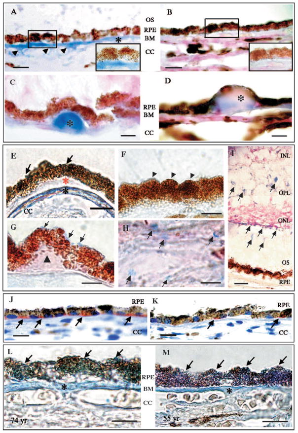
Methods: Evidence of complement activation and associated mechanisms was assessed in normal human retina (n = 52) using a panel of antibodies directed against membrane attack complex (C5b-9), microglia (CD11b), amyloid precursor protein (APP), scavenger receptor (CD36), and a phytolectin (RCA-I). Fifty-two eyes, categorized into two age groups, were used. Nineteen "younger" eyes (<56 years) and 33 "older" eyes (>69 years) with no history of ocular disease were processed between 4 and 22 hours, with a median delay of 14 hours postmortem.

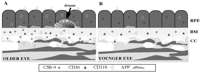
Results: Age-dependent expression was evident in C5b-9, APP, CD11b, and RCA-I, but not CD36, immunoreactivity. Immunoreactivity for C5b-9 was robust in Bruch membrane (BM) and the intercapillary pillars of Bruch. Immunoreactivity for APP was robust in the basal cytoplasm of the retinal pigment epithelium. Immunoreactivity for CD11b was robust on the surface of the retinal pigment epithelial cell, in the choriocapillaris, and in BM. Lectin binding of RCA-I was strong throughout the neuroretina.

Conclusions: Robust immunostaining for APP in older donor eyes suggested that amyloid beta peptides may be one of the triggers of complement activation during the normal aging process. Microglial markers CD11b and RCA-I also increase with age, suggesting a concomitant inflammatory response to C5b-9 deposits in the retinal pigment epithelium, BM, and CC. Immunoreactivity for CD36 was strong in both age groups; the lack of age dependence in this candidate receptor for amyloid beta suggested that complement activation may arise from interactions of amyloid beta with other candidate receptors in normal human retina.


Figures

Figure 1
Figure 1
(AD) Immunoreactivity for C5b-9 in the RPE–choroidal complex. (A) Representative immunoreactivity of tissues from an eye in the older donor group. Bright blue (vector blue) reaction product demarcates positive immunoreactivity for the membrane attack complex, C5b-9. Note that Bruch membrane (asterisk) and the intercapillary pillars of Bruch (arrowheads) label intensely. The basal aspect of the RPE cell was faintly immunoreactive for C5b-9 (enlargement). (B) Background immunolabeling pattern is seen in tissues from eyes in the younger donor group. Note faint blue staining in Bruch membrane. (C, D) Occasional drusen sites (asterisks) were observed in the tissues from eyes of the older donor group. All drusen were strongly immunoreactive for C5b-9. Note that the RPE cells do not exhibit positive (blue) immunoreactivity even at higher magnifications, as shown here. Cryosections were counterstained with neutral red, resulting in a light pink stain of the choroid and retinal pigment epithelium. The RPE layer is brown because of endogenous pigment. (EH) Immunoreactivity for CD11b in RPE–choroidal complex. Representative cryosections from eyes of the older donor group are immunoreactive for the activated microglial marker, CD11b. Positive immunoreactivity is bright blue (vector blue) and is present on the apical surfaces of some RPE cells (E, G, arrows) and in Bruch membrane (E, black asterisk). Not all RPE cells were immunoreactive for CD 11b (F, arrowheads). Basal laminar deposit (E, red asterisk) and drusen (G, black triangle) remain negative for CD11b. Several perivascular cells are CD11b immunoreactive in the choroid (H, arrows). (I) RCA-I binding in neural retina. Note vector blue–positive profiles scattered throughout the outer plexiform layer (OPL), outer segment layer (OS), and at the border between the outer nuclear layer (ONL) and OS. (JK) Immunoreactivity for APP in RPE–choroidal complex in paraffin-embedded sections. (J) Representative section from an eye of the older donor group demonstrating strong immunoreactivity for APP. Arrows point to the intense red reaction product (AEC) in the basal cytoplasmic compartment of the RPE cell. (K) Representative section from an eye of the younger donor group reveals a weaker immunoreactive labeling pattern. Two RPE cells (arrows) demonstrate APP immunoreactivity confined to the basal cytoplasm. Sections were counterstained with hematoxylin (blue). (L, M) Immunoreactivity for the scavenger receptor, CD36, in the RPE–choroidal complex. Immunoreactivity for CD36 did not demonstrate age dependence. Several examples indicated strong immunoreactivity for CD36 in older (L) and in younger (M) donor eyes, as shown by the bright blue reaction product (vector blue). CD36 was strongly immunoreactive on the retinal pigment epithelium (arrows), Bruch membrane (BM), and intercapillary pillars of Bruch (asterisk) and moderately immunoreactive in the choriocapillaris (CC). Scale bar: (A, B, IK) 20 μm; (C, D, EH, L, M) 10 μm.
Figure 2
Figure 2
Summary diagram depicting the distribution of C5b-9, CD36, CD11b, and APP immunoreactivity in the RPE–choroidal complex of older and younger eyes. Older eyes demonstrated significantly stronger immunoreactivity for C5b-9 in Bruch membrane and the intercapillary pillars of Bruch. Drusen, found in the older eyes in this study, were also immunoreactive for C5b-9. RPE cells demonstrating APP immunoreactivity were more numerous in older eyes. Activated microglia immunoreactivity was significantly greater in the RPE layer, the connective matrix of the choroid, and Bruch membrane of the older eyes. CD36 immunoreactivity was present in eye tissues of both age groups.

References

    1. Haines JL, Hauser MA, Schmidt S, et al. Complement factor H variant increases the risk of age-related macular degeneration. Science. 2005;308:419–421. - PubMed
    1. Edwards A, Ritter R, 3rd, Abel KJ, Manning A, Panhuysen C, Farrer LA. Complement factor H polymorphism and age-related macular degeneration. Science. 2005;308:421–424. - PubMed
    1. Klein RJ, Zeiss C, Chew Y, et al. Complement factor H polymorphism in age-related macular degeneration. Science. 2005;308:385–389. - PMC - PubMed
    1. Hageman GS, Anderson DH, Johnson LV, et al. A common haplotype in the complement regulatory gene factor H (HF1/CFH) predisposes individuals to age-related macular degeneration. Proc Natl Acad Sci USA. 2005;102:7227–7232. - PMC - PubMed
    1. Dentchev T, Milam AH, Lee VMY, Trojanowski JQ, Dunaief JL. Amyloid beta is found in drusen from some age-related macular degeneration retinas, but not in drusen from normal retinas. Mol Vis. 2003;9:184–190. - PubMed

Publication types

MeSH terms