. 2008 Feb;19(2):217-24.
doi: 10.1681/ASN.2005111155. Epub 2008 Jan 30.

ATF3 protects against renal ischemia-reperfusion injury

Affiliations

ATF3 protects against renal ischemia-reperfusion injury

Takumi Yoshida et al. J Am Soc Nephrol. 2008 Feb.

Abstract

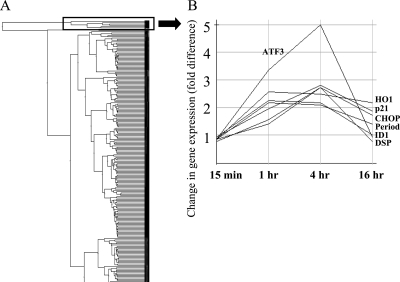
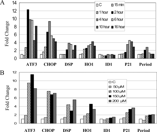
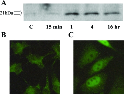
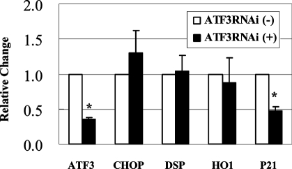
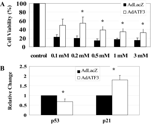
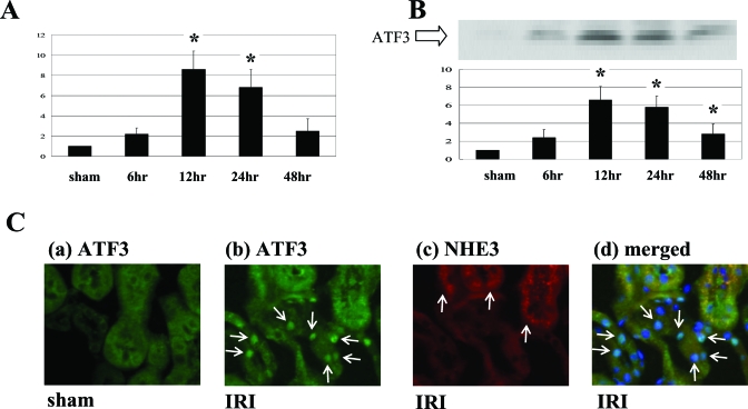
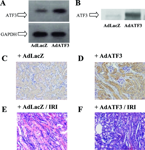
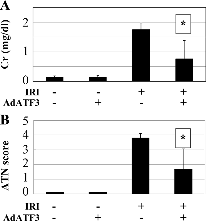
Oxidative stress-induced cell death plays a major role in the progression of ischemic acute renal failure. Using microarrays, we sought to identify a stress-induced gene that may be a therapeutic candidate. Human proximal tubule (HK2) cells were treated with hydrogen peroxide (H2O2) and RNA was applied to an Affymetrix gene chip. Five genes were markedly induced in a parallel time-dependent manner by cluster analysis, including activating transcription factor 3 (ATF3), p21(WAF1/CiP1) (p21), CHOP/GADD153, dual-specificity protein phosphatase, and heme oxygenase-1. H2O2 rapidly induced ATF3 approximately 12-fold in HK2 cells and approximately 6.5-fold in a mouse model of renal ischemia-reperfusion injury. Adenovirus-mediated expression of ATF3 protected HK2 cells against H2O2-induced cell death, and this was associated with a decrease of p53 mRNA and an increase of p21 mRNA. Moreover, when ATF3 was overexpressed in mice via adenovirus-mediated gene transfer, ischemia-reperfusion injury was reduced. In conclusion, ATF3 plays a protective role in renal ischemia-reperfusion injury and the mechanism of the protection may involve suppression of p53 and induction of p21.


Figures

Figure 1.
Figure 1.
Expression profile of H2O2 induced alteration of gene expression. (A) A tree of the clustering analysis is represented. (B) Expression profile of genes upregulated by H2O2. The markedly upregulated genes at 1 and 4 h include p21, ATF3, CHOP, DSP, HO1, ID1, and Period from the top at 4 h.
Figure 2.
Figure 2.
H2O2-Induced alterations of gene expression were confirmed by real-time RT-PCR. (A) Time course of gene expression in HK2 cells treated with 100 μM H2O2 at the indicated period were confirmed. ATF3, CHOP, DSP, HO1 and p21 were significantly upregulated. (B) Dose-response of genes in cells treated with the indicated concentrations of H2O2 for 4 h. Each experiment was performed in triplicate.
Figure 3.
Figure 3.
Induction of ATF3 by 100 μM H2O2 treatment. (A) Induction of ATF3 was clearly observed by Western blot analysis. Immunohistochemistry of ATF3 in the presence (B) and absence (C) of 100 μM H2O2 stimulation in HK2 cells for 4 h. Magnification: ×400.
Figure 4.
Figure 4.
siRNA silencing of ATF3 suppresses p21 induction, but not other genes in HK2 cells in real-time PCR (n = 4). Values are mean ± SEM. *P < 0.05 versus ATF3RNAi (−).
Figure 5.
Figure 5.
Effects of adenovirus-mediated gene transfer and overexpression of ATF3 (A). HK2 cells were significantly protected from H2O2 by AdATF3 at various doses of treatment with H2O2. A cell viability assay was performed using calcein AM (n = 6). (B) p53 mRNA was significantly decreased and p21 mRNA was significantly increased by treating with AdATF3 using real-time RT-PCR. (n = 4). Values are mean ± SEM. *P < 0.05 versus AdLacZ.
Figure 6.
Figure 6.
ATF3 was markedly induced by renal IRI in the mouse model. (A) Real-time PCR showed that ATF3 mRNA was significantly induced by IRI at 12 and 24 h after ischemia reperfusion compared with sham. (B) Representative Western blot analysis showed that ATF3 was significantly induced by IRI at 12 to 48 h after ischemia reperfusion compared with sham. (C-a) Immunofluorescent staining showed that ATF3 (green) was not detected in sham. (C-b) However, ATF3 (green) was induced by IRI for 24 h and was localized mainly in the nuclei of tubules (arrows). (C-c) NHE3 (red), the marker of proximal tubular cells (arrow), stained by immunofluorescent study. (C-d) Overlay picture of ATF3, NHE3, and DAPI (blue) is shown. ATF3 and DAPI were costained in the nuclei (arrow) of the distal tubule cells that were not stained for NHE3. For (A) and (B), values are mean ± SEM (n = 4). *P < 0.05 versus sham. Magnification for (C), ×200.
Figure 7.
Figure 7.
ATF3 protected tubular injury from ischemia reperfusion in mouse kidney. (A) RT-PCR showed that ATF3 mRNA was markedly induced by injection of AdATF3 in the kidney. (B) ATF3 induction by AdATF3 injection was confirmed by Western blotting. (C and D) Representative immunohistochemical stain for ATF3 is shown. ATF3 was upregulated mainly in the all-nephron segments in the mouse injected with AdATF3 (D). (E and F) Representative histochemical staining is shown. AdATF3 protected the tubular damage against IRI compared with AdLacZ (E). Magnification, ×200.
Figure 8.
Figure 8.
Protective effect of AdATF3 from renal IRI. Serum creatinine (A) and ATN score (B) in the sham (−) and IRI (+) treated mouse with AdATF3 or AdLacZ after 24 h reperfusion are presented. Both serum creatinine and ATN score were significantly ameliorated compared with treatment with AdLacZ. Values indicated by means ± SEM (n = 4 to 5). *P < 0.05 compared with AdLacZ.

References

    1. Waikar SS, Curhan GC, Wald R, McCarthy EP, Chertow GM: Declining mortality in patients with acute renal failure, 1988–2002. J Am Soc Nephrol 17: 1143–1150, 2006 - PubMed
    1. Star RA: Treatment of acute renal failure. Kidney Int 54: 1817–1831, 1998 - PubMed
    1. Mangano CM, Diamondstone LS, Ramsay JG, Aggarwal A, Herskowitz A, Mangano DT: Renal dysfunction after myocardial revascularization: Risk factors, adverse outcomes, and hospital resource utilization. The Multicenter Study of Perioperative Ischemia Research Group. Ann Intern Med 128: 194–203, 1998 - PubMed
    1. Kazmers A, Jacobs L, Perkins A: The impact of complications after vascular surgery in Veterans Affairs Medical Centers. J Surg Res 67: 62–66, 1997 - PubMed
    1. Tracey KJ, Beutler B, Lowry SF, Merryweather J, Wolpe S, Milsark IW, Hariri RJ, Fahey TJ 3rd, Zentella A, Albert JD, Shires GT, Cerami A. Shock and tissue injury induced by recombinant human cachectin. Science 234: 470–474, 1986 - PubMed

MeSH terms