. 2008 Jan 31;451(7178):596-9.
doi: 10.1038/nature06528.

Structural basis for the function and inhibition of an influenza virus proton channel

Affiliations

Structural basis for the function and inhibition of an influenza virus proton channel

Amanda L Stouffer et al. Nature. .

Erratum in

  • Nature. 2008 Mar 20;452(7185):380

Abstract

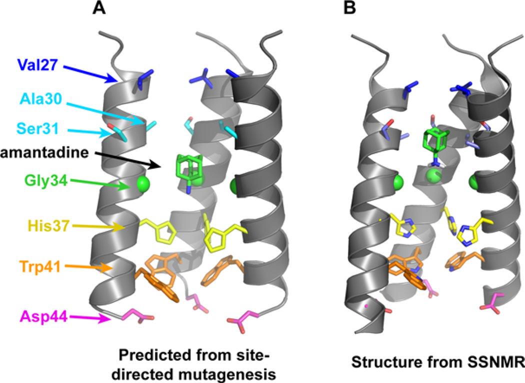
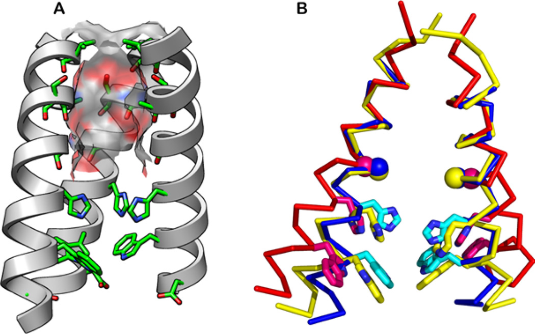
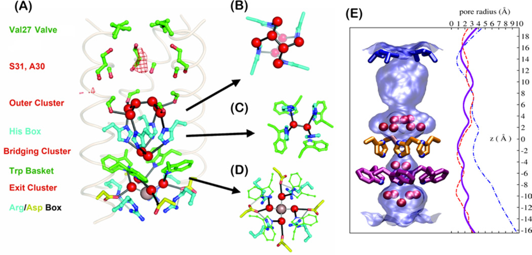
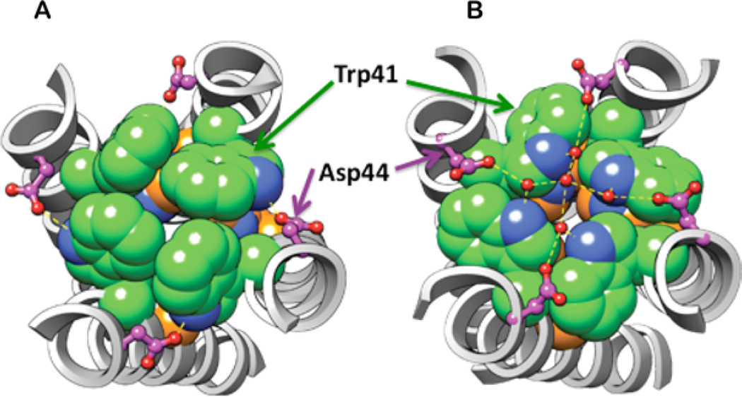
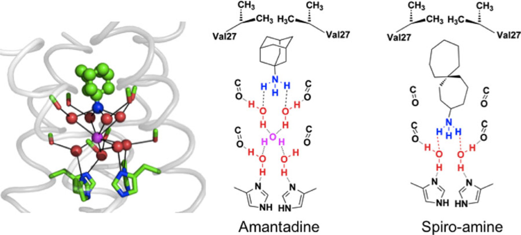
The M2 protein from influenza A virus is a pH-activated proton channel that mediates acidification of the interior of viral particles entrapped in endosomes. M2 is the target of the anti-influenza drugs amantadine and rimantadine; recently, resistance to these drugs in humans, birds and pigs has reached more than 90% (ref. 1). Here we describe the crystal structure of the transmembrane-spanning region of the homotetrameric protein in the presence and absence of the channel-blocking drug amantadine. pH-dependent structural changes occur near a set of conserved His and Trp residues that are involved in proton gating. The drug-binding site is lined by residues that are mutated in amantadine-resistant viruses. Binding of amantadine physically occludes the pore, and might also perturb the pK(a) of the critical His residue. The structure provides a starting point for solving the problem of resistance to M2-channel blockers.


Conflict of interest statement

Conflict of interest: WFD was the scientific founder of InfluMedix.

Figures

Figure 1
Figure 1
(A) Functional model of the TM domain of the M2 tetramer, showing the positions of crucial side chains and the drug amantadine. The model was obtained from cysteine scanning mutagenesis., (B) High-resolution structure of the amantadine-bound M2TM in DMPC bilayers obtained from SSNMR (PDB: 2KQT). The overall shape of the tetrameric bundle from the functional model is in excellent agreement with the high-resolution structure; however, specific differences exist such as the helix tilt angle and the conformations of several side chains (e.g., Ser31 and Trp41). In both images, the “front” helix has been removed for clarity.
Figure 2
Figure 2
Structures of the TM domain of M2. (A) The N-terminal pore is lined by the hydroxyl of Ser31 and backbone carbonyl groups (pictured structure is the 1.65Å crystal structure at pH 6.5, PDB: 3LBW). The molecular surface of the channel is color-coded with the oxygen atoms in red, carbon in gray, and nitrogen in blue. The “front” helix has been removed for clarity. (B) Superimposed solid-state NMR structure at pH 7.5 (2L0J, yellow), the 1.65-Å crystal structure at pH 6.5 (3LBW, blue), and the 3.5-Å crystal structure at pH 5.3 (3C9J, red). His37 and Trp41 side chains (sticks) and Gly34 Cα (ball) are shown.
Figure 3
Figure 3
(A) The proton conduction pathway seen in a 1.65-Å resolution crystal structure including three clusters of crystallographic waters. (B–D) A second perspective of the outer (B), bridging (C), and exit (D) clusters viewed normal to the membrane plane. (E) The surface of the pore (light blue shading) is shown along with the crystallographic waters (red spheres). Val27, His37, and Trp41 residues are rendered in blue, orange, and magenta, respectively. Pore radius profiles are plotted for the high-resolution crystal structure (3LBW, blue solid line), low-pH (3C9J, blue dash-dotted line), and amantadine-inhibited (red dashed line) structures.
Figure 4
Figure 4
The Trp41 rotamer and Trp41-Asp44 contact in the (A) high pH, drug-bound solution NMR structure (2RLF), and (B) the pH 6.5 X-ray crystal structure (3LBW). A larger pore radius at Trp41 is found in B due to the 180° χ2 angle change. The Trp41 side chains are shown in spheres for Trp41 (green for C, blue for N), and in ball-and-stick for Asp44 (pink C, blue O). His37 is shown as spheres with orange for C, blue for N. His37’s side chain is almost fully occluded by Trp41 in (A); in (B), it has slightly more accessibility. Crystallographically defined water molecules are shown in small red spheres in B, with hydrogen bonds shown in dashed lines.
Figure 5
Figure 5
Snapshot from a simulation of amantadine with WT (far left). Water molecules (red) associate with carbonyl groups (green/red sticks) in a square planar array. This array of water molecules can stabilize the bound ammonium group of amantadine (green and blue bound drug) or a centrally located water molecule (magenta). The remaining panels show vertical slices of the channel in schematic form, showing how a longer inhibitor than amantadine places its ammonium group deeper in the channel and displaces more water molecules.

Comment in

Similar articles

Cited by

References

    1. Hay AJ, Wolstenholme AJ, Skehel JJ, Smith MH. The molecular basis of the specific anti-influenza action of amantadine. EMBO J. 1985;4:3021–3024. - PMC - PubMed
    1. Park EK, Castrucci MR, Portner A, Kawaoka Y. The M2 ectodomain is important for its incorporation into influenza A virions. J Virol. 1998;72:2449–2455. - PMC - PubMed
    1. Pinto LH, Dieckmann GR, Gandhi CS, Papworth CG, Braman J, Shaughnessy MA, Lear JD, Lamb RA, DeGrado WF. A functionally defined model for the M2 proton channel of influenza A virus suggests a mechanism for its ion selectivity. Proc Natl Acad Sci USA. 1997;94:11301–11306. - PMC - PubMed
    1. Gandhi CS, Shuck K, Lear JD, Dieckmann GR, DeGrado WF, Lamb RA, Pinto LH. Cu(II) inhibition of the proton translocation machinery of the influenza A virus M2 protein. J Biol Chem. 1999;274:5474–5482. - PubMed
    1. Cady SD, Schmidt-Rohr K, Wang J, Soto CS, DeGrado WF, Hong M. Structure of the amantadine binding site of influenza M2 proton channels in lipid bilayers. Nature. 2010;463:689–692. - PMC - PubMed

Publication types

MeSH terms