

Review
doi: 10.1007/s12031-007-9027-5.
Gap junctions couple astrocytes and oligodendrocytes
Affiliations
- PMID: 18236012
- PMCID: PMC2650399
- DOI: 10.1007/s12031-007-9027-5
Item in Clipboard
Review
Gap junctions couple astrocytes and oligodendrocytes
J Mol Neurosci.
2008 May.
Abstract
In vertebrates, a family of related proteins called connexins form gap junctions (GJs), which are intercellular channels. In the central nervous system (CNS), GJs couple oligodendrocytes and astrocytes (O/A junctions) and adjacent astrocytes (A/A junctions), but not adjacent oligodendrocytes, forming a "glial syncytium." Oligodendrocytes and astrocytes each express different connexins. Mutations of these connexin genes demonstrate that the proper functioning of myelin and oligodendrocytes requires the expression of these connexins. The physiological function of O/A and A/A junctions, however, remains to be illuminated.
Figures
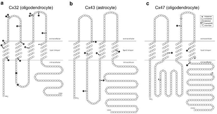
Cx32, Cx43, and Cx47 mutants associated with CNS disease. The structures of the connexin proteins are depicted according to Yeager and Nicholson (1996). Connexins have four transmembrane domains linked by two extracellular loops and one intracellular loop. Mutants associated with CNS disease are depicted in black circles. a The Cx32 mutants associated with a transient white matter disease in CMT1X are W24C, M34V, A39V, T55I, T55R, D66N, R75W, M93V, E102del, E109stop, R142W, R164W, R164Q, C168Y, R183H, and T191frame-shift (Lee et al. 2002; Taylor et al. 2003). b The Cx43 ODDD mutants associated with well-documented CNS findings are R76S, L90V, L113P, G138R, T154N, and V216L (Opjordsmoen and Nyberg-Hansen 1980; Norton et al. 1995; Paznekas et al. 2003; Shapiro et al. 1997; Stanislaw et al. 1998; Wiest et al. 2006). c The recessive Cx47 mutants that cause PMLD are P87S, P128frameshift, G233S, R237stop, Y269D, L278frameshift, M283T, P327frameshift (Uhlenberg et al. 2004; Bugiani et al. 2006)
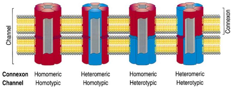
Diversity in GJ channel formation. This figure shows that two connexins (red and blue) can form GJ channels individually or in combination. Reprinted from (Kumar and Gilula 1996), with permission from Elsevier
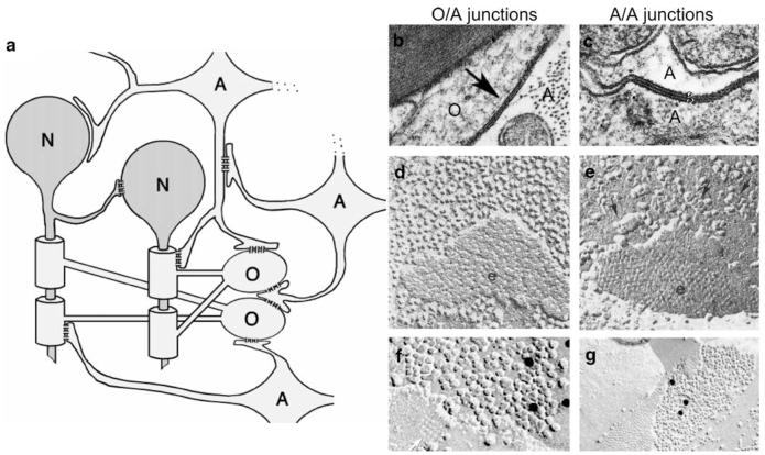
The “glial syncytium.” a In this schematic depiction of the “glial syncytium,” GJs join adjacent astrocytes, as well as adjacent astrocytes and oligodendrocytes, but not adjacent oligodendrocytes or adjacent neurons and glial cells. b—g Transmission (b and c)and freeze-fracture (d—g) electron microscopic images of GJs between adjacent glial cell membranes (arrow in b; g in c). b A GJ between an oligodendrocyte (O) and an astrocyte (A). c A GJ between two astrocytes. d An O/A GJ is composed of hundreds of individual channels in fractured apposed membranes. e An A/A junction; note that the pits of cleaved connexons are more densely packed in A/A (e)than in O/A junctions (d). f A FRIL image of an O/A junction immunogold-labeled for Cx47 (12-nm gold beads, black dots). g A FRIL image of an A/A junction immunogold-labeled for Cx43 (20-nm gold bead, black dots). Square arrays (orthogonal assemblies of aquaporin-4) identify astrocyte membranes in e (black arrow)and f (white arrow). Adapted from Rash et al. (2001; a), Mugnaini (1986; b, d, and e), Peters et al. (1991; c), Kamasawa et al. (2005; f), and Nagy et al. (2001; g), with permission from the Society for Neuroscience (a, copyright 2001), Elsevier (b, d, e, f), Oxford University Press (c), and Wiley—Liss (g)
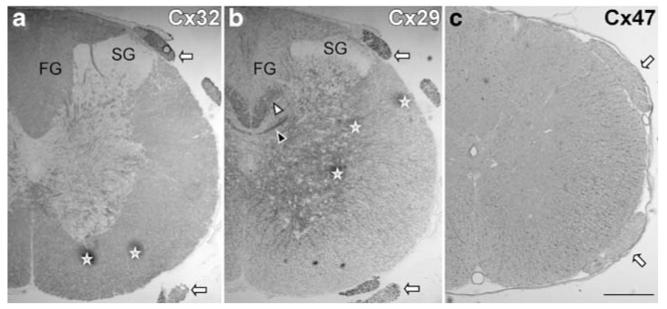
Differential expression of Cx32, Cx29, and Cx47. These are images of transverse sections of unfixed (a and b) or Zamboni-fixed (c) adult rat lumbar spinal cord using rabbit antisera against Cx32 (a), Cx29 (b), and Cx47 (c), visualized with a peroxidase-conjugated secondary antiserum. Cx32 immunoreactivity is mainly found in white matter tracts. Cx29 immunoreactivity is stronger in the gray matter and in fiber tracts that are enriched in small diameter fibers, such as the crossing posterior commissural fibers (black arrowhead) or corticospinal fibers (white arrowhead). Cx47 immunoreactivity is mainly present in oligodendrocyte perikarya, which are more numerous in the white matter. Cx32 and Cx29, but not Cx47, are strongly expressed in the spinal roots (arrows). SG Substantia gelatinosa and FG Fasciculus gracilis. Asterisks mark artifactual areas of staining. Scale bar, 200 μm. From Kleopa et al. (2004), with permission of Wiley—Liss
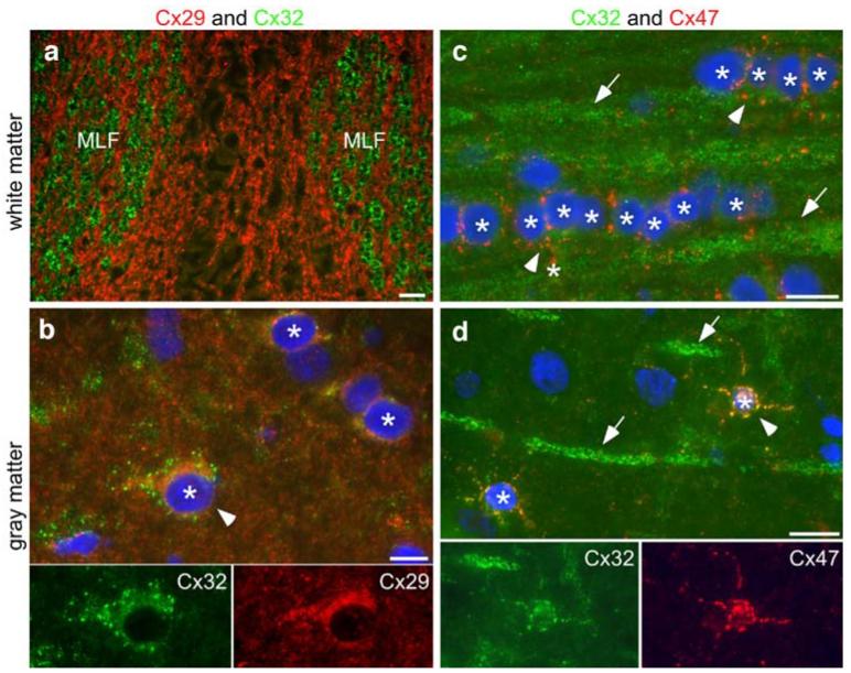
Differential distribution of Cx29, Cx32, and Cx47 in white and gray matter. These are images of unfixed rat CNS, immunostained with a rabbit antiserum to Cx29 (red; a—b), a mouse monoclonal antibody against Cx32 (green; a—d) or a rabbit antiserum to Cx47 (red; c—d), along with nuclear staining with 4′,6-diamidino-2-phenyl-indole (blue in b—d). In transverse sections of the pons (a), the large myelinated fibers of the medial longitudinal fasciculus (MLF) are Cx32-positive, whereas the surrounding smaller myelinated fibers are Cx29-positive. In the spinal cord gray matter (b), the same cell (arrowhead; also shown in the bottom two panels) has Cx29 and Cx32 immunoreactivity, but Cx29 is intracellular; it does not colocalize with Cx32 in GJ plaques. Oligodendrocyte perikarya (asterisks) in white matter (c; dorsal funiculus, longitudinal section) express Cx47 but not Cx32; those in gray matter (d; neocortex) express both Cx47 and Cx32. Note that there is substantial overlap between the Cx32 and Cx47 signals (arrowhead; also shown in the bottom two panels). Myelinated sheaths (arrows) express abundant Cx32 but little Cx47. Scale bars, 10 μm. These images are modified from Kleopa et al. (2004), with permission of Wiley—Liss
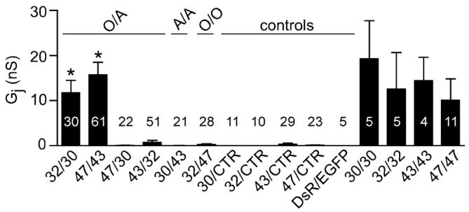
Cx32/Cx30 and Cx47/C43 form functional channels. Neuro2A cells were transiently transfected with pIRES2-EGFP or pIRES2-DsRed vectors containing human Cx30, Cx32, Cx43, Cx47, or no insert (vector alone control; CTR). After 24 h, EGFP- and DsRed-expressing cells were mixed in a 1:1 ratio; pairs were assessed by dual whole-cell patch clamping 6 to 48 h later. For each combination listed on the x-axis, the mean and standard error of the junctional conductance (Gj) and number of pairs tested are shown. Using Kruskal—Wallis test followed by Dunn’s multiple comparison test, only Cx32/Cx30 and Cx47/Cx43 channels have Gj that is significantly greater (p <0.001; asterisks) than those of the corresponding control pairs (32/CTR and 30/CTR or 43/CTR and 47/CTR). From Orthmann-Murphy et al. (2007b), copyright 2007 by the Society for Neuroscience
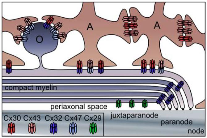
The gap junction network of astrocytes and oligodendrocytes. This drawing depicts a model for the GJ channels that connect astrocytes (A) to oligodendrocytes (O) and other astrocytes. A/A junctions contain Cx30/Cx30 and Cx43/Cx43 channels; O/A junctions contain Cx32/Cx30 and Cx47/Cx43 channels. Homotypic Cx32/Cx32 channels connect layers of myelin sheath at the paranode and incisures (not shown). Cx29 hemichannels are localized to the adaxonal membrane of oligodendrocytes, apposing the axon. From Orthmann-Murphy et al. (2007b), copyright 2007 by the Society for Neuroscience
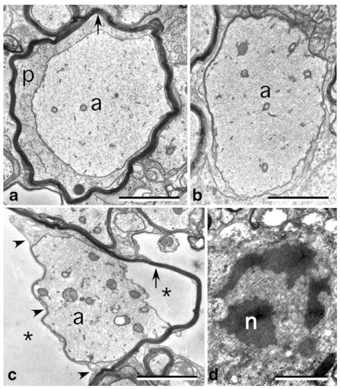
Pathological findings in Cx32/Cx47 double null mice. These are electron micrographs of the ventral funiculus from a postnatal day 30 Cx32/Cx47 double null mouse. a An axon (a) is surrounded by a periaxonal collar of cytoplasm (p) and a thin myelin sheath (arrow). b A demyelinated axon (a) has neurofilaments that are more tightly packed than in a. c An axon (a) is still partially surrounded by the adaxonal process of an oligodendrocyte (arrowheads) but is separated from its myelin sheath (arrow) by extracellular space (asterisks). d An apoptotic oligodendrocyte nucleus is shown. Scale bars: 1 μm. From Menichella et al. (2003), copyright 2003 by the Society for Neuroscience
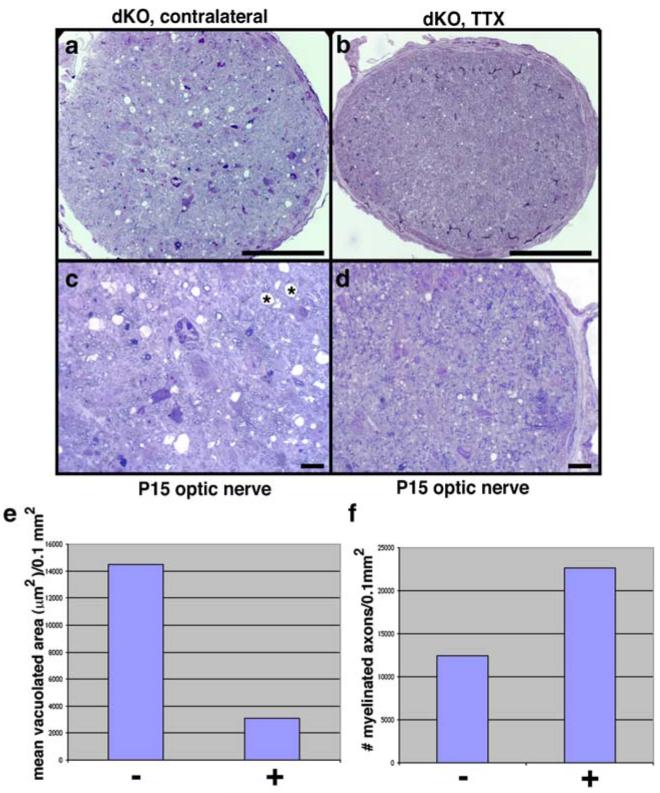
Suppressing axonal activity reduces vacuolation in optic nerves from Cx32/Cx47 double null mice. Intraocular injections of tetrodotoxin (TTX) were performed at P11 and P13; optic nerves were harvested at P15. (a—d) Semi-thin sections from the optic nerve associated with an injected eye reveal a dramatic reduction in vacuolation compared to the contralateral control nerve. Scale bars: top row (a, b), 100 μm; bottom row (c, d), 10 μm. (e—f) Quantification of small (<20 μm2) and large (>20 μm2) vacuoles, as well as the total vacuolated area (μm2/0.1 mm2) and the number of myelinated axons (per 0.1 mm2) in TTX injected (+) and uninjected/contralateral (−) optic nerves. TTX injections lead to fewer vacuoles and a reduction in total vacuolated area. Error bars indicate SEM. From Menichella et al. (2006), copyright 2006 by the Society for Neuroscience
References
-
- Abrams CK, Oh S, Ri Y, Bargiello TA. Mutations in connexin 32: The molecular and biophysical bases for the X-linked form of Charcot—Marie—Tooth disease. Brain Research Reviews. 2000;32:203–214. - PubMed
-
- Ahmad S, Chen SP, Sun JJ, Lin X. Connexins 26 and 30 are co-assembled to form gap junctions in the cochlea of mice. Biochemical and Biophysical Research Communications. 2003;307:362–368. - PubMed
Publication types
MeSH terms
Substances
Grants and funding
LinkOut - more resources
Full Text Sources
Miscellaneous
