

Effects of oxygen concentration on the proliferation and differentiation of mouse neural stem cells in vitro
- PMID: 18236013
- PMCID: PMC11515460
- DOI: 10.1007/s10571-007-9237-y
Effects of oxygen concentration on the proliferation and differentiation of mouse neural stem cells in vitro
Abstract
Background and purpose: Cerebral ischemia is known to elicit the activation of neural stem cells (NSCs); however its mechanism is not fully determined. Although oxygen concentration is known to mediate many ischemic actions, there has been little attention given to the role of pathological oxygen changes under cerebral ischemia on the activation of NSCs. We investigated the effects of various oxygen concentrations on mouse neural stem cells in vitro.
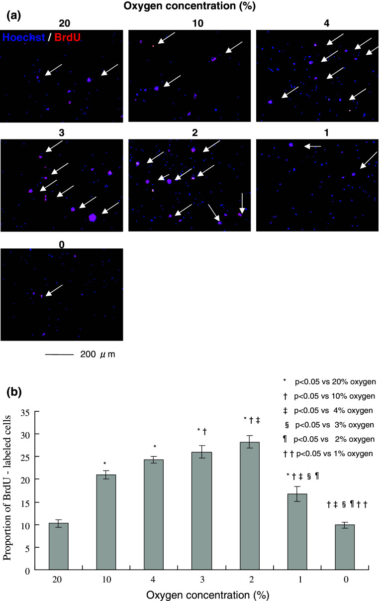
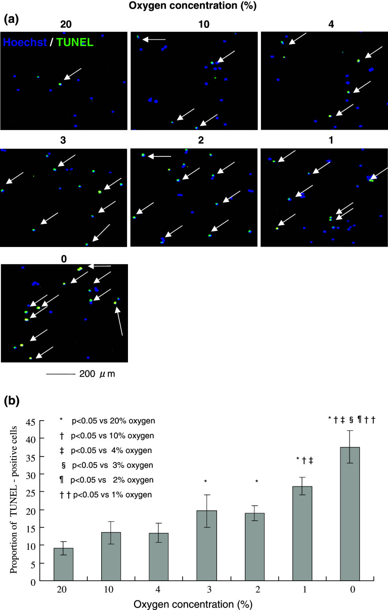
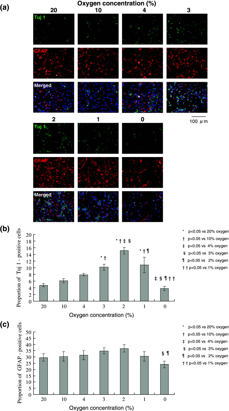
Methods: NSCs were cultured from the ganglionic eminence of fetal ICR mice on embryonic day 15.5 using a neurosphere method. The effects of oxygen concentrations on proliferation, differentiation, and cell death of NSCs were evaluated by bromodeoxyuridine (BrdU) incorporation, immunocytochemistry, and TUNEL assay, respectively.
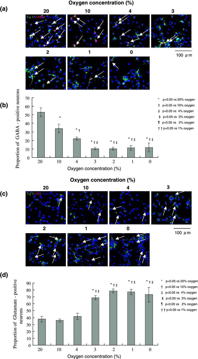
Results: The highest proliferation and the neuronal differentiation of the NSCs were observed in 2% oxygen, which yielded significantly higher proportions of both BrdU-labeled cells and Tuj1-positive cells when compared with 20% and 4% oxygen. On the other hand, the differentiation to the astrocytes was not affected by oxygen concentrations, except in the case of anoxia (0% oxygen). The cell death of the NSCs increased in lower oxygen conditions and peaked at anoxia. Furthermore, the switching of the neuronal subtype differentiation from GABA-positive to glutamate-positive neurons was observed in lower oxygen conditions.
Conclusions: These findings raise the possibility that reduced oxygen levels occurring with cerebral ischemia enhance NSC proliferation and neural differentiation, and that mild hypoxia (2% oxygen), which is known to occur in the ischemic penumbra, is suitable for abundant neuronal differentiation.
Figures
Similar articles
-
Hypoxia stimulates proliferation of rat neural stem cells with influence on the expression of cyclin D1 and c-Jun N-terminal protein kinase signaling pathway in vitro.Neuroscience. 2010 Feb 3;165(3):705-14. doi: 10.1016/j.neuroscience.2009.11.007. Epub 2009 Nov 10. Neuroscience. 2010. PMID: 19909792
-
Acute anoxia stimulates proliferation in adult neural stem cells from the rat brain.Exp Brain Res. 2008 Jun;188(1):33-43. doi: 10.1007/s00221-008-1336-6. Epub 2008 Mar 11. Exp Brain Res. 2008. PMID: 18330547
-
In vitro investigation of the mechanism underlying the effect of ginsenoside on the proliferation and differentiation of neural stem cells subjected to oxygen-glucose deprivation/reperfusion.Int J Mol Med. 2018 Jan;41(1):353-363. doi: 10.3892/ijmm.2017.3253. Epub 2017 Nov 13. Int J Mol Med. 2018. PMID: 29138802 Free PMC article.
-
A Tale of Two: When Neural Stem Cells Encounter Hypoxia.Cell Mol Neurobiol. 2023 Jul;43(5):1799-1816. doi: 10.1007/s10571-022-01293-6. Epub 2022 Oct 29. Cell Mol Neurobiol. 2023. PMID: 36308642 Free PMC article. Review.
-
New Insights into the Role of Mild Hypoxia in Regulating Neural Stem Cell Characteristics.Stem Cells Dev. 2024 Jul;33(13-14):333-342. doi: 10.1089/scd.2024.0020. Epub 2024 Jun 11. Stem Cells Dev. 2024. PMID: 38753713 Review.
Cited by
-
Neurogenesis continues in the third trimester of pregnancy and is suppressed by premature birth.J Neurosci. 2013 Jan 9;33(2):411-23. doi: 10.1523/JNEUROSCI.4445-12.2013. J Neurosci. 2013. PMID: 23303921 Free PMC article.
-
Post ischemia intermittent hypoxia induces hippocampal neurogenesis and synaptic alterations and alleviates long-term memory impairment.J Cereb Blood Flow Metab. 2013 May;33(5):764-73. doi: 10.1038/jcbfm.2013.15. Epub 2013 Feb 27. J Cereb Blood Flow Metab. 2013. PMID: 23443175 Free PMC article.
-
Low-oxygen culture conditions extend the multipotent properties of human retinal progenitor cells.Tissue Eng Part A. 2014 May;20(9-10):1465-75. doi: 10.1089/ten.TEA.2013.0361. Epub 2014 Jan 24. Tissue Eng Part A. 2014. PMID: 24320879 Free PMC article.
-
Oxygen levels and the regulation of cell adhesion in the nervous system: a control point for morphogenesis in development, disease and evolution?Cell Adh Migr. 2012 Jan-Feb;6(1):49-58. doi: 10.4161/cam.19582. Cell Adh Migr. 2012. PMID: 22647940 Free PMC article. Review.
-
Reduced Cerebral Oxygen Content in the DG and SVZ In Situ Promotes Neurogenesis in the Adult Rat Brain In Vivo.PLoS One. 2015 Oct 14;10(10):e0140035. doi: 10.1371/journal.pone.0140035. eCollection 2015. PLoS One. 2015. PMID: 26466323 Free PMC article.
References
-
- Arvidsson A, Collin T, Kirik D, Kokaia Z, Lindvall O (2002) Neuronal replacement from endogenous precursors in the adult brain after stroke. Nat Med 8:963–970 - PubMed
-
- Astrup J, Siesjo BK, Symon L (1981) Thresholds in cerebral ischemia—the ischemic penumbra. Stroke 12:723–725 - PubMed
-
- Brusselmans K, Bono F, Maxwell P, Dor Y, Dewerchin M, Collen D, Herbert JM, Carmeliet P (2001) Hypoxia-inducible factor-2alpha (HIF-2alpha) is involved in the apoptotic response to hypoglycemia but not to hypoxia. J Biol Chem 276:39192–39196 - PubMed
-
- Burke RE, Kenyon N (1991) The effect of neonatal hypoxia-ischemia on striatal cholinergic neurophil: a quantitative morphologic analysis. Exp Neurol 113:63–73 - PubMed
-
- Daadi MM (2002) In vitro assays for neural stem cell differentiation. Meth Mol Biol 198:149–155 - PubMed
MeSH terms
Substances
LinkOut - more resources
Full Text Sources
Medical
