. 2008 Jul;57(7):1017-27.
doi: 10.1007/s00262-007-0433-x.

A33 antigen displays persistent surface expression

Affiliations

A33 antigen displays persistent surface expression

Margaret E Ackerman et al. Cancer Immunol Immunother. 2008 Jul.

Abstract

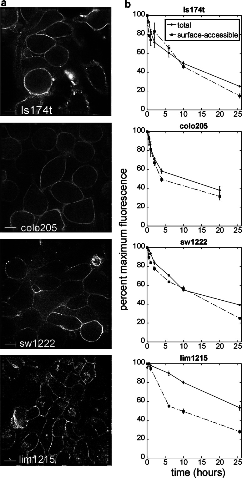
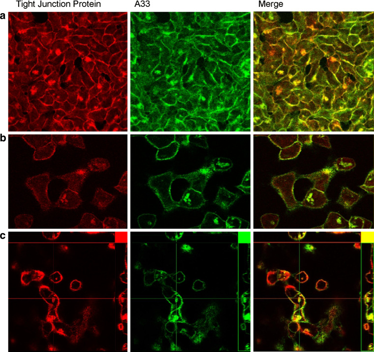
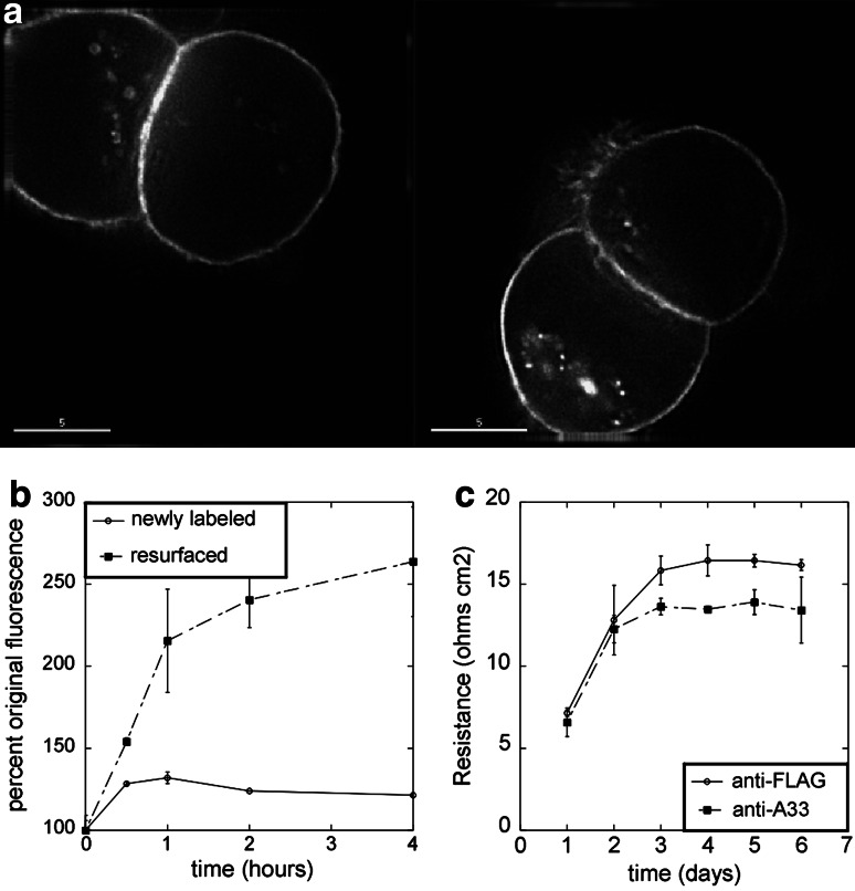
The A33 antigen is a cell surface glycoprotein of the small intestine and colonic epithelium with homology to tight junction-associated proteins of the immunoglobulin superfamily, including CAR and JAM. Its restricted tissue localization and high level of expression have led to its use as a target in colon cancer immunotherapy. Although the antigen is also present in normal intestine, radiolabeled antibodies against A33 are selectively retained by tumors in the gut as well as in metastatic lesions for as long as 6 weeks. Accordingly, we have studied the trafficking and kinetic properties of the antigen to determine its promise in two-step, pretargeted therapies. The localization, mobility, and persistence of the antigen were investigated, and this work has demonstrated that the antigen is both highly immobile and extremely persistent-retaining its surface localization for a turnover halflife of greater than 2 days. In order to explain these unusual properties, we explored the possibility that A33 is a component of the tight junction. The simple property of surface persistence, described here, may contribute to the prolonged retention of the clinically administered antibodies, and their uncommon ability to penetrate solid tumors.


Figures

Fig. 1
Fig. 1
Distribution of antibody-bound A33 antigen. a Cultured colon tumor epithelial cells contain little intracellular A33 antigen. Each of four cell lines were grown on coverslips, fixed, permeabilized, and A33 antigen was labeled with the monoclonal antibody m100.310*Alexa 488. Internal pools of antigen were seen only in LIM1215 cells. Bar represents 5 μm. b Antibody-bound A33 antigen shows persistent surface localization in a flow cytometric assay of dynamic internalization kinetics. Suspended cells were A33-labeled at 0°C and then incubated at 37°C, allowing labeled antigen to be internalized. After shifting cultures to 37°C, aliquots were removed and labeled with anti-mouse PE and analyzed flow cytometry in order to determine the fraction of labeled A33 that remained surface accessible over time. The level of surface-accessible A33 did not fall significantly below the level of total A33, indicating little internalization of the antibody-bound antigen in all cell lines tested except LIM1215. The decrease in total A33 signal is likely due to dissociation of the primary antibody, as the antigen is not shed
Fig. 2
Fig. 2
Colocalization profile of A33 antigen. Tight junction proteins are false-colored in red, and A33 antigen in green. a A33 partially colocalizes with the tight junction protein occludin. Immunofluorescence image of LIM1215 cells showing colocalization of A33 antigen and occludin. b A33 partially colocalizes with the actin-binding, tight junction-associated protein, ZO-1. Immunofluorescence image of LIM1215 cells showing colocalization of A33 antigen and ZO-1. c A33 partially colocalizes with the cytoskeleton. Immunofluorescence image of LIM1215 cells showing colocalization of A33 antigen and actin. Images have been false-colored for clarity
Fig. 3
Fig. 3
Investigation of A33 as a tight junction-associated protein. a Calcium chelation triggers A33 internalization. LS174T cells were labeled with m100.310*Alexa 488, and culture media was treated with 25 mM EGTA to chelate calcium. At 2 h, there were clear intracellular pools of antigen, a phenomenon common to junction-associated proteins. b Replacement of calcium restores A33 surface localization. When EGTA is washed out, previously labeled A33 returns to the surface. Cell aliquots were labeled with m100.310*Alexa 488 and then treated with EGTA for 2 h. Following media replacement, aliquots were removed and labeled with a secondary-PE conjugate, and then again with m100.310*Alexa 488, and subjected to flow cytometry. c Anti-A33 antibody decreases monolayer integrity. SW1222 cells were grown on transwell inserts, treated with antibodies 1 day after plating, and transepithelial electrical resistance was measured across the monolayer
Fig. 4
Fig. 4
Determination of the role of cytoskeleton in A33 localization. a Effect of disrupting actin on antigen distribution. Immunofluorescence images of A33-labeled LS174T cells after treatment with 100 nM latrunculin B for 2 h demonstrates internalization. b Effect of disrupting microtubules on antigen distribution. Immunofluorescence images of A33-labeled LS174T cells after treatment with 50 μM nocodazole for 2 h demonstrates internalization. c Disruption of the cytoskeleton prevents recovery of surface-expressed A33. LS174T cells were labeled and then incubated with EGTA. Following internalization, media was exchanged with either fresh media (washout), or fresh media containing latrunculin, nocodazole, or EGTA (continued EGTA). Surface localization was restored only in cells with intact cytoskeleton and normal calcium levels. d Possible functional homology between A33 and occludin intracellular domains. A33 and occludin share a highly acidic intracellular region and a negatively charged hinge found to be important for TJ localization. Hydrophobicity plot of A33 antigen, detailing the hinge region with high homology to occludin
Fig. 5
Fig. 5
Determination of the mobility and persistence of antibody-bound A33. a Over the course of 1 min, bleached plasma membrane has a near full recovery of fluorescence while very little A33 fluorescence recovers. Fraction of fluorescence recovered over time for bleached m100.310 antibody bound to A33 and the plasma membrane marker DiI. (N = 8, LS174T cells). b Even over much longer time scales, antibody-bound A33 fluorescence recovery is slow and incomplete. 35-min time course of antibody bound to A33 after photobleaching. (N = 6, LS174T cells). c The A33 antigen is persistent, having a surface expression halflife of 56 h in cultured monolayers. Cell surface proteins of cultured LS174T cells were pulse-biotinylated, extracted, and pulled down with streptavidin beads. The precipitated proteins were reduced off the beads, subjected to gel electrophoresis and blotted to detect A33 antigen. Signal intensity of two data sets is plotted against time post-biotinylation, and fit to an exponential decay. d While other immunotherapy targets are internalized, A33 antigen remains on the surface. LS174T cells were labeled with both m100.310*Alexa 488 and anti-CEA shMFE*594 for 2 h at 37°C before imaging on a deconvolution microscope

References

    1. DeNardo GL. Treatment of non-Hodgkin’s lymphoma (NHL) with radiolabeled antibodies (mAbs) Semin Nucl Med. 2005;35(3):202–211. doi: 10.1053/j.semnuclmed.2005.02.006. - DOI - PubMed
    1. Pagel JM, Hedin N, Subbiah K, Meyer D, Mallet R, Axworthy D, Theodore LJ, Wilbur DS, Matthews DC, Press OW. Comparison of anti-CD20 and anti-CD45 antibodies for conventional and pretargeted radioimmunotherapy of B-cell lymphomas. Blood. 2003;101(6):2340–2348. doi: 10.1182/blood-2002-03-0874. - DOI - PubMed
    1. Gruaz-Guyon A, Raguin O, Barbet J. Recent advances in pretargeted radioimmunotherapy. Curr Med Chem. 2005;12(3):319–338. - PubMed
    1. Boerman OC, van Schaijk FG, Oyen WJ, Corstens FH. Pretargeted radioimmunotherapy of cancer: progress step by step. J Nucl Med. 2003;44(3):400–411. - PubMed
    1. Johnstone CN, Tebbutt NC, Abud HE, White SJ, Stenvers KL, Hall NE, Cody SH, Whitehead RH, Catimel B, Nice EC, Burgess AW, Heath JK. Characterization of mouse A33 antigen, a definitive marker for basolateral surfaces of intestinal epithelial cells. Am J Physiol Gastrointest Liver Physiol. 2000;279(3):G500–G510. - PubMed

Publication types

LinkOut - more resources