. 2008 Jan 31:9:9.
doi: 10.1186/1471-2121-9-9.

Identification of a novel Leucine-rich repeat protein and candidate PP1 regulatory subunit expressed in developing spermatids

Affiliations

Identification of a novel Leucine-rich repeat protein and candidate PP1 regulatory subunit expressed in developing spermatids

Rong Wang et al. BMC Cell Biol. .

Abstract

Background: Spermatogenesis is comprised of a series of highly regulated developmental changes that transform the precursor germ cell into a highly specialized spermatozoon. The last phase of spermatogenesis, termed spermiogenesis, involves dramatic morphological change including formation of the acrosome, elongation and condensation of the nucleus, formation of the flagella, and disposal of unnecessary cytoplasm. A prominent cytoskeletal component of the developing spermatid is the manchette, a unique microtubular structure that surrounds the nucleus of the developing spermatid and is thought to assist in both the reshaping of the nucleus and redistribution of spermatid cytoplasm. Although the molecular motor KIFC1 has been shown to associate with the manchette, its precise role in function of the manchette and the identity of its testis specific protein partners are unknown. The purpose of this study was to identify proteins in the testis that interact with KIFC1 using a yeast 2 hybrid screen of a testis cDNA library.

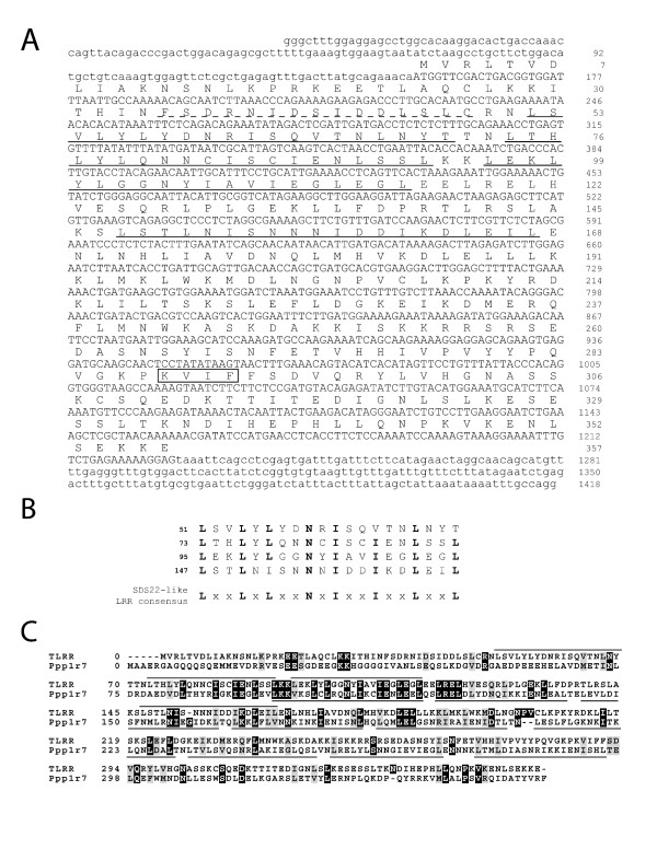
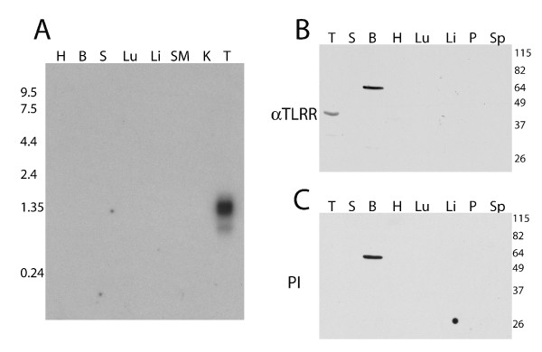
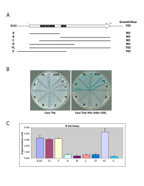
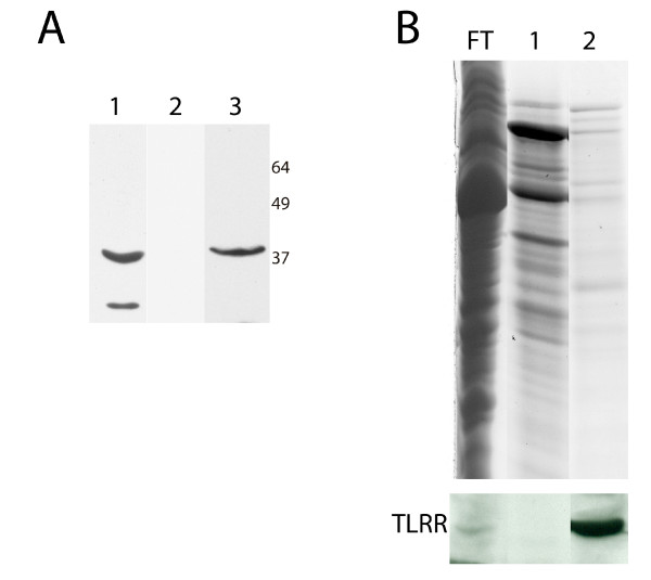
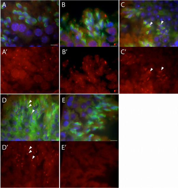
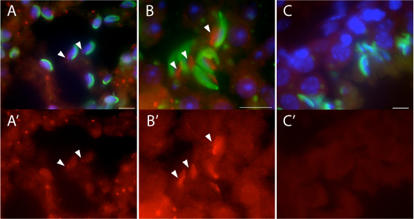
Results: Thirty percent of the interacting clones identified in our screen contain an identical cDNA encoding a 40 kD protein. This interacting protein has 4 leucine-rich repeats in its amino terminal half and is expressed primarily in the testis; therefore we have named this protein testis leucine-rich repeat protein or TLRR. TLRR was also found to associate tightly with the KIFC1 targeting domain using affinity chromatography. In addition to the leucine-rich repeats, TLRR contains a consensus-binding site for protein phosphatase-1 (PP1). Immunocytochemistry using a TLRR specific antibody demonstrates that this protein is found near the manchette of developing spermatids.

Conclusion: We have identified a previously uncharacterized leucine-rich repeat protein that is expressed abundantly in the testis and associates with the manchette of developing spermatids, possibly through its interaction with the KIFC1 molecular motor. TLRR is homologous to a class of regulatory subunits for PP1, a central phosphatase in the reversible phosphorylation of proteins that is key to modulation of many intracellular processes. TLRR may serve to target this important signaling molecule near the nucleus of developing spermatids in order to control the cellular rearrangements of spermiogenesis.


Figures

Figure 1
Figure 1
TLRR is a leucine-rich repeat protein. (A) The DNA sequence of the interacting clone (IC#2) is shown with the translated sequence above the nucleotide sequence. The leucine-rich repeats (LRRs) are indicated with solid underlining and the peptide used for preparation of antibody indicated with dashed underlining. The putative PP1 binding site is boxed. (B) The TLRR repeats are aligned with the LRR consensus sequence for the sds22 subfamily of LRR proteins. (C) An alignment between TLRR and Ppp1r7 is shown with the LRRs indicated with overlines (TLRR) and underlines (Ppp1r7). Multiple sequence alignment was done using ClustalW 1.82 and the results displayed using BOXSHADE 3.31. LRRs in TLRR and Ppp1r7 were identified using the Pfam protein domain database [35].
Figure 2
Figure 2
TLRR is expressed primarily in testis. (A) A multiple tissue northern blot was hybridized with radiolabeled probe specific for the TLRR mRNA as described in Methods. Each lane contains 2 μg mouse poly(A+) RNA separated on a denaturing formaldehyde 1.0% agarose gel and transferred to nylon. Lanes 1–8 contain: H, heart; B, whole brain; S, spleen; Lu, lung; Li, liver; SM, skeletal muscle; K, kidney; T, testis. The migration and size of RNA markers is shown on the left. (B and C) Total protein (75 μg) from each of the indicated tissues was separated by PAGE, transferred to membrane, and incubated with the affinity purified TLRR antibody (B), or preimmune serum (C). Lanes 1–8 in both B and C contain: T, testis; S, spleen; B, whole brain; H, heart; Lu, lung; Li, liver; P, pancreas; Sp, epidydimal sperm.
Figure 3
Figure 3
LRR domains are not sufficient for interaction with KIFC1 tail sequence. (A) A schematic is shown of the KIFC1 interacting clone with the 357 amino acid open reading frame indicated with an arrow. The LRR domains are indicated with black boxes and below are shown the regions tested for interaction with the KIFC1 bait plasmid and the results of interaction tests. (B) Plate assay of interaction of TLRR deletion constructs with KIFC1bait. The plate on the left selects only for yeast harboring both bait and prey plasmids; all cotransformants are able to grow. The plate on the right also lacks adenine and histidine and is therefore selective for transactivation of the reporter genes. Transactivation of the MEL1 reporter gene is detected by incorporation of X-α-gal into the media resulting in blue colonies. Positive control for protein interaction (+C) is yeast cotransformed with pGADT7-T and pGBKT7-53 while cells containing pGADT7 and pGBKT7-Lam (-C) is negative control. (C) Liquid assay of interaction of TLRR deletion constructs with KIFC1bait. The β-galactosidase activity of cultures of TLRR/KIFC1 bait cotransformants was determined as described in Methods. Each assay was repeated at least three times. Standard error of the mean for each transformant is indicated by bracketed lines in panel C.
Figure 4
Figure 4
TLRR associates tightly with the KIFC1 targeting sequence. (A) 200 μg testis lysate (lane 1 and 2) or an aliquot of recombinant HIS6 tagged TLRR (lane 3) were resolved by SDS-PAGE and blotted with TLRR antibody (lane 1 and 3) or preimmune serum (lane 2). (B) Testis lysate was loaded onto a column consisting of KIFC1 targeting peptide-linked resin. Flow through (FT) was collected, the column washed and bound complexes eluted with glycine, pH 2.5 (lane 1) or glycine plus 0.5 M NaCl (lane 2). Coomassie stained gel is shown at top with TLRR western below.
Figure 5
Figure 5
TLRR localizes to the manchette of developing spermatids. (A-D) Merged images of adult mouse testis sections triple stained with anti-TLRR (red), anti-α-tubulin (green, manchette), and DAPI (blue, nuclei) as described in the Methods section. (A'-D') TLRR signal corresponding to each of the merged images shown in A-D. Increasingly elongated spermatids are shown from panels A, A' through D, D'. TLRR is associated with the manchette in mid stage spermatids (arrowheads, panels C and C') and this staining narrows with the elongation of the manchette in approximately step 15 spermatids (arrowheads, panels D and D'). (E, E') Negative control for these experiments where TLRR specific primary antibody was omitted. Bar represents 10 μm.
Figure 6
Figure 6
TLRR localizes near the distal pole of the spermatid nucleus. (A-B) Merged images of adult mouse testis sections triple stained with anti-TLRR (red), peanut agglutinin (green, acrosome) and DAPI (blue, nuclei). (A'-B') TLRR signal corresponding to each of the merged images shown in A-B. TLRR is found near the nuclear membrane at a site opposite the acrosome and is most prominent in later step spermatids (arrowheads, panels B and B'). (C, C') Control staining for this series where TLRR antibody was replaced with normal rabbit IgG. Bar represents 10 μm.

References

    1. Clermont Y. Kinetics of spermatogenesis in mammals: seminiferous epithelium cycle and spermatogonial renewal. Physiol Rev. 1972;52:198–236. - PubMed
    1. Navolanic PM, Sperry AO. Identification of isoforms of a mitotic motor in mammalian spermatogenesis. Biol Reprod. 2000;62:1360–1369. doi: 10.1095/biolreprod62.5.1360. - DOI - PubMed
    1. Lawrence CJ, Dawe RK, Christie KR, Cleveland DW, Dawson SC, Endow SA, Goldstein LS, Goodson HV, Hirokawa N, Howard J, Malmberg RL, McIntosh JR, Miki H, Mitchison TJ, Okada Y, Reddy AS, Saxton WM, Schliwa M, Scholey JM, Vale RD, Walczak CE, Wordeman L. A standardized kinesin nomenclature. J Cell Biol. 2004;167:19–22. doi: 10.1083/jcb.200408113. - DOI - PMC - PubMed
    1. Yang WX, Sperry AO. C-terminal kinesin motor KIFC1 participates in acrosome biogenesis and vesicle transport. Biol Reprod. 2003;69:1719–1729. doi: 10.1095/biolreprod.102.014878. - DOI - PubMed
    1. Russell LD, Russell JA, MacGregor GR, Meistrich ML. Linkage of manchette microtubules to the nuclear envelope and observations of the role of the manchette in nuclear shaping during spermiogenesis in rodents. Am J Anat. 1991;192:97–120. doi: 10.1002/aja.1001920202. - DOI - PubMed

Publication types

MeSH terms