

Bioluminescence in vivo imaging of autoimmune encephalomyelitis predicts disease
- PMID: 18237444
- PMCID: PMC2267451
- DOI: 10.1186/1742-2094-5-6
Bioluminescence in vivo imaging of autoimmune encephalomyelitis predicts disease
Abstract
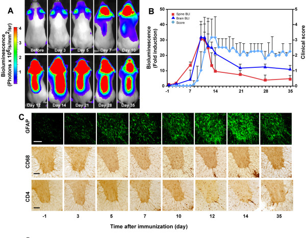
Background: Experimental autoimmune encephalomyelitis is a widely used animal model to understand not only multiple sclerosis but also basic principles of immunity. The disease is scored typically by observing signs of paralysis, which do not always correspond with pathological changes.
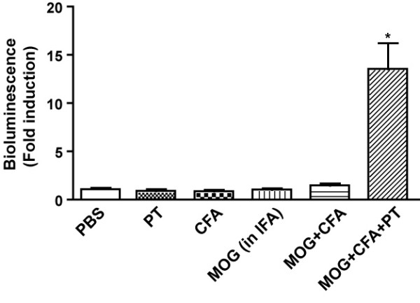
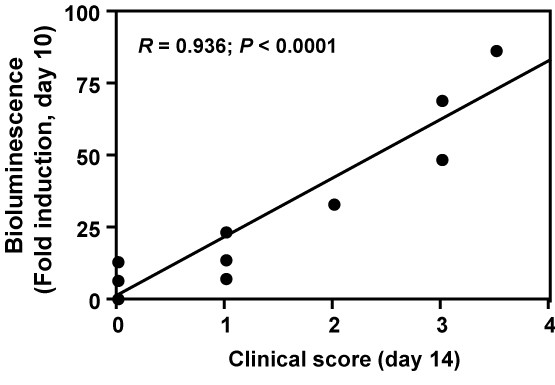
Methods: Experimental autoimmune encephalomyelitis was induced in transgenic mice expressing an injury responsive luciferase reporter in astrocytes (GFAP-luc). Bioluminescence in the brain and spinal cord was measured non-invasively in living mice. Mice were sacrificed at different time points to evaluate clinical and pathological changes. The correlation between bioluminescence and clinical and pathological EAE was statistically analyzed by Pearson correlation analysis.
Results: Bioluminescence from the brain and spinal cord correlates strongly with severity of clinical disease and a number of pathological changes in the brain in EAE. Bioluminescence at early time points also predicts severity of disease.
Conclusion: These results highlight the potential use of bioluminescence imaging to monitor neuroinflammation for rapid drug screening and immunological studies in EAE and suggest that similar approaches could be applied to other animal models of autoimmune and inflammatory disorders.
Figures
References
-
- Kallen B, Nilsson O. Dissociation between histological and clinical signs of experimental auto-immune encephalomyelitis. Acta Pathol Microbiol Immunol Scand [A] 1986;94:159–164. - PubMed
Publication types
MeSH terms
Substances
Grants and funding
LinkOut - more resources
Full Text Sources
Other Literature Sources
Medical
Molecular Biology Databases
Miscellaneous
