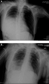
Minimally invasive repair of traumatic right-sided diaphragmatic hernia with delayed diagnosis
- PMID: 18237515
- PMCID: PMC3015846
Minimally invasive repair of traumatic right-sided diaphragmatic hernia with delayed diagnosis
Abstract
Background: Traumatic diaphragmatic hernias are a diagnostic and therapeutic challenge due to variable presentations. Early repair is important because of risks of incarceration and strangulation of abdominal contents along with respiratory and cardiovascular compromise. Minimally invasive techniques have been useful for diagnosis and treatment of diaphragmatic hernias in both blunt and penetrating trauma.
Method: We present the case of a 54-year-old victim of a motor vehicle crash who presented with a delayed diagnosis of a right-sided traumatic diaphragmatic hernia. By using a 4-port technique and intracorporeal suturing, the hernia was repaired. This case highlights the difficulties associated with diagnosing diaphragmatic hernias and the role of minimally invasive techniques to repair them.
Conclusion: Minimally invasive surgical techniques are being increasingly used to both diagnose and repair traumatic diaphragmatic injuries with excellent results.
Figures


Similar articles
-
Laparoscopic repair of traumatic diaphragmatic hernias.Surg Endosc. 2000 Nov;14(11):1010-4. doi: 10.1007/s004640000206. Surg Endosc. 2000. PMID: 11116407 Review.
-
Laparoscopic repair of chronic traumatic diaphragmatic hernia using biologic mesh with cholecystectomy for intrathoracic gallbladder.JSLS. 2011 Oct-Dec;15(4):546-9. doi: 10.4293/108680811X13176785204472. JSLS. 2011. PMID: 22643514 Free PMC article. Review.
-
Laparoscopic repair of a traumatic intrapericardial diaphragmatic hernia.JSLS. 2014 Apr-Jun;18(2):333-7. doi: 10.4293/108680813X13753907290955. JSLS. 2014. PMID: 24960502 Free PMC article. Review.
-
Delayed hepatothorax due to right-sided traumatic diaphragmatic rupture.Gen Thorac Cardiovasc Surg. 2007 Oct;55(10):434-6. doi: 10.1007/s11748-007-0158-y. Gen Thorac Cardiovasc Surg. 2007. PMID: 18018610 Review.
-
Traumatic Right Diaphragmatic Hernia; A Delayed Presentation.J Ayub Med Coll Abbottabad. 2016 Jul-Sep;28(3):625-626. J Ayub Med Coll Abbottabad. 2016. PMID: 28712253
Cited by
-
Laparoscopic repair and total gastrectomy for delayed traumatic diaphragmatic hernia complicated by intrathoracic gastric perforation with tension empyema: a case report.Surg Case Rep. 2022 Jun 20;8(1):117. doi: 10.1186/s40792-022-01477-8. Surg Case Rep. 2022. PMID: 35718811 Free PMC article.
-
Systemic inflammatory response syndrome following laparoscopic repair of diaphragmatic injury: A case report.J Minim Access Surg. 2010 Jan;6(1):16-8. doi: 10.4103/0972-9941.62530. J Minim Access Surg. 2010. PMID: 20585489 Free PMC article.
-
Laparoscopic repair in simultaneous occurrence of recurrent chronic traumatic diaphragmatic hernia and transdiaphragmatic intercostal hernia.Arq Bras Cir Dig. 2015;28(1):90-2. doi: 10.1590/S0102-67202015000100024. Arq Bras Cir Dig. 2015. PMID: 25861081 Free PMC article. No abstract available.
-
Traumatic diaphragmatic hernia: tertiary centre experience.Hernia. 2010 Apr;14(2):159-64. doi: 10.1007/s10029-009-0579-x. Epub 2009 Nov 12. Hernia. 2010. PMID: 19908108
-
Laparoscopic management of traumatic diaphragmatic rupture with herniation of abdominal contents into left hemithorax.Clin Case Rep. 2023 May 18;11(5):e7385. doi: 10.1002/ccr3.7385. eCollection 2023 May. Clin Case Rep. 2023. PMID: 37215976 Free PMC article.
References
-
- Eren S, Kantarci M, Okur A. Imaging of diaphragmatic rupture after trauma. Clin Radiol. 2006;61(6):467–477 - PubMed
-
- Kaw LL, Jr., Potenza BM, Coimbra R, Hoyt DB. Traumatic diaphragmatic hernia. J Am Coll Surg. 2004;198(4):668–669 - PubMed
-
- Sirbu H, Busch T, Spillner J, Schachtrupp A, Autschbach R. Late bilateral diaphragmatic rupture: challenging diagnostic and surgical repair. Hernia. 2005;9(1):90–92 - PubMed
-
- Baglaj M, Dorobisz U. Late-presenting congenital diaphragmatic hernia in children: a literature review. Pediatr Radiol. 2005;35(5):478–488 - PubMed
-
- Christie D, Chapman J, Wynne J, Ashley D. Delayed right-sided diaphragmatic hernia and chronic herniation of unusual abdominal contents. J Am Coll Surg. 2007;204(1):176. - PubMed
Publication types
MeSH terms
LinkOut - more resources
Full Text Sources
