. 2008 Feb 12;105(6):2040-5.
doi: 10.1073/pnas.0711619105. Epub 2008 Jan 31.

Structure and specificity of lamprey monoclonal antibodies

Affiliations

Structure and specificity of lamprey monoclonal antibodies

Brantley R Herrin et al. Proc Natl Acad Sci U S A. .

Abstract

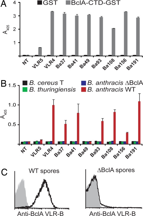
Adaptive immunity in jawless vertebrates (lamprey and hagfish) is mediated by lymphocytes that undergo combinatorial assembly of leucine-rich repeat (LRR) gene segments to create a diverse repertoire of variable lymphocyte receptor (VLR) genes. Immunization with particulate antigens induces VLR-B-bearing lymphocytes to secrete antigen-specific VLR-B antibodies. Here, we describe the production of recombinant VLR-B antibodies specific for BclA, a major coat protein of Bacillus anthracis spores. The recombinant VLR-B antibodies possess 8-10 uniform subunits that collectively bind antigen with high avidity. Sequence analysis, mutagenesis, and modeling studies show that antigen binding involves residues in the beta-sheets lining the VLR-B concave surface. EM visualization reveals tetrameric and pentameric molecules having a central core and highly flexible pairs of stalk-region "arms" with antigen-binding "hands." Remarkable antigen-binding specificity, avidity, and stability predict that these unusual LRR-based monoclonal antibodies will find many biomedical uses.


Conflict of interest statement

The authors declare no conflict of interest.

Figures

Fig. 1.
Fig. 1.
Production of monoclonal VLR-B antibodies specific for BclA-CTD of B. anthracis. (A) VLR-B antibody clones expressed by HEK-293T cells detect recombinant BclA-CTD by ELISA. NT indicates supernatant from nontransfected cells. (B and C) VLR-B antibody clones specifically detect B. anthracis spores by ELISA (B) and flow cytometry (C). ΔBclA, BclA-deficient B. anthracis spores.
Fig. 2.
Fig. 2.
A model of the putative antigen binding surface of a BclA-CTD-specific VLR-B antibody. (A) Homology-based model of the tertiary structure of clone VLR4 constructed by using the crystal structure coordinates of hagfish VLR-B (PDB ID codes 2O6R and 2O6S) as a template. (Upper) Frontal view. (Lower) Side view. Hypervariable positions of each LRR module are indicated by color: LRR-NT (blue), LRR1 (light blue), LRRV (green), LRRVe (light green), CP (yellow), and LRR-CT (orange). (B) Alignment of the LRR-CT sequences of VLR-B clones specific for BclA-CTD. Invariant residues are shaded black, and variable residues are shaded orange.
Fig. 3.
Fig. 3.
Quaternary structure of secreted VLR-B antibodies. (A) Molecular mass estimate of a purified recombinant VLR-B antibody (VLR4) by Ferguson plot analysis. Blue dots, Molecular mass standards; red line, VLR4. (B) Western blot of a partially reduced VLR-B antibody; monomeric (*) and dimeric (**) subunits of the partially reduced antibody. (C) Negative stain TEM of VLR4. (Upper) Pentameric arrangement of VLR-B subunits. (Lower) Flexibility of subunits. (D) A model of VLR-B antibody quaternary structure with red lines representing disulfide bonds. (E) MS/MS sequencing of tryptic fragments of reduced VLR4. Ions matching the sequence of the C terminus of VLR4 are labeled in the spectrograph. CAM indicates carboxyamidomethylation of cysteine by iodoacetamide. (F) Cartoon depicting the location of the cysteine-rich peptide in the VLR-B antibody. The predicted GPI cleave site (bold text) is indicated by an arrow, cysteines that may form intermolecular disulfide bonds are highlighted red, and the peptide detected in E is indicated by a line above the text.
Fig. 4.
Fig. 4.
The primary antigen binding site of VLR-B is located on the concave surface. (A) Sequence alignment of high-avidity (Ba41, Ba191, VLR4) and low-avidity (VLR5) VLR-B antibodies specific for BclA-CTD. Hypervariable positions are shaded black. Amino acids in VLR5 that differ from the high-avidity clones are shaded in color if they are in hypervariable positions or gray if located elsewhere, except V97/N98 (yellow). (B) Homology-based model of VLR5 structure. Amino acids in hypervariable positions are color-coded as in A. (C) Surface plasmon resonance measurement of the interaction between VLR4WT, VLR5WT, or VLR5 mutant antibodies with BclA-CTD. VLR5 residues in hypervariable positions that differ from the high-avidity clones were mutated to the consensus amino acid of the high-avidity clones at that position. Response curves are color-coded with respect to amino acid positions depicted in A and B.

References

    1. Pancer Z, et al. Somatic diversification of variable lymphocyte receptors in the agnathan sea lamprey. Nature. 2004;430:174–180. - PubMed
    1. Pancer Z, et al. Variable lymphocyte receptors in hagfish. Proc Natl Acad Sci USA. 2005;102:9224–9229. - PMC - PubMed
    1. Rogozin IB, et al. Evolution and diversification of lamprey antigen receptors: evidence for involvement of an AID-APOBEC family cytosine deaminase. Nat Immunol. 2007;8:647–656. - PubMed
    1. Alder MN, et al. Diversity and function of adaptive immune receptors in a jawless vertebrate. Science. 2005;310:1970–1973. - PubMed
    1. Nagawa F, et al. Antigen-receptor genes of the agnathan lamprey are assembled by a process involving copy choice. Nat Immunol. 2007;8:206–213. - PubMed

Publication types

LinkOut - more resources