. 2008 Feb 12;105(6):1949-54.
doi: 10.1073/pnas.0707900105. Epub 2008 Jan 31.

Dicer-dependent pathways regulate chondrocyte proliferation and differentiation

Affiliations

Dicer-dependent pathways regulate chondrocyte proliferation and differentiation

Tatsuya Kobayashi et al. Proc Natl Acad Sci U S A. .

Abstract

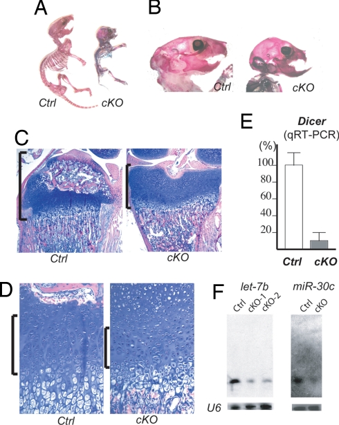
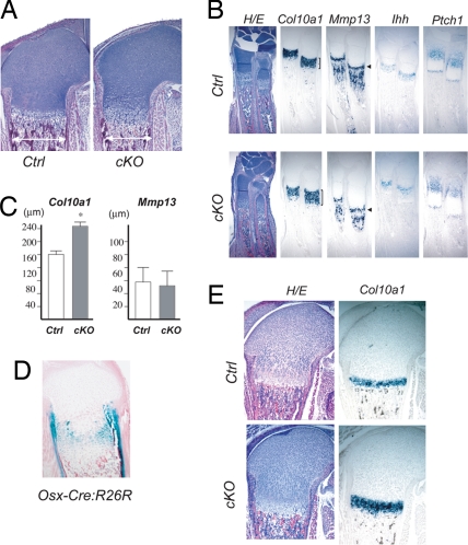
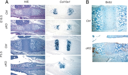
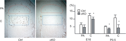
Small noncoding RNAs, microRNAs (miRNAs), bind to messenger RNAs through base pairing to suppress gene expression. Despite accumulating evidence that miRNAs play critical roles in various biological processes across diverse organisms, their roles in mammalian skeletal development have not been demonstrated. Here, we show that Dicer, an essential component for biogenesis of miRNAs, is essential for normal skeletal development. Dicer-null growth plates show a progressive reduction in the proliferating pool of chondrocytes, leading to severe skeletal growth defects and premature death of mice. The reduction of proliferating chondrocytes in Dicer-null growth plates is caused by two distinct mechanisms: decreased chondrocyte proliferation and accelerated differentiation into postmitotic hypertrophic chondrocytes. These defects appear to be caused by mechanisms downstream or independent of the Ihh-PTHrP signaling pathway, a pivotal signaling system that regulates chondrocyte proliferation and differentiation. Microarray analysis of Dicer-null chondrocytes showed limited expression changes in miRNA-target genes, suggesting that, in the majority of cases, chondrocytic miRNAs do not directly regulate target RNA abundance. Our results demonstrate the critical role of the Dicer-dependent pathway in the regulation of chondrocyte proliferation and differentiation during skeletal development.


Conflict of interest statement

The authors declare no conflict of interest.

Figures

Fig. 1.
Fig. 1.
Dicer is required for normal skeletal development. (A) Skeletal preparation of 9-day-old mice. Col2-Cre:Dicer fl/fl mice (cKO) show postnatal growth retardation compared with control littermates (Ctrl). (B) cKO mice at postnatal day 17 show growth defect of the maxilla, causing a relative overgrowth of the mandible. (C and D) Hematoxylin/eosin-stained 12-day-old tibiae. The epiphysis (brackets) of cKO mice is flattened, and formation of the secondary ossification center is delayed (C). The number of column-forming proliferating chondrocytes (brackets) in cKO mice is reduced (D). (E) qRT-PCR analysis using RNA isolated from microdissected hindlimb cartilage of 3-day-old mice shows a reduction in Dicer mRNA level by 87% in cKO mice. (F) Northern blot analysis on RNA of hindlimb cartilage of 3-day-old mice shows reduced expression of let-7b and miR-30c in cKO mice.
Fig. 2.
Fig. 2.
Acceleration of hypertrophic differentiation of Dicer-null chondrocytes. (A) The bone width (double arrows) is reduced, and the hypertrophic region of the growth plate is expanded in neonatal Col2-Cre:Dicer fl/fl (cKO) mice. (B) In situ hybridization analysis demonstrates the expansion of the hypertrophic region in E18.5 cKO growth plates of the radius, as indicated by the enlargement of the Col10a1 domain, whereas there is no expansion of the Mmp13 domain that marks terminally differentiated hypertrophic chondrocytes in the growth plate (arrowheads), suggesting that acceleration of hypertrophic differentiation, rather than reduction of cartilage resorption, is responsible for the expansion of the hypertrophic region. Mmp13 is also expressed in osteoblasts in the bone marrow. Expression of mRNAs for Indian hedgehog (Ihh) and its transcriptional target, Ptch1 is preserved. (C) The longitudinal length of the Col10a1 domain was significantly increased in cKO mice by 35% (ANOVA; n = 3 P < 0.05), whereas the Mmp13 domain was not expanded in the cKO growth plate. (D and E) Deletion of Dicer in late-proliferating chondrocytes using Osx-Cre mice results in expansion of the hypertrophic region. Cre activity of Osx-Cre transgenic mice, assessed by R26R reporter assay (54), is observed in late proliferating chondrocytes in addition to cells in the osteoblast lineage (C). Neonatal Osx-Cre:Dicer fl/fl mice (cKO) show expansion of hypertrophic region marked with Col10a1 (D).
Fig. 3.
Fig. 3.
Reduced number of proliferating chondrocytes in the basisphenoidal-baisoccipital growth plate. (A) Hematoxylin/eosin (H/E) staining and Col10a1 expression in the basiosphenoidal/basioccipital growth plate at E18.5 and P3.5. Col2-Cre:Dicer fl/fl mice (cKO) show a progressive reduction in the width of the growth plate and the proliferating chondrocytes flanked by Col10a1-positive hypertrophic chondrocytes. Asterisks indicate the pituitary gland. (B) Reduced BrdU-positive chondrocytes in the cKO growth plate at P3.5.
Fig. 4.
Fig. 4.
Reduced chondrocyte proliferation in Col2-Cre:Dicer fl/fl mice (cKO). The BrdU-labeling assay demonstrates a dramatic reduction in chondrocyte proliferation in cKO growth plates. The BrdU-labeling index was calculated in the periarticular (PA) and columnar proliferating (C) region of the proximal growth plate of the tibia at the indicated ages. Representative BrdU-staining pictures of P0.5 tibiae are shown. The BrdU-labeling index in cKO mice was significantly reduced in the columnar region at E16.5, and in both periarticular and columnar regions at P0.5. Asterisks indicate statistical significance with the P < 0.05.
Fig. 5.
Fig. 5.
miRNA expression in chondrocytes. (A) Relative expression levels of the 30 most-abundant miRNAs in neonatal hindlimb cartilage (chondrocytes) and calvarial cells (osteoblasts). Many abundant miRNAs are similarly expressed in chondrocytes and osteoblasts. (B) Thirty miRNAs preferentially expressed in chondrocytes. The expression of muscle-specific miRNAs, mir-206, -133, and -1 could be overestimated because of possible contamination of muscle tissues to cartilage specimens. (C) Expression of 30 miRNAs in control and Dicer-deficient chondrocytes. RNA isolated from neonatal hindlimb cartilage was subjected to miRNA profiling. Expression levels of most of miRNAs were reduced by 60–70% in Dicer-deficient chondrocytes. Complete data are shown in SI Table 2.

References

    1. Rodriguez A, Vigorito E, Clare S, Warren MV, Couttet P, Soond DR, van Dongen S, Grocock RJ, Das PP, Miska EA, et al. Science. 2007;316:608–611. - PMC - PubMed
    1. Thai TH, Calado DP, Casola S, Ansel KM, Xiao C, Xue Y, Murphy A, Frendewey D, Valenzuela D, Kutok JL, et al. Science. 2007;316:604–608. - PubMed
    1. van Rooij E, Sutherland LB, Qi X, Richardson JA, Hill J, Olson EN. Science. 2007;316:575–579. - PubMed
    1. Zhao Y, Ransom JF, Li A, Vedantham V, von Drehle M, Muth AN, Tsuchihashi T, McManus MT, Schwartz RJ, Srivastava D. Cell. 2007;129:303–317. - PubMed
    1. Bernstein E, Kim SY, Carmell MA, Murchison EP, Alcorn H, Li MZ, Mills AA, Elledge SJ, Anderson KV, Hannon GJ. Nat Genet. 2003;35:215–217. - PubMed

Publication types