Review
. 2008 May 1;111(9):4446-55.
doi: 10.1182/blood-2007-08-019729. Epub 2008 Jan 31.

Telomere maintenance and human bone marrow failure

Affiliations
Review

Telomere maintenance and human bone marrow failure

Rodrigo T Calado et al. Blood. .

Abstract

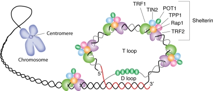
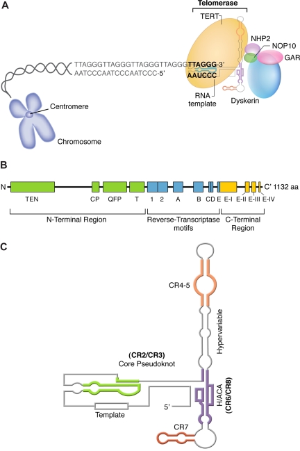
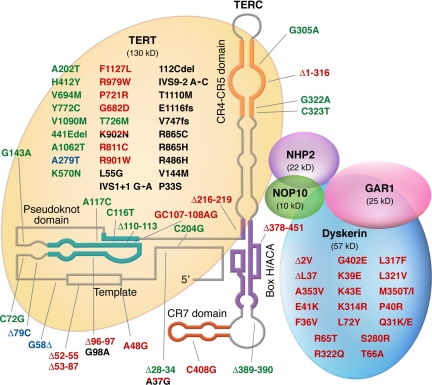
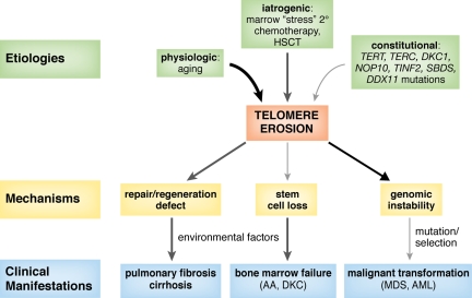
Acquired and congenital aplastic anemias recently have been linked molecularly and pathophysiologically by abnormal telomere maintenance. Telomeres are repeated nucleotide sequences that cap the ends of chromosomes and protect them from damage. Telomeres are eroded with cell division, but in hematopoietic stem cells, maintenance of their length is mediated by telomerase. Accelerated telomere shortening is virtually universal in dyskeratosis congenita, caused by mutations in genes encoding components of telomerase or telomere-binding protein (TERT, TERC, DKC1, NOP10, or TINF2). About one-third of patients with acquired aplastic anemia also have short telomeres, which in some cases associate with TERT or TERC mutations. These mutations cause low telomerase activity, accelerated telomere shortening, and diminished proliferative capacity of hematopoietic progenitors. As in other genetic diseases, additional environmental, genetic, and epigenetic modifiers must contribute to telomere erosion and ultimately to disease phenotype. Short telomeres also may cause genomic instability and malignant progression in these marrow failure syndromes. Identification of short telomeres has potential clinical implications: it may be useful in dyskeratosis congenita diagnosis, in suggesting mutations in patients with acquired aplastic anemia, and for selection of suitable hematopoietic stem cell family donors for transplantation in telomerase-deficient patients.


Figures

Figure 1
Figure 1
Schematic representation of telomere structure. Telomeres are at the extremities of chromosome DNA. The telomeric 3′ end terminates as a single-stranded, G-rich overhang able to form the t-loop, in which the overhang invades the telomeric double helix, remodeling the DNA into a circle. Telomeres are capped by at least 6 proteins (TRF1, TRF2, TPP1, POT1, TIN2, and Rap1), collectively known as shelterin, that physically shield the DNA. TRF1, TRF2, and TPP1 specifically recognize and bind to double-stranded TTAGGG repeats; POT1 binds to the single-stranded telomeric overhang,; TIN2 and Rap1 complete the shelterin complex. Shelterin allows discrimination of telomeres from double-stranded DNA breaks; lack of shelterin allows telomeres to be identified as double-stranded DNA breaks and triggers DNA-damage pathways.
Figure 2
Figure 2
Structure and function of the telomerase complex. (A) TERT enzymatically adds TTAGGG nucleotide repeats to the 3′ end of telomere's leading strand using TERC as a template. Other proteins (dyskerin, NOP10, NHP2, and GAR) also bind to TERC and stabilize the complex. (B) Linear structure of TERT, which is highly conserved among eukaryotes and consists of the central reverse transcriptase (RT) motifs (1, 2, A, B, C, D, and E), a large N-terminal region, and a short C-terminal region, all necessary for telomerase enzymatic function. The N-terminal region comprises a telomerase-essential N-terminal domain (TEN), the CP, and the QFP domains, required for RNA interaction, and a telomerase-specific T motif. The C-terminal region contains 4 conserved domains (E-I to E-IV). (C) Secondary structure of human TERC, which contains 7 conserved regions (CRs), a pseudoknot important for interaction with TERT (CR2/CR3), and a template used by TERT for telomere elongation (CR1). TERC also encloses a small nucleolar H/ACA motif; box H/ACA refers to a tail region of small nucleolar RNAs (snoRNAs) carrying a conserved H motif (AnAnnA) and consensus ACA triplet positioned 3 nucleotides before the 3′ end of the RNA that characterize a major snoRNA family involved in pseudouridylation of pre-rRNAs. TERC binds to other proteins, such as dyskerin, GAR, NHP2, and NOP10, through box H/ACA.
Figure 3
Figure 3
Mutations in telomerase complex genes and human disease. Mutations in green were described in patients with acquired aplastic anemia; mutations in red were described in patients with dyskeratosis congenita; mutations in black were described in patients with pulmonary fibrosis; and polymorphisms are represented in blue. Mutations found in more than one disease type are double-colored.
Figure 4
Figure 4
Proposed model for the role of dysfunctional and short telomeres in the pathogenesis of human disease. Aging, bone marrow stress, and genetic factors, such as mutations in telomerase complex genes (TERT, TERC, DKC1, and NOP10), shelterin components (TINF2, TERF1, and TERF2), or in other genes (SBDS and DDX11) produce progressive telomere erosion. Excessive telomere shortening results in defective cell proliferation, senescence, apoptosis, and genomic instability. Environmental factors, such as viruses, drugs, smoking, or asbestos exposure, may contribute to telomere shortening as well as injury to an organ with limited regeneration capacity, thus triggering disease development (aplastic anemia, pulmonary fibrosis, and hepatic cirrhosis). Short telomeres also promote genomic instability, breakage-fusion-bridge cycles, and aneuploidy, which can lead to myelodysplasia (MDS) or leukemia (AML).

References

    1. Ball SE, Gibson FM, Rizzo S, et al. Progressive telomere shortening in aplastic anemia. Blood. 1998;91:3582–3592. - PubMed
    1. Brummendorf TH, Maciejewski JP, Young NS, Lansdorp PL. Telomere length in leukocyte subpopulations of patients with aplastic anemia. Blood. 2001;97:895–900. - PubMed
    1. Lee JJ, Kook H, Chung IJ, et al. Telomere length changes in patients with aplastic anaemia. Br J Haematol. 2001;112:1025–1030. - PubMed
    1. Leteurtre F, Li X, Guardiola P, et al. Accelerated telomerase shortening and telomerase activation in Fanconi's anemia. Bone Marrow Transplant. 1999;23:S30.
    1. li X, Leteurtre F, Rocha V, et al. Abnormal telomere metabolism in Fanconi's anaemia correlates with genomic instability and the probability of developing severe aplastic anaemia. Br J Haematol. 2003;120:836–845. - PubMed

Publication types