. 2008 May 1;111(9):4511-22.
doi: 10.1182/blood-2007-07-102848. Epub 2008 Jan 31.

Stat5 activation enables erythropoiesis in the absence of EpoR and Jak2

Affiliations

Stat5 activation enables erythropoiesis in the absence of EpoR and Jak2

Florian Grebien et al. Blood. .

Abstract

Erythropoiesis requires erythropoietin (Epo) and stem cell factor (SCF) signaling via their receptors EpoR and c-Kit. EpoR, like many other receptors involved in hematopoiesis, acts via the kinase Jak2. Deletion of EpoR or Janus kinase 2 (Jak2) causes embryonic lethality as a result of defective erythropoiesis. The contribution of distinct EpoR/Jak2-induced signaling pathways (mitogen-activated protein kinase, phosphatidylinositol 3-kinase, signal transducer and activator of transcription 5 [Stat5]) to functional erythropoiesis is incompletely understood. Here we demonstrate that expression of a constitutively activated Stat5a mutant (cS5) was sufficient to relieve the proliferation defect of Jak2(-/-) and EpoR(-/-) cells in an Epo-independent manner. In addition, tamoxifen-induced DNA binding of a Stat5a-estrogen receptor (ER)* fusion construct enabled erythropoiesis in the absence of Epo. Furthermore, c-Kit was able to enhance signaling through the Jak2-Stat5 axis, particularly in lymphoid and myeloid progenitors. Although abundance of hematopoietic stem cells was 2.5-fold reduced in Jak2(-/-) fetal livers, transplantation of Jak2(-/-)-cS5 fetal liver cells into irradiated mice gave rise to mature erythroid and myeloid cells of donor origin up to 6 months after transplantation. Cytokine- and c-Kit pathways do not function independently of each other in hematopoiesis but cooperate to attain full Jak2/Stat5 activation. In conclusion, activated Stat5 is a critical downstream effector of Jak2 in erythropoiesis/myelopoiesis, and Jak2 functionally links cytokine- with c-Kit-receptor tyrosine kinase signaling.


Figures

Figure 1
Figure 1. Expression of cS5 rescues proliferation and differentiation of EpoR−/− and Jak2−/− erythroid cells
(A) E12.5 WT, EpoR−/−, and Jak2−/− fetal liver cells infected with retroviruses encoding GFP (left panel) or cS5 (right panel) were cultivated in proliferation medium and cumulative cell numbers calculated after daily determination of growth rates. Data plotted for 1 typical experiment of 4. (B) Cytospins were prepared at day 6 from erythroid cultures shown in (A) and stained for hemoglobin (brownish color) plus histologic dyes. WT, Jak2−/−, and EpoR−/− fetal liver cells expressing cS5 or GFP were subjected to CFU-E- (C) or BFU-E assays (D), and acid benzidine–positive colonies were scored at day 2 (CFU-E) or day 8 (BFU-E), respectively. (E) Cytospins of cells retrieved from the CFU-E assays in (C), stained with hematoxylin/eosin and for hemoglobin (brownish color).
Figure 2
Figure 2. cS5 expression allows erythroid development in the absence of Epo
(A) E12.5 WT, EpoR−/−, and Jak2−/− fetal liver cells were infected with retroviruses encoding GFP or cS5 and cultivated in proliferation medium without Epo for 6 days. Cumulative cell numbers are shown for one representative experiments of 3. (B) Cytospins were prepared from cultures shown in panel A at day 4 (→ in A) and stained with hematoxylin/eosin and benzidine. → indicates apoptotic cells; ▶, pyknotic nuclei. (C) WT fetal liver cells expressing cS5 or GFP, or Jak2−/− and EpoR−/− cells expressing cS5, were subjected to CFU-E assays in the absence of Epo and acid benzidine–positive colonies scored at day 2. (D) Cytospins of cells retrieved from the CFU-E assays in (C), stained with hematoxylin/eosin and for hemoglobin (brownish).
Figure 3
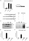
Figure 3. cS5 allows Epo-independent induction of a Stat5-responsive reporter construct but requires tyrosine phosphorylation and DNA-binding for activity
(A) Self renewing WT GFP and cS5-expressing WT and Jak2−/− erythroblasts were transfected with IL-2R-α-Luc. Before harvesting, cells were stimulated with Epo for 5 hours (+) or left untreated (−). Luciferase expression was measured 12 hours after transfection. Right, schematic representation of the promoter enhancer element from the IL-2R-α gene containing 2 Stat5 response elements (Stat5-RE) fused to the luciferase gene (IL-2R-α-Luc). (B) Schematic representation of the mutants used. 293T cells were transiently transfected with different cS5 constructs, together with a murine EpoR cDNA. Twenty-four hours later, cells were left untreated or treated for 30 minutes with Epo. Extracts were analyzed for tyrosine-phosphorylated Stat5 and total Stat5 by Western blot (C) and DNA-binding of transfected Stat5 constructs by EMSAs on a β-casein-specific promoter sequence (D). Jak2−/− (E) and EpoR−/− (F) fetal liver cells were infected with retroviruses encoding GFP, cS5, cS5-EE/AA, or cS5-Y694F and subjected to CFU-E assays. Acid benzidine–positive colonies were scored at day 2.
Figure 4
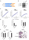
Figure 4. 4-Hydroxy-tamoxifen-induced Stat5a-ER* activation replaced Epo in erythropoiesis
(A) Scheme of the 4-OH-T-inducible Stat5-ER* constructs used, encoding fusion proteins of WT Stat5a, cS5, or Stat5aΔ749 with ER* (see “Methods”). (B) Western blot analysis of phosphorylated Stat5 (P-Y-Stat5), and total Stat5 protein in WT fetal liver erythroblasts expressing GFP or Stat5a-ER*. The larger protein recognized by P-Y-Stat5 and Stat5 antibodies corresponds to Stat5a-ER*. Actin, loading control. (C) Cumulative cell numbers (1 representative experiment of 3) of proliferating primary erythroblast cultures expressing Stat5a-ER* or GFP determined in the presence of Epo (+ EPO, −4-OH-T, left, normal self-renewal conditions), the presence of 4-OH-T (5 nM) instead of Epo (−Epo, + 4-OH-T, middle) and without Epo and 4-OH-T (right). (D) Percentage of apoptotic cells of cultures in panel C at day 6 (arrows in panel C) as analyzed by annexin V staining. (E) WT fetal liver cells expressing Stat5a-ER* or GFP were subjected to CFU-E assays in the presence of Epo (left panels) or 4-OH-T (50 nM) instead of Epo (right panels). Acid benzidine–positive colonies were scored at day 2. (F) Cytospins of cells retrieved from the CFU-E assays in panel E and stained with hematoxylin/eosin and for hemoglobin (brownish color).
Figure 5
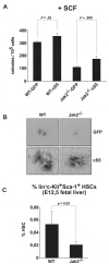
Figure 5. c-Kit signaling depends on Jak2 and is modulated by Stat5
(A) WT and Jak2−/− fetal liver cells transduced with GFP or cS5 were subjected to colony assays supplemented with SCF (50 ng/mL). Colonies were scored at day 8. (B) Photographs of colonies from (A). Representative pictures of 4 for each condition are shown. (C) E12.5 WT and Jak2−/− fetal livers analyzed for HSC content (linc-Kit+Sca-1+ cells) by flow cytometry. The percentage of HSCs is depicted (n = 4).
Figure 6
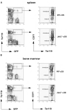
Figure 6. Jak2−/− cells expressing cS5 undergo erythroid differentiation in vivo
Equal numbers of cS5-transduced E12.5 WT and Jak2−/− fetal liver cells were injected into sublethally irradiated mice. Six months after transplantation, spleen (A) and bone marrow (B) of engrafted animals were monitored for GFP-positive erythroid cells. FACS plots for Ter119 and GFP (left) indicate transplant-derived double-positive erythroid cells. Gated cells (boxed) were further analyzed for CD71 and Ter119 (right) to discriminate between immature (CD71highTer119pos) and mature (CD71lowTer119pos) erythroid cells.
Figure 7
Figure 7. Jak kinase(s) and c-Kit cooperate in cS5 activation
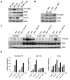
(A) Lysates from self-renewing WT GFP, WT cS5, Jak2−/−-cS5, and EpoR−/−-cS5 cultures were analyzed for P-Y-Stat5, total Stat5, Jak2, and EpoR protein levels. Actin, loading control. (B) cS5-expressing WT erythroblasts were starved for 3 hours (−) and subsequently stimulated with Epo, SCF, or Epo + SCF for 10 minutes. Lysates were analyzed for P-Y-Stat5 and total Stat5 protein. Erk1/2, loading control. (C) WT erythroblasts expressing GFP or cS5, and Jak2−/− and EpoR−/− cells expressing cS5 were starved for 3 hours (−) and subsequently stimulated with Epo, SCF, or SCF + imatinib (10 μmol/L) for 10 minutes. Lysates were analyzed for P-Y-Stat5 and total Stat5 protein. Erk1/2, loading control. (D) WT GFP, WT cS5, Jak2−/−-cS5, and EpoR−/−-cS5 cultures were starved for 3 hours (−) and subsequently restimulated with SCF for 2 hours (+). Expression of the Stat5 target genes oncostatinM, cyclin D2, and c-myc was assessed by real-time PCR.

Similar articles

Cited by

References

    1. Richmond TD, Chohan M, Barber DL. Turning cells red: signal transduction mediated by erythropoietin. Trends Cell Biol. 2005;15:146–155. - PubMed
    1. Klingmüller U, Wu H, Hsiao JG, et al. Identification of a novel pathway important for proliferation and differentiation of primary erythroid progenitors. Proc Natl Acad Sci U S A. 1997;94:3016–3021. - PMC - PubMed
    1. Chen C, Sytkowski AJ. Erythropoietin regulation of Raf-1 and MEK: evidence for a Ras-independent mechanism. Blood. 2004;104:73–80. - PubMed
    1. von Lindern M, Parren-van Amelsvoort M, van Dijk T, et al. Protein kinase C alpha controls erythropoietin receptor signaling. J Biol Chem. 2000;275:34719–34727. - PubMed
    1. Tong Q, Chu X, Cheung JY, et al. Erythropoietin-modulated calcium influx through TRPC2 is mediated by phospholipase Cgamma and IP3R. Am J Physiol Cell Physiol. 2004;287:C1667–1678. - PubMed

Publication types