. 2008 Jan;20(1):101-23.
doi: 10.1105/tpc.107.052001. Epub 2008 Jan 31.

Rab-A2 and Rab-A3 GTPases define a trans-golgi endosomal membrane domain in Arabidopsis that contributes substantially to the cell plate

Affiliations

Rab-A2 and Rab-A3 GTPases define a trans-golgi endosomal membrane domain in Arabidopsis that contributes substantially to the cell plate

Cheung-Ming Chow et al. Plant Cell. 2008 Jan.

Abstract

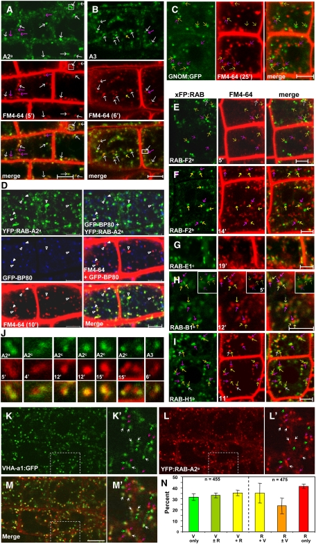
The Ypt3/Rab11/Rab25 subfamily of Rab GTPases has expanded greatly in Arabidopsis thaliana, comprising 26 members in six provisional subclasses, Rab-A1 to Rab-A6. We show that the Rab-A2 and Rab-A3 subclasses define a novel post-Golgi membrane domain in Arabidopsis root tips. The Rab-A2/A3 compartment was distinct from but often close to Golgi stacks and prevacuolar compartments and partly overlapped the VHA-a1 trans-Golgi compartment. It was also sensitive to brefeldin A and accumulated FM4-64 before prevacuolar compartments did. Mutations in RAB-A2a that were predicted to stabilize the GDP- or GTP-bound state shifted the location of the protein to the Golgi or plasma membrane, respectively. In mitosis, KNOLLE accumulated principally in the Rab-A2/A3 compartment. During cytokinesis, Rab-A2 and Rab-A3 proteins localized precisely to the growing margins of the cell plate, but VHA-a1, GNOM, and prevacuolar markers were excluded. Inducible expression of dominant-inhibitory mutants of RAB-A2a resulted in enlarged, polynucleate, meristematic cells with cell wall stubs. The Rab-A2/A3 compartment, therefore, is a trans-Golgi compartment that communicates with the plasma membrane and early endosomal system and contributes substantially to the cell plate. Despite the unique features of plant cytokinesis, membrane traffic to the division plane exhibits surprising molecular similarity across eukaryotic kingdoms in its reliance on Ypt3/Rab11/Rab-A GTPases.


Figures

Figure 1.
Figure 1.
At RAB-A2, At RAB-A3, and Ps RAB-A3 Label the Same Compartment, Which Is Distinct from the Golgi and the GFP-BP80–Labeled PVC. (A) CLSM analysis of seedling root tip epidermis coexpressing GFP:PsRAB-A3 (green) with YFP:RAB-A3 or YFP:RAB-A2a (fusions to Arabidopsis Rab proteins) as indicated (each red). White arrows indicate examples of structures with extensive overlap of GFP and YFP signals. Bars = 5 μm. (B) Protein gel blot analysis of total protein extracted from roots of 14-d-old seedlings of wild-type or transgenic plants expressing the indicated YFP:AtRab fusion and probed with desalted affinity-purified anti-RAB-A2a (1:1000) (top panel) or anti-GFP (1:1000) (bottom panel). Open arrows indicate the expected sizes of YFP:AtRAB fusions (50 kD); the closed arrow indicates the expected size of endogenous At RAB-A2a (26 kD). (C) Sequences of At RAB-A2a peptide antigen and the At RAB-A2d C terminus are dissimilar. The sequences were manually aligned to maximize the number of consecutive shared amino acids. Underlining indicates the conserved Cys residue that is expected to be geranylgeranylated in the native protein and was used for coupling the peptide. The At RAB-A2a peptide and At RAB-A2d share only eight residues (red), and none of these residues form a contig of three residues or more. (D) and (E) Immunolocalization of At RAB-A2a (red) in root tip cells expressing YFP:AtRAB-A2d (green). The 4′,6-Diamidino-2-phenylindole (DAPI)–stained nuclei in (E) are in blue. Arrowheads indicate examples of colocalizing punctate structures; arrows indicate labeling at the cell periphery. Bars = 5 μm. (F) CLSM analysis of seedling root tip epidermal cells coexpressing YFP:AtRAB-A2a or YFP:AtRAB-A3 (red) with ST-GFP or GFP-BP80 (each green) as indicated. Yellow arrows point to the Golgi or PVC (green), which are not close to any YFP:AtRAB-A–labeled structure (red puncta). Pink arrows point to the YFP:AtRAB-A–labeled structures, which are not close to the Golgi or PVC. White arrows point to the YFP:RAB-A–labeled punctate structures, which are closely associated with the Golgi or PVCs. Occasionally, YFP:RAB-A–labeled structures and GFP-BP80-labeled PVCs partially overlap (e.g., the boxed region). Bars = 5 μm.
Figure 2.
Figure 2.
The Rab-A2/A3 Compartment Is Rapidly Labeled by FM4-64 and Overlaps the VHA-a1 TGN. CLSM analysis of epidermal cells in seedling root tips. Bars = 5 μm (A) to (C) and (E) to (I) Roots expressing YFP:RAB-A2a (A), YFP:RAB-A3 (B), GNOM:GFP (C), YFP:RAB-F2a (E), mRFP:RAB-F2b (F), YFP:RAB-E1c (G), YFP:RAB-B1b (H), or YFP:RAB-H1b (I) (each green) incubated with FM4-64 (red) for the times indicated. Yellow arrows indicate xFP-labeled regions without detectable FM4-64 labeling; pink arrows indicate FM4-64–labeled regions without detectable xFP signal; white arrows indicate regions with extensive overlap of xFP and FM4-64 signals. Enlarged images of the boxed areas in (A) and (B) are presented in the first and last columns in (J). (D) Seedlings coexpressing YFP:RAB-A2a (green) and GFP-BP80 (blue) incubated with FM4-64 (red) for 10 min. YFP:RAB-A2a– but not GFP-BP80–labeled compartments are extensively colabeled with FM4-64. Left column, single channels; right column, overlay of two or three channels as indicated. Open arrowheads indicate YFP:RAB-A2a; arrowheads indicate GFP-BP80. (J) Examples of punctate structures with different degrees of overlap between FM4-64 and YFP:RAB-A2/A3. Green indicates YFP fusions to members of Arabidopsis Rab-A2 or Rab-A3 subclasses as indicated; red indicates FM4-64 with incubation times shown. (K) to (M) Seedlings coexpressing VHA-a1:GFP (green) and YFP:AtRAB-A2a (red). K′ to M′ show enlarged areas boxed in (K) to (M). Pink or green arrowheads indicate structures labeled only by YFP:RAB-A2a or VHA-a1:GFP, respectively; white arrowheads indicate structures with both signals in various ratios. (N) Quantitative assessment of the number of colocalizing VHA-a1 and Rab-A2/A3 compartments. V, VHA-a1:GFP; R, Rab-A2/A3. A total of 455 VHA-a1 compartments in images such as those in (M) were assigned to three categories designated V only, V ± R, and V + R, indicating that they had either no, weak, or similar intensity YFP:RAB-A2a signals, respectively. The reciprocal analysis was done on 475 YFP:RAB-A2a–labeled compartments, and the percentage in each category is shown. Error bars indicate sd values for data from three images.
Figure 3.
Figure 3.
RAB-A2/A3 Proteins Localize to Cell Plates. CLSM analysis of seedlings. (A) to (F) RAB-A2/A3 targeted xFP to cell plate–like structures, which were rapidly labeled with FM4-64. Meristemic tissues of root tips ([A] and [C] to [F]) or young true leaves (B) of Arabidopsis seedlings expressing fluorescent protein fusions to At RAB-A2a ([A] and [E]), At RAB-A2c (B), At RAB-A2d (F), At RAB-A3 (C), or Ps RAB-A3 (D). xFP:RAB-A is shown in white or green; FM4-64 is shown in red. xFP:RAB signals were seen to concentrate in linear structures in single optical sections ([A] to [D]; bars = 10 μm). (E) Top two panels, three-dimensional reconstruction at two different angles from serial optical sections of YFP:AtRAB-A2a–labeled discs; bottom panel, single optical section showing that these structures label with FM4-64 (red) but with a higher relative abundance of YFP:AtRAB-A2a at the periphery of the incomplete disc (arrow). Bars = 5 μm. (F) YFP:AtRAB-A2d (green) is concentrated at the periphery (arrows) of FM4-64 (red)–labeled cell plates. The three-dimensional reconstruction from serial sections of the dotted area is shown as an inset. Note that some of the YFP- and FM4-64–labeled cell plates have open ends. Bar = 5 μm. (G) to (J) Immunolocalization of α-tubulin (G) or KN ([H] to [J]) in meristemic tissues of root tips of 5-d-old Arabidopsis seedlings expressing YFP:AtRAB-A2a ([G], [I], and [J]) or YFP:AtRAB-A2c (H). Microtubules or KN are shown in red; DAPI is shown in blue; YFP is shown in green. White arrows indicate extensive overlap between YFP:AtRAB-A and KN signals; yellow arrows indicate YFP:AtRAB-A signal alone; pink arrows indicate KN signal alone. In (H) to (J), three-dimensional reconstruction images of serial optical sections are shown as merged images below the single optical sections in the top three rows. Bars = 5 μm. (K) Endogenous At RAB-A2a localizes to the cell plate. Double immunolocalization of α-tubulin (green) and RAB-A2a (red) in wild-type root tips of 5-d-old Arabidopsis seedlings stained with DAPI (blue). Affinity-purified anti-RAB-A2a (1:3000) was used to probe the native Rab-A2a proteins at different stages of mitosis and cytokinesis, including metaphase and telophase. Note that RAB-A2a only concentrated in the midzone of microtubule arrays in telophase. First to fourth columns, single section images; fifth column, three-dimensional reconstruction from serial images along the z axis. Bars = 5 μm.
Figure 4.
Figure 4.
Rab-A2/A3 Membranes Colocalize with KN in Mitosis and Maintain Their Identity during Cytokinesis. CSLM analysis of mitotic and dividing root tip cells expressing various Golgi and endosomal or prevacuolar markers. Three-dimensional reconstruction images are shown in (A) to (C), while single section images are shown in (D) to (M). Bars = 5 μm. (A) to (D) and (F) Immunolocalization of KN (red) at metaphase ([A] to [C]) or telophase ([D] and [F]) in roots expressing YFP:AtRAB-A2d, Nag:EGFP, GFP-BP80, or N-ST-YFP (green) stained with DAPI (blue). Enlarged images of boxed areas in (A) to (C) are shown at left of each image as single channels and merged images. The dashed line in (C) indicates the boundary of the mitotic cell. (E) and (G) Dividing root tip cells coexpressing ST-GFP or GFP-BP80 with YFP:AtRAB-A2c or -A2b. White arrows indicate YFP:AtRAB-A2 proteins, which are close to ST-GFP or GFP-BP80. (H) to (M) Dividing root tip cells expressing RFP:AtRAB-F2b, YFP:AtRAB-A2a, GNOM:GFP (GN:GFP), or VHA-a1:GFP as indicated, stained with FM4-64. In (F) and (H) to (L), white arrows indicate colocalizing structures and yellow or pink arrows indicate structures that have only green or red signal, respectively. The arrow in (M) indicates a cell plate labeled by YFP:AtRAB-2a but not VHA-a1.
Figure 5.
Figure 5.
Localization of Mutant At RAB-A2a Proteins. YFP-tagged At RAB-A2a wild type (WT), S26N (SN) mutant, or Q71L (QL) mutant (each green) with either the Golgi marker ST-RFP or FM4-64 (each red) as indicated. (C) shows cells after treatment with BFA. (D) shows dividing cells with labeled cell plates (arrows in merged images). All images are confocal sections of seedling root tips. Bars = 4 μm.
Figure 6.
Figure 6.
Dominant-Inhibitory At RAB-A2a Mutants Block Cytokinesis. At RAB-A2a wild type and S26N (SN), Q71L (QL), or N125I (NI) mutants were expressed in seedlings from a dexamethasone-inducible promoter. (A) and (B) Eleven-day-old seedlings (A) and their primary root tips (B) germinated and grown in the presence of 20 μM dexamethasone (+Dex) or the equivalent quantity of DMSO solvent (−Dex). (C) to (M) Confocal images of propidium iodide–stained nuclei (red) and Calcofluor-stained cell walls (green) in 7-d-old seedlings ([C] to [H]) or 14-d-old seedlings ([I] to [M]) grown with (+Dex) or without (−Dex) dexamethasone. Arrowheads in (F), (J), (L), and (M) indicate multiple nuclei in single cells; arrows indicate cell wall stubs. Serial confocal sections of the cells marked by asterisks in (F) and (G) revealed that they had four and six nuclei, respectively.
Figure 7.
Figure 7.
Schematic Models of Endosomal and Prevacuolar/Lysosomal Compartments in Arabidopsis Root Tips, Yeast, and a Nonpolarized Mammalian Cell. (A) Model for the organization of the endosomal system in Arabidopsis root tips based on the results of this study; the Rab-A2/A3 compartment overlaps the VHA-a1 TGN compartment and is an EE on the FM4-64 uptake pathway (red arrows and compartments). The proposed pathway of At RAB-A2a cycling is shown in blue arrows, with dashed arrows indicating cycling on and off membranes. Markers for each compartment are indicated in green. Dotted arrows are hypothetical. In accordance with Ueda et al. (2004), the Rab-F1 PVC is placed downstream of the overlapping Rab-F2 PVC, which is a multivesicular body. The position of the GNOM recycling compartment is unclear. FYVE is a PI(3)P binding marker. ER, endoplasmic reticulum. Circled numbers in all panels designate transport steps referred to in the text. (B) and (C) Schematic representations of the mammalian and S. cerevisiae endosomal systems after Gruenberg and Stenmark (2004), Prescianotto-Baschong and Riezman (2002), and Pelham (2002). Markers of each compartment are shown in red letters, with their Arabidopsis homolog in green. The mammalian EE shares several markers with the Arabidopsis PVC and yeast prevacuolar endosome (PVE), which are both later endosomes. The Arabidopsis Rab-A2/A3 and VHA-a1 compartments share markers with the closely connected yeast late-Golgi and post-Golgi endosome (PGE), which is also an EE. Light (LV) and dense (DV) vesicles mediate alternative routes to the cell surface in S. cerevisiae (Harsay and Schekman, 2002). The orthologous Rab and SNARE proteins label the mammalian recycling endosome, which lies downstream of the EE, on the secretory pathway for many proteins. (D) The cell plate shares markers specifically with the Rab-A2/A3 membrane domain but also apparently accumulates both GDP- and GTP-bound forms of RAB-A2a; the position of the Rab-A2/A3 domain on the exocytic and endocytic routes can account for the appearance of endocytosed molecules such as FM4-64 in the cell plate.

References

    1. Albertson, R., Riggs, B., and Sullivan, W. (2005). Membrane traffic: A driving force in cytokinesis. Trends Cell Biol. 15 92–101. - PubMed
    1. Anai, T., Hasegawa, K., Watanabe, Y., Uchimiya, H., Ishizaki, R., and Matsui, M. (1991). Isolation and analysis of cDNAs encoding small GTP-binding proteins of Arabidopsis thaliana. Gene 108 259–264. - PubMed
    1. Ausubel, F., Brent, R., Kingston, R.E., Moore, J.G., Seidman, J.G., Smith, J.A., and Struhl, J.G. (1999). Current Protocols in Molecular Biology. (New York: John Wiley & Sons).
    1. Baluska, F., Liners, F., Hlavacka, A., Schlicht, M., Van Cutsem, P., McCurdy, D.W., and Menzel, D. (2005). Cell wall pectins and xyloglucans are internalized into dividing root cells and accumulate within cell plates during cytokinesis. Protoplasma 225 141–155. - PubMed
    1. Baluska, F., Menzel, D., and Barlow, P.W. (2006). Cytokinesis in plant and animal cells: Endosomes ‘shut the door.’ Dev. Biol. 294 1–10. - PubMed

Publication types

MeSH terms

Associated data