. 2008 Jul;39(1):7-18.
doi: 10.1165/rcmb.2007-0342OC. Epub 2008 Jan 31.

Disruption of p21 attenuates lung inflammation induced by cigarette smoke, LPS, and fMLP in mice

Affiliations

Disruption of p21 attenuates lung inflammation induced by cigarette smoke, LPS, and fMLP in mice

Hongwei Yao et al. Am J Respir Cell Mol Biol. 2008 Jul.

Abstract

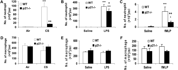
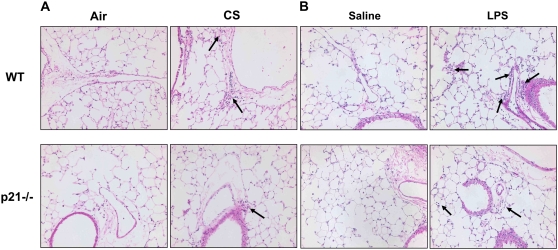
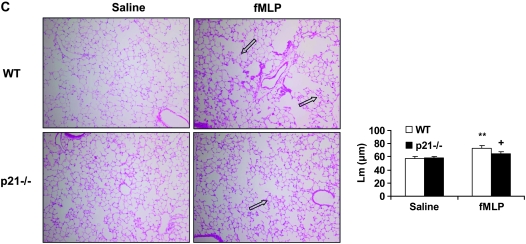
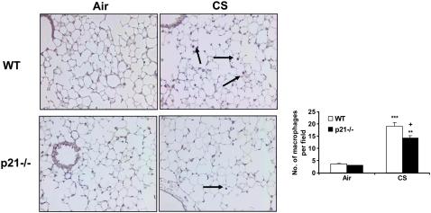
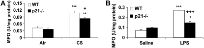
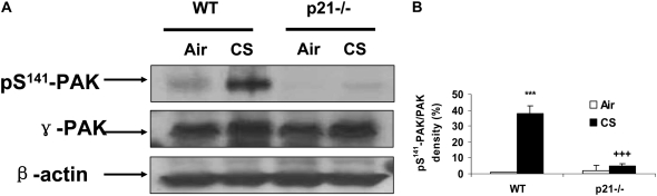
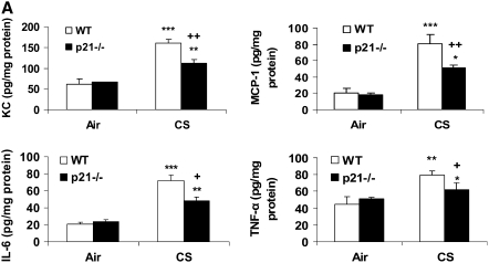
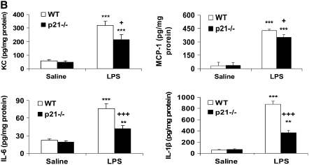
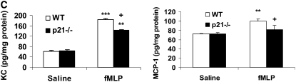
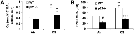
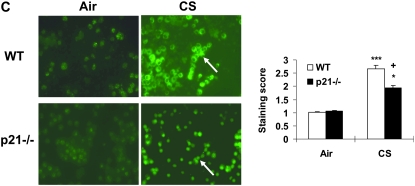
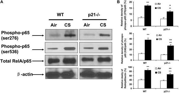
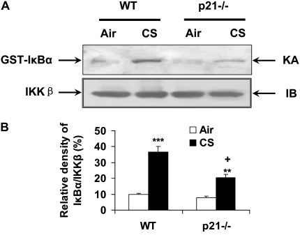
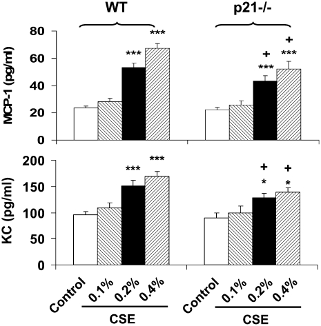
The cyclin-dependent kinase inhibitor p21(CIP1/WAF1/SDI1) (p21) is an important inhibitory checkpoint regulator of cell cycle progression in response to oxidative and genotoxic stresses. It is known that p21 potentiates inflammatory response and inhibits apoptosis and proliferation, leading to cellular senescence. However, the role of endogenous p21 in regulation of lung inflammatory and injurious responses by cigarette smoke (CS) or other pro-inflammatory stimuli is not known. We hypothesized that p21 is an important modifier of lung inflammation and injury, and genetic ablation of p21 will confer protection against CS and other pro-inflammatory stimuli (lipopolysacchride [LPS] and N-formyl-methionyl-leucyl-phenylalanine [fMLP])-mediated lung inflammation and injury. To test this hypothesis, p21-deficient (p21-/-) and wild-type mice were exposed to CS, LPS, or fMLP, and the lung oxidative stress and inflammatory responses as well as airspace enlargement were assessed. We found that targeted disruption of p21 attenuated CS-, LPS-, or fMLP-mediated lung inflammatory responses in mice. CS-mediated oxidative stress and fMLP-induced airspace enlargement were also decreased in lungs of p21-/- mice compared with wild-type mice. The mechanism underlying this finding was associated with decreased NF-kappaB activation, and reactive oxygen species generation by decreased phosphorylation of p47(phox) and down-modulating the activation of p21-activated kinase. Our data provide insight into the mechanism of pro-inflammatory effect of p21, and the loss of p21 protects against lung oxidative and inflammatory responses, and airspace enlargement in response to multiple pro-inflammatory stimuli. These data may have ramifications in CS-induced senescence in the pathogenesis of chronic obstructive pulmonary disease/emphysema.


Figures

<b>Figure 1.</b>
Figure 1.
The number of neutrophils, but not macrophages, was decreased in bronchoalveolar lavage (BAL) fluid of p21−/− mice in response to cigarette smoke (CS) exposure, lipopolysaccharide (LPS), or N-formyl-methionyl-leucyl-phenylalanine (fMLP) aerosolization. Mice were exposed to CS, LPS, or fMLP as described in Materials and Methods. After killing, the lungs were lavaged and cytospin slides were prepared. Total cells in BAL fluid were counted using a hemocytometer. At least 400 cells were counted in a blinded manner to determine the differential cell count using cytospin slides stained with Diff-Quick. The number of neutrophils in BAL fluid was decreased in p21−/− mice in response to (A) CS exposure, (B) LPS, or (C) fMLP aerosolization compared with respective wild-type (WT) mice. However, the number of macrophages in BAL fluid was not altered in p21−/− mice compared with WT mice in response to (D) CS exposure, (E) LPS, or (F) fMLP aerosolization. Data are shown as mean ± SEM (n = 4–5 per group). *P < 0.05, ***P < 0.001, significant compared with respective air- or saline-exposed groups; +P < 0.05, ++P < 0.01, significant compared with CS-, LPS-, or fMLP-exposed WT mice.
<b>Figure 2.</b>
Figure 2.
Reduced inflammatory cell influx or injurious response in lungs of p21−/− mice exposed to CS, LPS, or fMLP aerosolization. Mice were exposed to CS, LPS, or fMLP as described in Materials and Methods. After sacrificing, the left lung was fixed, embedded in paraffin, and stained with hematoxylin and eosin (H&E). Genetic ablation of p21 decreased inflammatory cell influx into the lungs in response to (A) CS exposure or (B) LPS aerosolization. In addition, fMLP-induced enlargement of airspace and destruction of alveolar septa were attenuated in p21−/− mice as compared with WT mice (C). The H&E-stained pictures represent three separate experiments. Solid arrows indicate inflammatory cells, and open arrows indicate enlargement of airspace with destruction of alveolar septa. Original magnification: A and B, ×200; C, ×100. **P < 0.01, significant compared with respective saline-exposed groups; +P < 0.05, significant compared with fMLP-exposed WT mice.
<b>Figure 3.</b>
Figure 3.
Macrophage influx into the lungs was reduced in p21−/− mice exposed to CS. Macrophages were detected with immunohistochemical staining using anti-mouse Mac-3 antibody in lung sections from CS-exposed mice. The number of macrophages in the lungs was decreased in p21−/− mice compared with WT mice exposed to CS. The pictures represent at least three staining experiments (n = 4–5 per group). Arrows indicate macrophages. Original magnification: ×200. **P < 0.01, ***P < 0.001, significant compared with respective air-exposed groups; +P < 0.05, significant compared with CS-exposed WT mice.
<b>Figure 4.</b>
Figure 4.
Myeloperoxidase (MPO) activity was decreased in lungs of p21−/− mice exposed to CS or aerosolized to LPS. Lung sections from mice exposed to CS (A) or aerosolized to LPS (B) were homogenized, and the MPO activity was determined spectrophotometrically. Targeted ablation of p21 decreased the MPO activity in the lungs in response to CS and LPS exposure. Data are shown as mean ± SEM (n = 3–4 per group). *P < 0.05, **P < 0.01, ***P < 0.001, significant compared with respective air- or saline-exposed group; +P < 0.05, +++P < 0.001, significant compared with CS- or LPS-exposed WT mice.
<b>Figure 5.</b>
Figure 5.
Phosphorylated level of p21-activated kinase (PAK) was decreased in lung cytosolic protein of p21−/− mice in response to CS exposure. The protein levels of γ-PAK and its phosphorylation on ser141 residue from air- and CS-exposed mice were analyzed by Western blotting as described in Materials and Methods, and β-actin was used as an indicator for equal protein loading. (A) Representative blotting bands. (B) Graphic summary of densitometric analysis of Western blotting bands from three independent experiments (n = 3–4 per group). The level of phosphorylated PAK was decreased in lung cytosolic protein of p21−/− mice exposed to CS. ***P < 0.001, significant compared with respective air-exposed group; +++P < 0.001, significant compared with CS-exposed WT mice.
<b>Figure 6.</b>
Figure 6.
Levels of pro-inflammatory mediators were decreased in lungs of p21−/− mice in response to CS exposure, LPS, and fMLP aerosolization. Mice were exposed to CS, LPS, and fMLP as described in Materials and Methods. Pro-inflammatory mediators in the lungs were measured by luminex assay in response to (A) CS exposure, (B) LPS, and (C) fMLP aerosolization. The levels of pro-inflammatory mediators in the lungs were increased in WT mice, but were decreased in p21−/− mice as compared to WT mice exposed to CS, LPS, and fMLP. Data are shown as mean ± SEM (n = 3–4 per group). *P < 0.05, **P < 0.01, ***P < 0.001, significant compared with respective air- or saline-exposed group; +P < 0.05, ++P < 0.01, +++P < 0.001, significant compared with CS-, LPS-, or fMLP-exposed WT mice.
<b>Figure 7.</b>
Figure 7.
Levels of oxidative stress markers (reactive oxygen species [ROS] release in BAL cells and lipid peroxidation products in lungs) were reduced in p21−/− mice in response to CS exposure. (A) BAL cells from air- or CS-exposed mice were incubated with or without PMA. Release of superoxide anion was determined by the reduction of ferricytochrome c after 80 minutes of stimulation as described in Materials and Methods. (B) Mice were exposed to CS and levels of 4-HNE plus MDA were measured spectrophotometrically in the lungs as described in Materials and Methods. Data are shown as mean ± SEM (n = 3–4 per group). (C) BAL cells on the cytospin slides from CS-exposed mice were incubated with DCF-DA. After being rinsed with PBS, the slides were observed under fluorescent microscope. The figures are representative of three independent staining experiments (n = 3–4 per group). Arrows indicate ROS in BAL cells. Original magnification: ×400. Genetic ablation of p21 attenuated ROS release in BAL cells and lipid peroxidation products in lungs in response to CS exposure. *P < 0.05, **P < 0.01, ***P < 0.001, significant compared with respective air-exposed mice; +P < 0.05, +++P < 0.001, significant compared with CS-exposed WT mice; #P < 0.05, compared with air-exposed WT mice.
<b>Figure 8.</b>
Figure 8.
Phosphorylated and total levels of RelA/p65 were decreased in lung nuclear protein of p21−/− mice in response to CS exposure. The levels of RelA/p65 and its phosphorylation on ser276 and ser536 residues in the lungs from air- and CS-exposed mice were analyzed by Western blotting as described in Materials and Methods, and β-actin was used as an indicator for equal protein loading. (A) Representative blotting bands. (B) Graphic summary of densitometric analysis of Western blotting bands from three independent experiments (n = 3–4 per group). The levels of phosphorylated and total RelA/p65 were reduced in the lungs of p21−/− mice compared with WT mice in response to CS. **P < 0.01, ***P < 0.001, significant compared with respective air-exposed mice; +P < 0.05, ++P < 0.01, significant compared with CS-exposed WT mice.
<b>Figure 9.</b>
Figure 9.
IKKβ activity was attenuated in lungs of p21−/− mice in response to CS exposure. Whole cell extracts from mouse lung tissue were immunoprecipitated with an antibody against IKKβ, and the immunoprecipitates were subjected to kinase assay (KA) using GST-IκBα as substrate. The samples were subjected to SDS-PAGE and transferred onto PVDF membranes and were exposed to a film. Equal amount of the immunoprecipitated kinase complex were confirmed by immunoblotting (IB) for IKKβ. (A) Representative blotting bands. (B) Graphic summary of densitometric analysis of blotting bands from three independent experiments (n = 3–4 per group). The IKKβ activity was attenuated in lungs of p21−/− mice in response to CS. **P < 0.01, ***P < 0.001, significant compared with respective air-exposed group; +P < 0.05, significant compared with CS-exposed WT mice.
<b>Figure 10.</b>
Figure 10.
Decreased levels of monocyte chemotactic protein (MCP)-1 and keratinocyte chemoattractant (KC) in peritoneal macrophages from p21−/− mice in response to CSE treatment. Peritoneal macrophages were isolated from WT and p21−/− mice, and treated with CSE (0.1, 0.2, and 0.4%). CSE induced the release of MCP-1 and KC from peritoneal macrophages of WT mice; this release was attenuated in peritoneal macrophages from p21−/− mice. Data are shown as mean ± SEM (n = 4–5 per group). *P < 0.05, ***P < 0.001, significant compared with respective control group; +P < 0.05, significant compared with respective CSE-treated peritoneal macrophages from WT mice.

Similar articles

Cited by

References

    1. Hogg JC, Chu F, Utokaparch S, Woods R, Elliott WM, Buzatu L, Cherniack RM, Rogers RM, Sciurba FC, Coxson HO, et al. The nature of small-airway obstruction in chronic obstructive pulmonary disease. N Engl J Med 2004;350:2645–2653. - PubMed
    1. Gan WQ, Man SF, Senthilselvan A, Sin DD. Association between chronic obstructive pulmonary disease and systemic inflammation: a systematic review and a meta-analysis. Thorax 2004;59:574–580. - PMC - PubMed
    1. Jeffery PK. Remodeling and inflammation of bronchi in asthma and chronic obstructive pulmonary disease. Proc Am Thorac Soc 2004;1:176–183. - PubMed
    1. Church DF, Pryor WA. Free-radical chemistry of cigarette smoke and its toxicological implications. Environ Health Perspect 1985;64:111–126. - PMC - PubMed
    1. Thatcher TH, Maggirwar SB, Baglole CJ, Lakatos HF, Gasiewicz TA, Phipps RP, Sime PJ. Aryl hydrocarbon receptor-deficient mice develop heightened inflammatory responses to cigarette smoke and endotoxin associated with rapid loss of the nuclear factor-kappaB component RelB. Am J Pathol 2007;170:855–864. - PMC - PubMed

Publication types

MeSH terms