

Persistent association of nailfold capillaroscopy changes and skin involvement over thirty-six months with duration of untreated disease in patients with juvenile dermatomyositis
- PMID: 18240225
- PMCID: PMC2830145
- DOI: 10.1002/art.23299
Persistent association of nailfold capillaroscopy changes and skin involvement over thirty-six months with duration of untreated disease in patients with juvenile dermatomyositis
Abstract
Objective: To determine the association of changes on nailfold capillaroscopy with clinical findings and genotype in children with juvenile dermatomyositis (DM), in order to identify potential differences in disease course over 36 months.
Methods: At diagnosis of juvenile DM in 61 children prior to the initiation of treatment, tumor necrosis factor alpha (TNFalpha) -308 allele and DQA1*0501 status was determined, juvenile DM Disease Activity Scores (DAS) were obtained, and nailfold capillaroscopy was performed. The disease course was monitored for 36 months. Variations within and between patients were assessed by regression analysis.
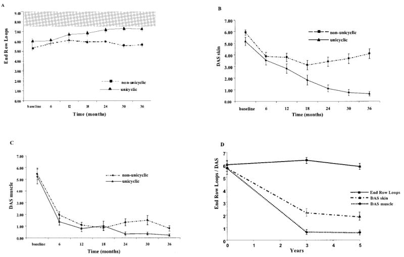
Results: At diagnosis, shorter duration of untreated disease (P = 0.05) and a lower juvenile DM skin DAS (P = 0.035) were associated with a unicyclic disease course. Over 36 months, end-row loop (ERL) regeneration was associated with lower skin DAS (P < 0.001) but not muscle DAS (P = 0.98); ERL regeneration and decreased bushy loops were associated with a shorter duration of untreated disease (P = 0.04 for both). At 36 months, increased ERL regeneration (P = 0.007) and improvement of skin DAS (P < 0.001) and muscle DAS (P = 0.025) were associated with a unicyclic disease course.
Conclusion: Early treatment of juvenile DM may lead to a unicyclic disease course. The non-unicyclic disease course usually involves continuing skin manifestations with persistent nailfold capillaroscopy changes. The correlation of nailfold capillaroscopy results with cutaneous but not with musculoskeletal signs of juvenile DM over a 36-month period suggests that the cutaneous and muscle vasculopathies have different pathophysiologic mechanisms. These findings indicate that efforts to identify the optimal treatment of cutaneous features in juvenile DM require greater attention.
Figures
References
-
- Tezak Z, Hoffman EP, Lutz JL, Fedczyna TO, Stephan D, Bremer EG, et al. Gene expression profiling in DQA1*0501+ children with untreated dermatomyositis: a novel model of pathogenesis. J Immunol. 2002;168:4154–63. - PubMed
-
- Pachman LM, Lipton R, Ramsey-Goldman R, Shamiyeh E, Abbott K, Mendez EP, et al. History of infection before the onset of juvenile dermatomyositis: results from the National Institute of Arthritis and Musculoskeletal and Skin Diseases Research Registry. Arthritis Rheum. 2005;53:166–72. - PubMed
-
- Pachman LM, Litt DL, Rowley AH, Hayford JR, Caliendo J, Heller S, et al. Lack of detection of enteroviral RNA or bacterial DNA in magnetic resonance imaging–directed muscle biopsies from twenty children with active untreated juvenile dermatomyositis. Arthritis Rheum. 1995;38:1513–8. - PubMed
-
- Pachman LM, Liotta-Davis MR, Hong DK, Kinsella TR, Mendez EP, Kinder JM, et al. TNFα-308A allele in juvenile dermatomyositis: association with increased production of tumor necrosis factor α, disease duration, and pathologic calcifications. Arthritis Rheum. 2000;43:2368–77. - PubMed
Publication types
MeSH terms
Substances
Grants and funding
LinkOut - more resources
Full Text Sources
Research Materials
