. 2008 Mar;7(2):207-24.
doi: 10.1111/j.1474-9726.2007.00363.x. Epub 2008 Jan 28.

Plasticity of hippocampal stem/progenitor cells to enhance neurogenesis in response to kainate-induced injury is lost by middle age

Affiliations

Plasticity of hippocampal stem/progenitor cells to enhance neurogenesis in response to kainate-induced injury is lost by middle age

Bharathi Hattiangady et al. Aging Cell. 2008 Mar.

Abstract

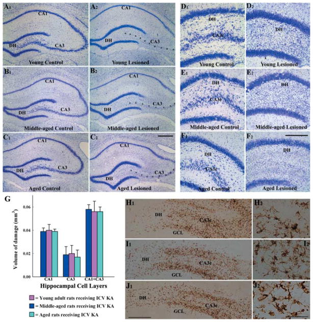
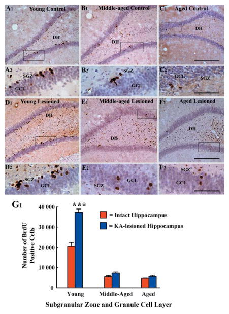
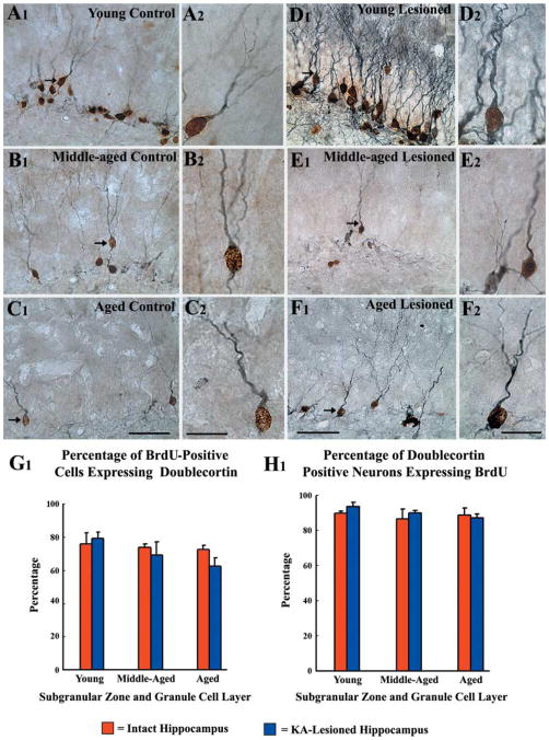
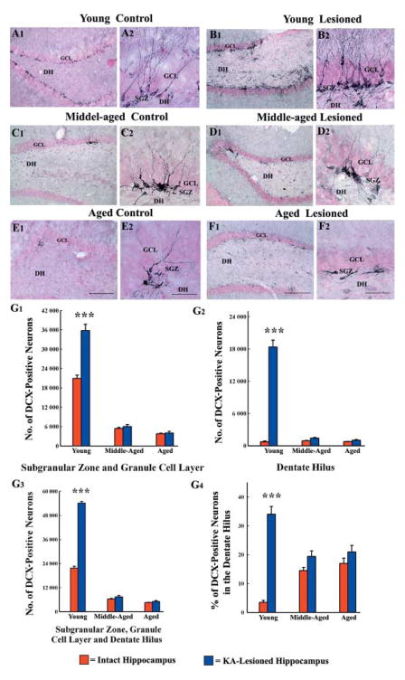
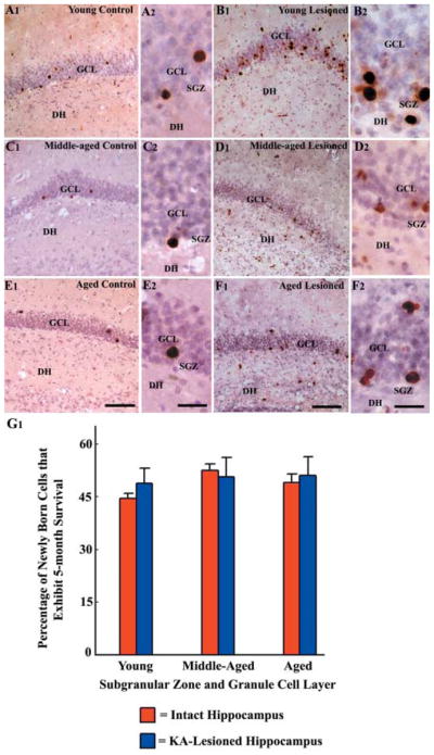
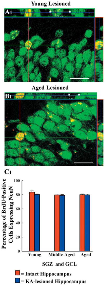
A remarkable up-regulation of neurogenesis through increased proliferation of neural stem/progenitor cells (NSCs) is a well-known plasticity displayed by the young dentate gyrus (DG) following brain injury. To ascertain whether this plasticity is preserved during aging, we quantified DG neurogenesis in the young adult, middle-aged and aged F344 rats after kainic acid induced hippocampal injury. Measurement of new cells that are added to the dentate granule cell layer (GCL) between post-injury days 4 and 15 using 5'-bromodeoxyuridine labeling revealed an increased addition of new cells in the young DG but not in the middle-aged and aged DG. Quantification of newly born neurons using doublecortin immunostaining also demonstrated a similar trend. Furthermore, the extent of ectopic migration of new neurons into the dentate hilus was dramatically increased in the young DG but was unaltered in the middle-aged and aged DG. However, there was no change in neuronal fate-choice decision of newly born cells following injury in all age groups. Similarly, comparable fractions of new cells that are added to the GCL after injury exhibited 5-month survival and expressed the mature neuronal marker NeuN, regardless of age or injury at the time of their birth. Thus, hippocampal injury does not adequately stimulate NSCs in the middle-aged and aged DG, resulting in no changes in neurogenesis after injury. Interestingly, rates of both neuronal fate-choice decision and long-term survival of newly born cells remain stable with injury in all age groups. These results underscore that the ability of the DG to increase neurogenesis after injury is lost as early as middle age.


Figures

Fig 1
Fig 1
Pattern of hippocampal neurodegeneration at 16 days after an unilateral intracerebroventricular (ICV) kainic acid (KA) administration, visualized by Nissl staining. Left panel: A1, B1, and C1 illustrate examples of the hippocampus from naïve young adult (A1), middle-aged (B1) and aged (C1) rats, whereas A2, B2, and C2 show examples of the hippocampus ipsilateral to the ICV KA administration from age-matched KA-treated young adult (A2), middle-aged (B2) and aged (C2) rats. Note an extensive loss of CA3 pyramidal neurons (indicated by asterisks) in all three age groups. Right panel: D1, E1, and F1 illustrate examples of the dentate gyrus from naïve young adult (A1), middle-aged (B1) and aged (C1) rats, whereas D2, E2, and F2 show examples of the dentate gyrus ipsilateral to the ICV KA administration from age-matched KA-treated young adult (D2), middle-aged (E2) and aged (F2) rats. Note an extensive loss of neurons in the dentate hilus (DH) in all three age groups. Scale bar: A1–C2, 400 μm; D1–F2, 200 μm. The bar chart (G) illustrates the volume of damage in CA1 and CA3 cell layers of different age groups of rats after ICV KA administration. H1–J2 show the distribution of ED-1 immunopositive activated microglial cells in the dentate gyrus at 16 days after the KA administration in the three age groups of animals. Note that the distribution and density of ED-1 immunopositive activated microglial cells in the DH and the CA3c subregion is comparable in the young adult (H1), middle-aged (I1), and aged (J1) rats. H2, I2 and J2 are magnified views of regions from H1, I1 and J1 demonstrating the morphology of ED-1 immunopositive activated microglial cells in the three age groups. The morphology appears similar across the three age groups. GCL, granule cell layer; scale bar: H1, I1, J1 = 250 μm; H2, I2, J2 = 50 μm.
Fig 2
Fig 2
Distribution of newly generated cells in the dentate gyrus (DG) at 24 h after 12 daily injections of 5′-bromodeoxyuridine (BrdU) in different groups, visualized with BrdU immunostaining and hematoxylin counterstaining. The groups include the dentate gyrus of naïve young adult (A1, A2), middle-aged (B1, B2) and aged (C1, C2) rats, and the dentate gyrus of hippocampi ipsilateral to kainic acid (KA) administration from young adult (D1, D2), middle-aged (E1, E2) and aged (F1, F2) rats. A2, B2, C2, D2, E2, and F2 are magnified views of regions from A1, B1, C1, D1, E1, and F1. Note that the density of newly born cells in the subgranular zone-granule cell layer (SGZ-GCL) increases in the young adult rat after KA administration (D1, D2), in comparison to age-matched naïve rat (A1, A2). In contrast, the density of newly born cells in these regions is unchanged in the middle-aged (E1, E2) and aged (F1, F2) rats after similar KA administration in comparison to respective naïve groups (B1, B2, C1, C2). The dentate hilus and CA3c subregion show increased density of BrdU immunopositive cells in KA-treated animals regardless of the age (D1, E1, F1). The bar chart in G1 compares the absolute numbers of newly born cells added over a period of 12 days to the SGZ-GCL in different groups of rats. Note that, KA administration considerably increases the numbers of newly born cells in the SGZ-GCL in young adult rats but not in middle-aged and aged rats. Scale bar: A1, B1, C1 D1, E1, F1 = 200 μm; A2, B2, C2, D2, E2, F2 = 50 μm.
Fig 3
Fig 3
Neuronal differentiation of newly born cells in the subgranular zone-granule cell layer (SGZ-GCL) of different groups at 24 h after the last of 12 daily injections of 5′-bromodeoxyuridine (BrdU) and visualized through BrdU and doublecortin (DCX) immunostaining. These include SGZ-GCL of naïve young adult (A1, A2), middle-aged (B1, B2) and aged (C1, C2) rats, and SGZ-GCL of hippocampi ipsilateral to kainic acid (KA) administration from young adult (D1, D2), middle-aged (E1, E2) and aged (F1, F2) rats at 16 days post-KA administration. A2, B2, C2, D2, E2, and F2 are magnified views of regions from A1, B1, C1, D1, E1, and F1. Note that a majority of newly born (BrdU +) cells (brown nuclei) are positive for DCX (blue-grey reaction product in the cytoplasm of the soma and dendrites) in all groups. Furthermore, majority of DCX + neurons are positive for BrdU in allthree age groups. Figure G1 shows that the percentages of BrdU + cells that are positive for DCX are comparable in different groups, suggesting that the rate of neuronal differentiation remains stable across the three age groups under both intact and lesioned conditions. The bar chart H1 illustrates that similar percentages (83–94%) of DCX + cells are immunoreactive for BrdU in different groups, implying that a vast majority of neurons visualized with DCX immunostaining in the SGZ-GCL of both intact and KA-treated young adult, middle-aged and aged rats are new granule cells that were generated during the preceding 12 days. Scale bar: A1, B1, C1, D1, E1, F1 = 50 μm; A2, B2, C2, D2, E2, F2 = 20 μm.
Fig 4
Fig 4
Distribution of newly born neurons in the subgranular zone-granule cell layer (SGZ-GCL) of different groups, visualized through doublecortin (DCX) immunostaining. The groups include DG of naïve young adult (A1, A2), middle-aged (C1, C2) and aged (E1, E2) rats, and SGZ-GCL of hippocampi ipsilateral to kainic acid (KA) administration from young adult (B1, B2), middle-aged (D1, D2) and aged (F1, F2) rats at 16 days post-KA administration. A2, B2, C2, D2, E2, and F2 are magnified views of regions from A1, B1, C1, D1, E1, and F1. Note that the density of newly born neurons in the SGZ-GCL increases in the young adult rat after KA administration (B1, B2), in comparison to age-matched naïve rat (A1, A2). In contrast, the density of newly born cells in these regions is unchanged in the middle-aged (D1, D2) and aged (F1, F2) rats after similar KA administration in comparison to respective naïve groups (C1, C2 and E1, E2). The dentate hilus (DH) shows an increased density of ectopically migrated newly born neurons in the KA-treated young animal (B1, B2), whereas the KA-treated middle-aged and aged animals show only a few ectopically migrated neurons (D1, F1). Scale bar: A1, B1, C1, D1, E1, F1 = 200 μm; A2, B2, C2, D2, E2, F2 = 50 μm. The bar charts in G1–G3 compare the absolute numbers of newly born neurons added over 12 days to the SGZ-GCL (G1), the DH (G2), and the SGZ-GCL plus the DH (G3) in different groups of rats. Note that, KA administration considerably increases the numbers of newly born cells in all these regions in young adult rats but not in middle-aged and aged rats. The bar chart in G4 compares the percentages of newly born neurons that have migrated ectopically into the DH in different groups of rats. Note that KA administration considerably increases the ectopic migration of newly born cells in young adult rats but not in middle-aged and aged rats.
Fig 5
Fig 5
Distribution of newly born cells in the subgranular zone-granule cell layer (SGZ-GCL) of different groups at 5 months after 12 daily injections of 5′-bromodeoxyuridine (BrdU), visualized through BrdU immunostaining and hematoxylin counterstaining. The groups include the dentate gyrus of naïve young adult (A1, A2), middle-aged (C1, C2) and aged (E1, E2) rats, and the dentate gyrus of hippocampi ipsilateral to kainic acid (KA) administration from young adult (B1, B2), middle-aged (D1, D2) and aged (F1, F2) rats. A2, B2, C2, D2, E2, and F2 are magnified views of regions from A1, B1, C1, D1, E1, and F1. Note that newly added cells exhibit long-term survival and migrate up into the GCL in all groups. Scale bar: A1, B1, C1, D1, E1, F1 = 200 μm; A2, B2, C2, D2, E2, F2 = 50 μm. The bar chart G1 illustrates that similar percentages (44–53%) of newly added cells exhibit 5-month survival in different groups, suggesting that the long-term survival of newly added cells to the SGZ-GCL is independent of age as well as injury.
Fig 6
Fig 6
Orthogonal views of newly added cells that differentiated into neuron-specific nuclear antigen (NeuN) positive mature neurons in the granule cell layer of kainic acid-treated young adult (A1) and aged (B1) rats. The analysis was done at 5 months after 12 daily injections of 5′-bromodoxyuridine (BrdU) through BrdU and NeuN dual immunofluorescence and confocal microscopy. Scale bar: 20 μm. The bar chart in C1 demonstrates that 79–84% of new cells that persist in the granule cell layer differentiate into NeuN + positive neurons in different groups, implying that majority of new cells that exhibit long-term survival are neurons in all groups. SGZ, subgranular zone; GCL, granule cell layer.

Similar articles

Cited by

References

    1. Abdel-Rahman A, Rao MS, Shetty AK. Nestin expression in hippocampal astrocytes after injury depends on the age of the hippocampus. Glia. 2004;47:299– 313. - PubMed
    1. Aimone JB, Wiles J, Gage FH. Potential role for adult neurogenesis in the encoding of time in new memories. Nat Neurosci. 2006;9:723– 727. - PubMed
    1. Altman J, Das GD. Autoradiographic and histological evidence of postnatal hippocampal neurogenesis in rats. J Comp Neurol. 1965;124:319– 335. - PubMed
    1. Bengzon J, Kokaia Z, Elmer E, Nanobashvili A, Kokaia M, Lindvall O. Apoptosis and proliferation of dentate gyrus neurons after single and intermittent limbic seizures. Proc Natl Acad Sci U S A. 1997;94:10432–10437. - PMC - PubMed
    1. Bernal GM, Peterson DA. Neural stem cells as therapeutic agents for age-related brain repair. Aging Cell. 2004;3:345– 351. - PubMed

Publication types

MeSH terms