. 2008 Jun;198(6):651.e1-7.
doi: 10.1016/j.ajog.2007.11.032. Epub 2008 Feb 1.

Posterior compartment anatomy as seen in magnetic resonance imaging and 3-dimensional reconstruction from asymptomatic nulliparas

Affiliations

Posterior compartment anatomy as seen in magnetic resonance imaging and 3-dimensional reconstruction from asymptomatic nulliparas

Yvonne Hsu et al. Am J Obstet Gynecol. 2008 Jun.

Abstract

Objective: The objective of the study was to identify characteristic anatomical features of the posterior compartment using magnetic resonance (MR) cross-sectional anatomy and 3-dimensional (3-D) modeling.

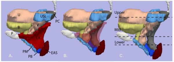
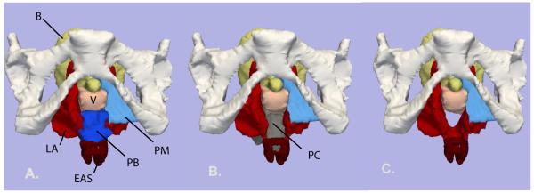
Study design: Supine, static proton-density MR images of 20 nulliparas were analyzed. MR images were used to create models in a selected exemplar.

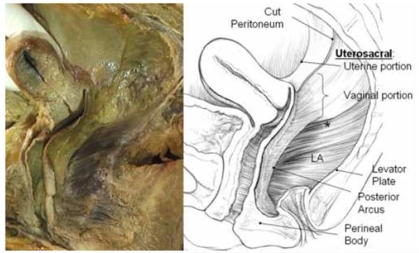
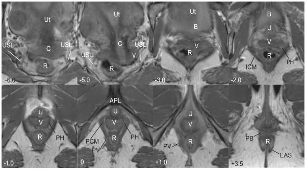
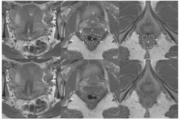
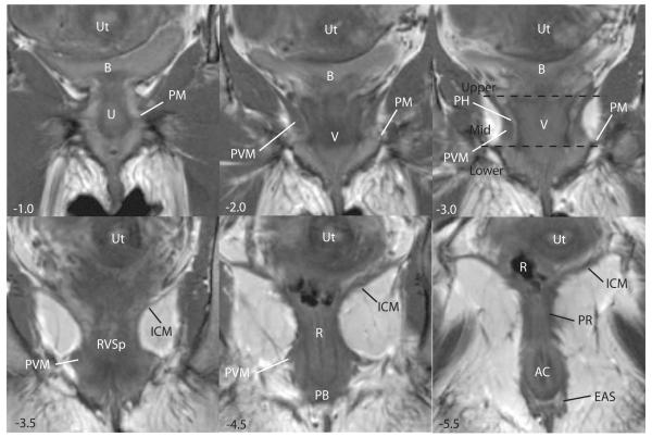
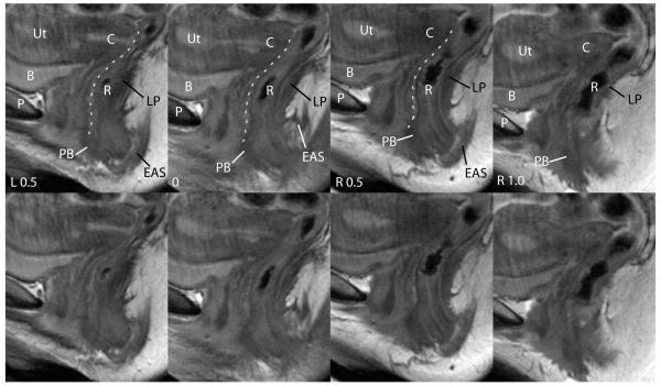
Results: The compartment's upper, mid, and lower segments are best seen in the axial plane. It is bounded inferiorly by the perineal body, ventrally by the posterior vaginal wall, and dorsally by the levator ani muscles and coccyx. In the upper portion, the compartment is bordered laterally by the uterosacral ligaments, whereas in the middle portion, there is more direct contact with the lateral levator ani muscles. In the lower portion, the contact becomes obliterated because the vagina and levator ani muscles become fused to each another and to the perineal body.

Conclusion: The posterior compartment has characteristic anatomic features in MR cross-sectional anatomy that can be further elucidated and integrated with 3-D anatomy.


Figures

Figure 1
Figure 1
Cadaver dissection (left) and illustration (right) of posterior compartment of a 56 year old multipararous female showing structural relationships after the rectum has been removed. Note the apical connections of the upper posterior vagina to the inside of the pelvic wall in a retroperitoneal position. These lie below the peritoneum and are dorsal and caudal to what is traditionally referred to as the uterosacral ligament. These structures are continuous with the posterior arcus tendineus fascia pelvis. At the distal end of the vagina the wall merges into the top of the perineal body. The lateral and dorsal margins of the compartment are formed by the levator ani muscles (LA) and the levator plate. The asterisk (*) denotes the region of the sacrospinous ligament overlain by the coccygeus muscle. © DeLancey 2007.
Figure 2
Figure 2
Axial scan of a nulliparous female showing various features of the posterior compartment. Images are presented cephalad (negative numbers) to caudad (positive numbers) in centimeters relative to the arcuate pubic ligament (APL). In the upper portion of the compartment (−6.0 to −2.0), there is loss of distinct separation of vagina (V) and surrounding structures; the bladder (B), iliococcygeus muscle (ICM) and uterus (Ut) are visible. At −6.0, the course of the uterosacral ligaments (USL) and the cervix (C) are visible. The course of the uterosacral ligaments (USL) is further delineated in −5.0. At −2.0 to 0.0 in the mid-portion of the compartment, the vagina has a “W” shape and is surrounded by a perivascular halo (PH); the pubococcygeus (PCM) and pubovaginalis (PV) are also visible. At +1.0, the pubovaginal division (PV) of the levator ani is still visible and the urethra (U) and arcuate pubic ligament (APL) become visible. In the lower portion of the compartment (+1.0) the vagina (V) takes on a “U” shape and fuses with the levator ani muscles. At +3.5 the perineal body (PB) is seen anterior to the rectum (R) and the external anal sphincter (EAS). © DeLancey 2007
Figure 3
Figure 3
Left lateral view of the 3-D model. The outline of the mid-sagittal pubic bone (P) is shown. Bladder (B) – yellow; uterus (U) and vagina (V) – pink; uterosacral ligament (USL) and perineal membrane (PM) - turquoise blue; levator ani muscle (LA) – red; perineal body (PB) - royal blue; external anal sphincter (EAS) - dark red; posterior compartment (PC) - gray. Image A: all organs are shown. B: levator ani muscles have been faded to show underlying structures. C: levator ani muscles have been removed. The locations of the upper, mid and lower axial cross-sections in figure 2.2 are shown. © DeLancey 2007.
Figure 4
Figure 4
Axial MR cross-sections of the levels of the posterior compartment. The compartment is outlined in a white dashed line. Upper: uterus (Ut), cervix (C), vagina (V), rectum (R), uterosacral ligaments (black arrowheads). Mid: urethra (Ur), perivascular halo (white unfilled arrows), pubococcygeus muscle (white filled arrows), bladder (B), urethra (Ur). Lower: pubovaginalis muscle (white filled arrows), rectovaginal space (grey unfilled arrow). The same images without labels are also shown. © DeLancey 2007.
Figure 5
Figure 5
Coronal scans of the same subject are depicted. Images are presented ventral (negative numbers) to dorsal (positive numbers) in centimeters relative to the APL . The bladder (B), uterus (Ut), urethra (Ur), vagina (V) are seen from −1.0 to −3.0. The fibers of the pubovisceral muscle (PVM) are seen in −2.0 to −4.5. At −1.0 to −3.0, the lateral portion of the perineal membrane (PM) is seen. At −3.0 the peri-vascular halo of the vagina is visible. At −3.5 the rectovaginal space (RVSp) and iliococcygeus muscle (ICM) are seen. The perineal body (PB) becomes visible at −4.5. At −5.5, the external anal sphincter (EAS) and puborectalis (PR) can also be seen as well as the anal canal (AC). The division of the upper, mid and lower portions of the posterior compartment are shown in panel −3.0. © DeLancey 2007.
Figure 6
Figure 6
Sagittal scans of the same patient showing features of posterior compartment. The midline sagittal image is labeled 0 and other images left (L) or right (R) relative to it. The pubic symphysis (P), bladder (B), uterus (Ut), cervix (C) and rectum (R) are seen. The perineal body (PB) is visible anterior to the rectum and the external anal sphincter (EAS) as a low signal intensity triangular structure. The posterior wall of the vagina is outlined in white. The levator plate (LP) is also visible, forming the dorsal boundary of the posterior compartment. The same images without labels are also shown. © DeLancey 2007.
Figure 7
Figure 7
Inferior view of 3-D model with the pubic bones in place. A: Levator ani (LA) – red; Perineal membrane (PM) - turquoise blue; Perineal body (PB) – royal blue; External anal sphincter (EAS) – dark red; Bladder (B) – yellow; Vagina (V) – light pink. B: Bladder and perineal body have been removed to show the posterior compartment (PC) - grey. C: Posterior compartment model removed to show the space it occupies. © DeLancey 20

References

    1. Boyles SH, Weber AM, Meyn L. Procedures for pelvic organ prolapse in the united states, 1979-1997. Am J Obstet Gynecol. 2003;188(1):108–15. - PubMed
    1. Silva WA, Pauls RN, Segal JL, Rooney CM, Kleeman SD, Karram MM. Uterosacral ligament vault suspension: Five-year outcomes. Obstet Gynecol. 2006;108(2):255–63. - PubMed
    1. DeLancey JO. Anatomic aspects of vaginal eversion after hysterectomy. Am J Obstet Gynecol. 1992;166:1717–1728. - PubMed
    1. Umek WH, Morgan DM, Ashton-Miller JA, DeLancey JO. Quantitative analysis of uterosacral ligament origin and insertion points by magnetic resonance imaging. Obstet Gynecol. 2004;103(3):447–51. - PMC - PubMed
    1. Leffler KS, Thompson JR, Cundiff GW, Buller JL, Burrows LJ, Ybarra MA Schon. Attachment of the rectovaginal septum to the pelvic sidewall. Am J Obstet Gynecol. 2001;185(1):41–3. - PubMed

Publication types