. 2008 Feb;134(2):511-22.
doi: 10.1053/j.gastro.2007.11.058. Epub 2007 Dec 4.

A molecular signature of gastric metaplasia arising in response to acute parietal cell loss

Affiliations

A molecular signature of gastric metaplasia arising in response to acute parietal cell loss

Koji Nozaki et al. Gastroenterology. 2008 Feb.

Abstract

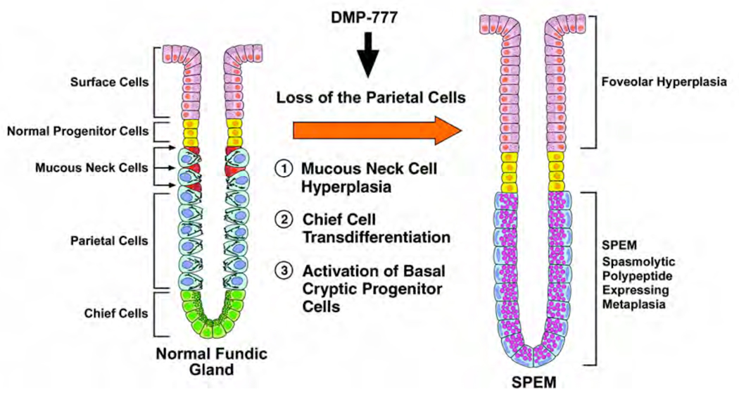
Background & aims: Loss of gastric parietal cells is a critical precursor to gastric metaplasia and neoplasia. However, the origin of metaplasia remains obscure. Acute parietal cell loss in gastrin-deficient mice treated with DMP-777 leads to the rapid emergence of spasmolytic polypeptide/trefoil factor family 2 (TFF2)-expressing metaplasia (SPEM) from the bases of fundic glands. We now sought to characterize more definitively the pathway for emergence of SPEM.

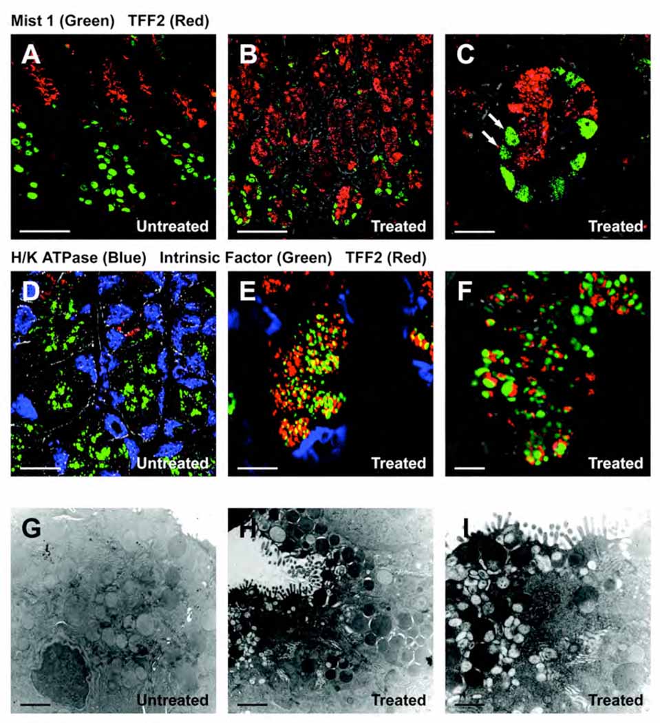
Methods: Emerging SPEM lineages in gastrin-deficient mice treated with DMP-777 were examined for immunolocalization of TFF2, intrinsic factor, and Mist1, and morphologically with electron microscopy. Emerging SPEM was isolated with laser-capture microdissection and RNA was analyzed using gene microarrays. Immunohistochemistry in mouse and human samples was used to confirm up-regulated transcripts.

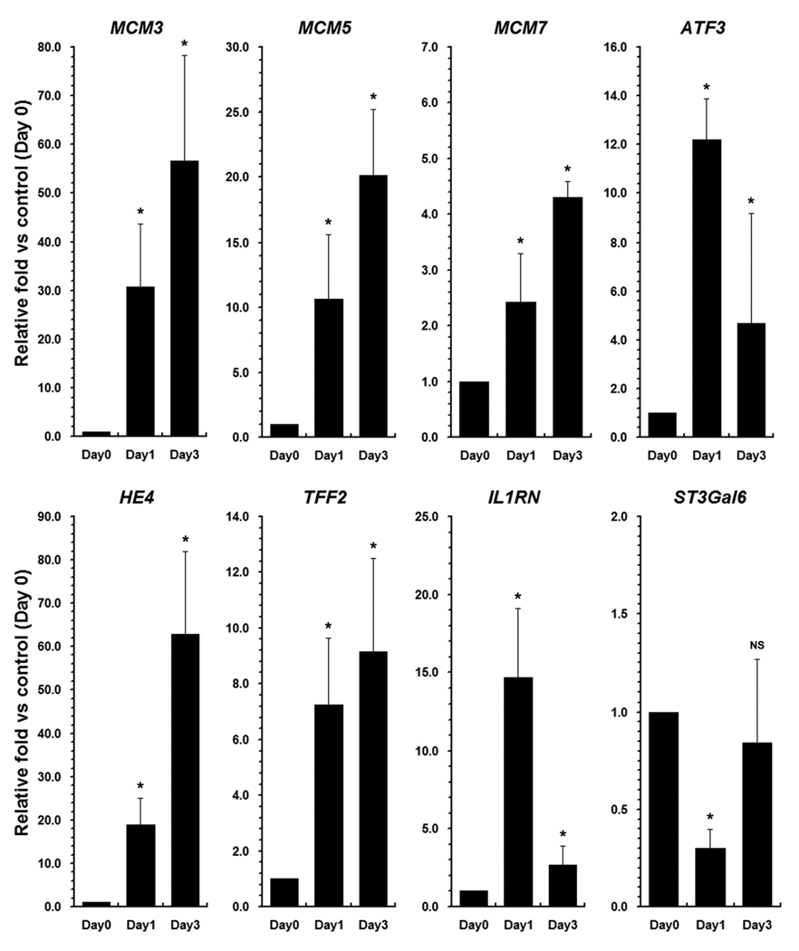
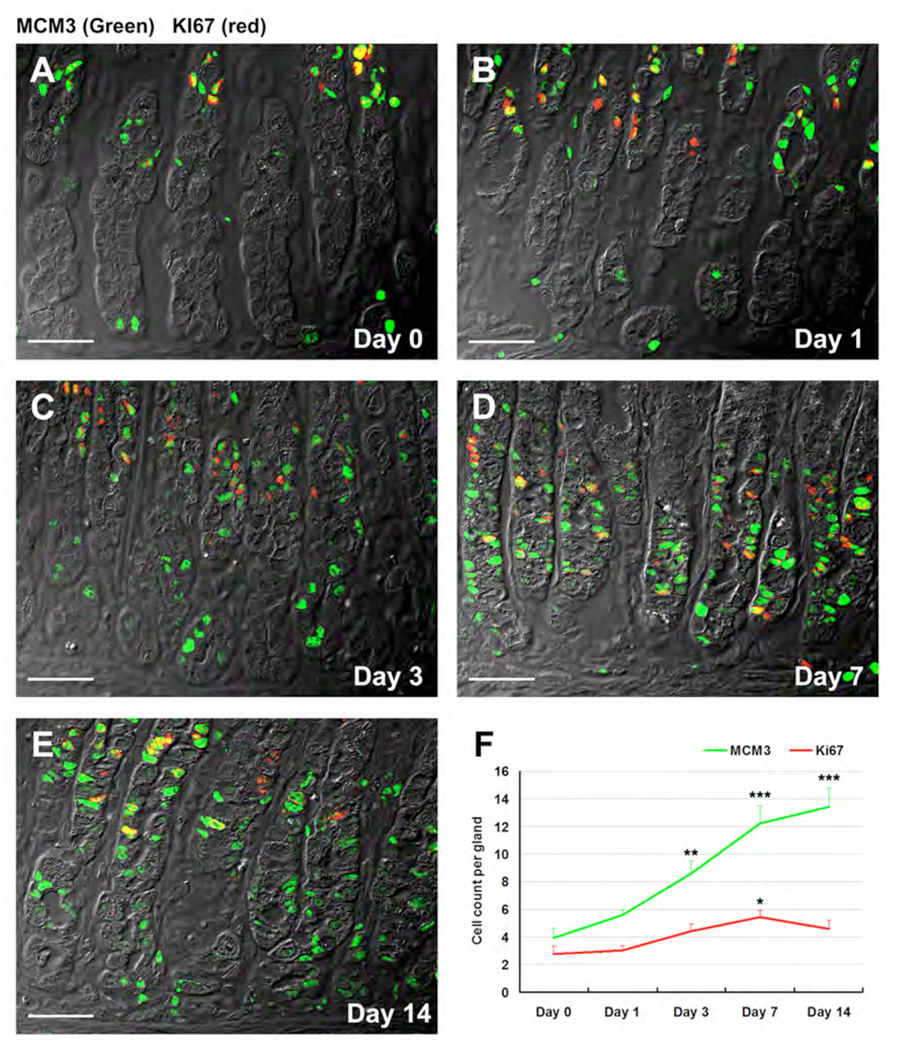
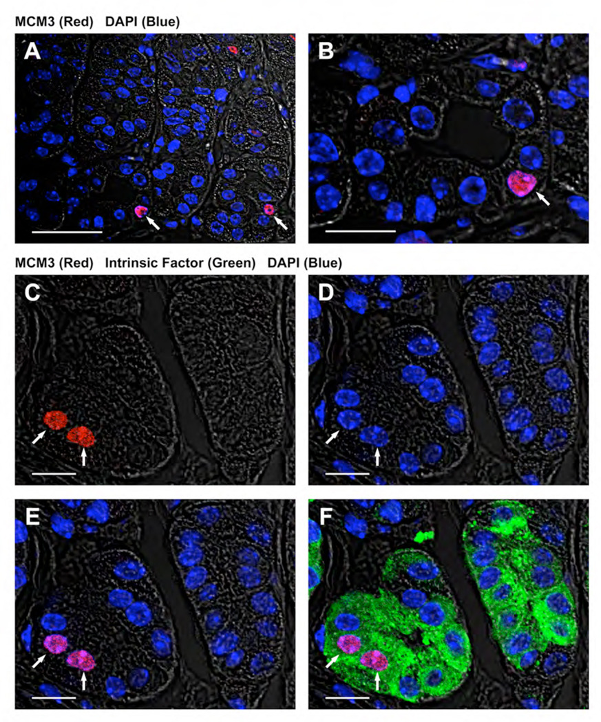
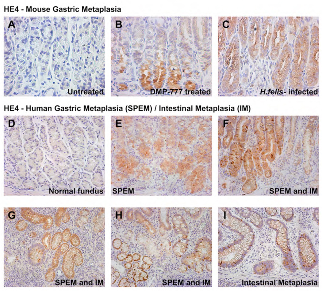
Results: DMP-777-induced SPEM was immunoreactive for TFF2 and the differentiated chief cell markers, Mist1 and intrinsic factor, suggesting that SPEM derived from transdifferentiation of chief cells. Microarray analysis of microdissected SPEM lineages induced by DMP-777 showed up-regulation of transcripts associated with G1/S cell-cycle transition including minichromosome maintenance deficient proteins, as well as a number of secreted factors, including human epididymis 4 (HE4). HE4, which was absent in the normal stomach, was expressed in SPEM of human and mouse and in intestinal metaplasia and gastric cancer in human beings.

Conclusions: Although traditionally metaplasia was thought to originate from normal mucosal progenitor cells, these studies indicate that SPEM evolves through either transdifferentiation of chief cells or activation of a basal cryptic progenitor. In addition, induction of metaplasia elicits the expression of secreted factors, such as HE4, relevant to gastric preneoplasia.


Conflict of interest statement

Conflict of interest: The authors have declared that no conflicts of interest exist.

Figures

Figure 1
Figure 1. Characterization of emerging SPEM
(A) Fundic mucosa from an untreated gastrin deficient mouse was dual immunostained for Mist1 (green) and TFF2 (red). Mist1 antibodies stained the nuclei of mature chief cells at the base of fundic glands, while TFF2 was positive in mucous neck cells in the neck region of the fundic gland. No discernible dual staining cells were observed (Scale bar: 50 µm). (B, C) Mist1 and TFF2 expression in a gastrin-deficient mouse treated with DMP-777 for 3 days. The number of Mist1 immunoreactive cells (green) decreased in the bottom of glands, and a number of the Mist1 positive cells were dual immunoreactive for TFF2 (red). (Scale bar: B, 50 µm; C, 10 µm) The high power view in C. demonstrates the presence of cells dual labeled for Mist1 and TFF2 (arrows). (D) Mucosa from an untreated mouse stained with antibodies for the H/K-ATPase (blue), TFF2 (red) and Intrinsic factor (green). Note that there was no overlap in the staining of the three lineages (Scale bar: 20 µm). (E, F) Mucosa from a mouse treated with DMP-777 for 3 days stained with antibodies for the H/K ATPase (blue), TFF2 (red) and Intrinsic factor (green). While parietal cells only stained with H/K-ATPase antibodies, SPEM cells at the base of the glands stained for both intrinsic factor (green) and TFF2 (red). The higher power view in F. demonstrates the presence of two separate populations of vesicles in the SPEM cells. (Scale bar: E, 10 µm; F, 4 µm). (G, H, I) Electron micrographs of chief cells from an untreated mouse (G) and emerging SPEM cells from a 3 day DMP-777-treated mouse (H, I). (G) Chief cells showed uniformly staining zymogen granules (Scale bar: 2 µm). (H, I) SPEM cells at the base of fundic glands of a mouse treated with DMP-777 for 3 days contained a heterogeneous set of granule morphologies. (Scale bar: G, H, 2 µm; I, 1 µm).
Figure 2
Figure 2. Quantitative PCR analysis of transcripts during the emergence of SPEM
Expression of selected up-regulated or down-regulated transcripts in microdissected lineages was examined using quantitative PCR to validate the results of the gene microarray studies. Results are represented as fold change in expression compared with levels in untreated chief cells (n=3, +/− standard error of the mean, SE). (* p<0.05 by Mann-Whitney U; NS, not significant)
Figure 3
Figure 3. MCM3 expression during acute oxyntic atrophy
Sections from an untreated mouse (A) or mice treated with DMP-777 for 1 (B), 3 (C), 7 (D) or 14 (E) days were dual stained with antibodies against MCM3 (green) and antibodies against Ki67 (red). With increasing amounts of treatment DMP-777, we observed an increase in MCM3 staining in cells at the bases of glands (Scale bar: 40 µm). (F) Quantification of MCM3 and Ki-67 staining showed a significant increase in MCM3 staining at days 3–14 of treatment (green line, ** p<0.01; *** p<0.001). Ki-67 staining showed a smaller increase, which was only significant at 7 days of treatment (* p<0.05).
Figure 4
Figure 4. MCM3 positive cells at the bases of fundic glands in untreated mice are chief cells
Section from an untreated mouse stained with rabbit antibody against MCM3 (red), goat antibody against Intrinsic factor (green) and DAPI (blue). (A–B) Nuclear MCM3 expression observed in cells at the bases of fundic glands (Scale bar: A, 40 µm; B, 20 µm). (C–F) MCM3 positive cells at the bases of fundic glands were also stained for Intrinsic factor, indicating their identification as chief cells. (C) MCM3 positive cells (arrows) in the bottom of the fundic gland, (D) DAPI staining, (E) dual overlay image with MCM3 and DAPI, (F) triple overlay image with MCM3, Intrinsic Factor and DAPI. (Scale bar: C–F, 10 µm).
Figure 5
Figure 5. Up-regulation of HE4 in both SPEM and goblet cell intestinal metaplasia
We examined HE4 expression in gastric metaplasias from mice (A–C) and humans (D–I). (A) Little immunoreactivity for HE4 was detectable in the normal gastric mucosa of untreated gastrin-deficient mice. (B) Prominent staining for HE4 in SPEM in a gastrin deficient mouse treated with DMP-777. (C) Strong staining for HE4 was observed in SPEM in the mucosa of a C57BL/6 mouse infected with Helicobacter felis for 9 months. (D) No staining for HE4 was observed in the normal human gastric fundic mucosa. (E) SPEM from a human patient stained strongly with HE4. (F) SPEM and goblet cell intestinal metaplasia both stained for HE4 in a region of metaplastic transition. (G, H) In sections containing both SPEM and intestinal metaplasia, HE4 immunoreactivity was present in both metaplasias. (I) Intestinal metaplasia showing strong positivity for HE4.
Figure 6
Figure 6. Immunostaining for HE4 in human gastric adenocarcinoma
HE4 antibodies were used to stain sections of gastric adenocarcinomas with immunohistochemical detection with alkaline phosphatase conjugated secondary antibody and Vector Red chromogen (A–I). (A–D) Well to moderately differentiated gastric cancers. Note the prominent expression of HE4 in intestinal type gastric adenocarcinoma. (E–G) Poorly-differentiated gastric cancers. Although the majority of poorly differentiated or diffuse cancers did not show immunoreactivity, a minority of tumors did show positivity. (H, I) Signet ring-cell type gastric adenocarcinomas. The strongest HE4 positivity was observed in signet ring adenocarcinomas. The positivity for HE4 in gastric cancers was also confirmed with immunofluorescence staining using Alexa488-anti-rabbit secondary antibodies (green) (J–L), supporting the immunohistochemical staining results for HE4 in gastric cancers.
Figure 7
Figure 7. Hypothetical pathways for development of SPEM from gastric fundic glands
Three hypothetical pathways for the development of SPEM arising after acute parietal cell loss: 1, Mucous neck cell hyperplasia; 2, Transdifferentiation of chief cells; and 3, Activation of basal cryptic progenitor cells.

References

    1. Pisani P, Bray F, Parkin DM. Estimates of the world-wide prevalence of cancer for 25 sites in the adult population. Int J Cancer. 2002;97:72–81. - PubMed
    1. Parsonnet J, Friedman GD, Vandersteen DP, Chang Y, Vogelman JH, Orentreich N, Sibley RK. Helicobacter pylori infection and the risk of gastric cancer. New Eng.J.Med. 1991;325:1127–1131. - PubMed
    1. Stepan V, Ramamoorthy S, Nitsche H, Zavros Y, Merchant JL, Todisco A. Regulation and function of the sonic hedgehog signal transduction pathway in isolated gastric parietal cells. J Biol Chem. 2005;280:15700–15708. - PubMed
    1. Beauchamp RD, Barnard JA, McCutchen CM, Cherner JA, Coffey RJ., Jr Localization of transforming growth factor alpha and its receptor in gastric mucosal cells. J.Clin.Invest. 1989;84:1017–1023. - PMC - PubMed
    1. Schmidt PH, Lee JR, Joshi V, Playford RJ, Poulsom R, Wright NA, Goldenring JR. Identification of a metaplastic cell lineage associated with human gastric adenocarcinoma. Lab.Invest. 1999;79:639–646. - PubMed

Publication types

MeSH terms

Substances