Comparative Study
. 2008 Apr 1;75(7):1472-81.
doi: 10.1016/j.bcp.2007.12.005. Epub 2007 Dec 27.

Acetylation of prostaglandin H2 synthases by aspirin is inhibited by redox cycling of the peroxidase

Affiliations
Comparative Study

Acetylation of prostaglandin H2 synthases by aspirin is inhibited by redox cycling of the peroxidase

Manju Bala et al. Biochem Pharmacol. .

Abstract

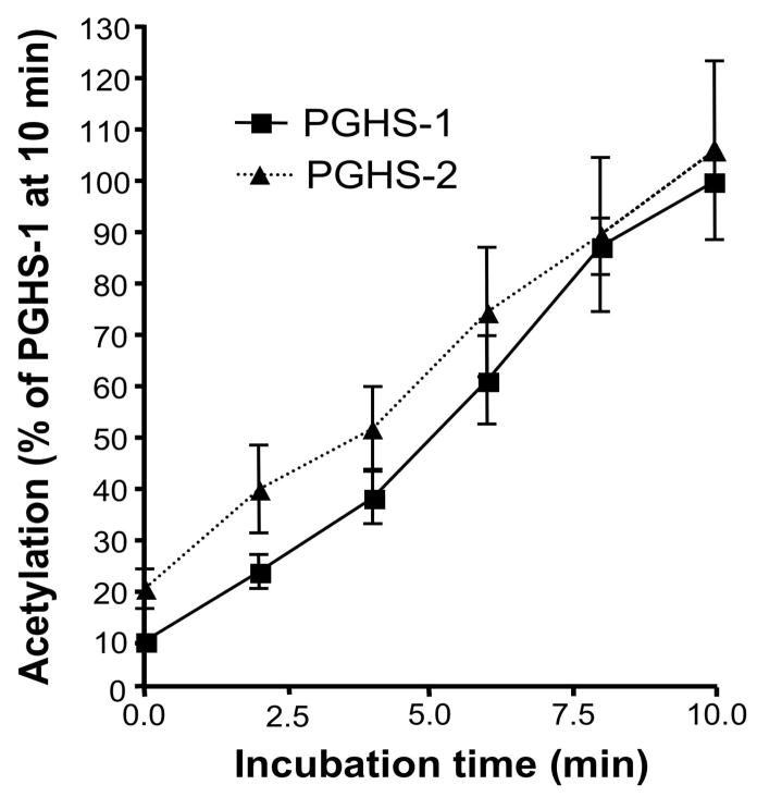
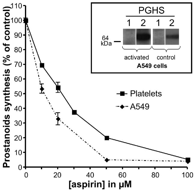
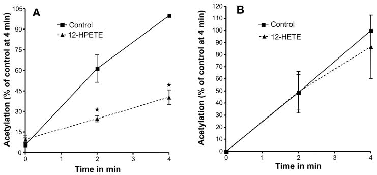
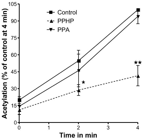
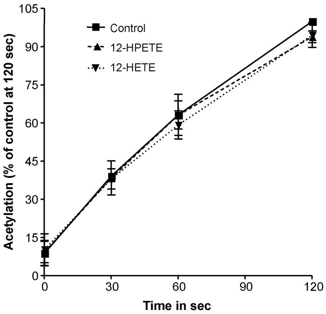
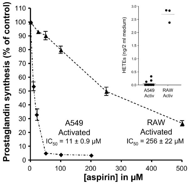
Aspirin exerts its unique pharmacological effects by irreversibly acetylating a serine residue in the cyclooxygenase site of prostaglandin-H2-synthases (PGHSs). Despite the irreversibility of the inhibition, the potency of aspirin varies remarkably between cell types, suggesting that molecular determinants could contribute to cellular selectivity. Using purified enzymes, we found no evidence that aspirin is selective for either of the two PGHS isoforms, and we showed that hydroperoxide substrates of the PGHS peroxidase inhibited the rate of acetylation of PGHS-1 by 68%. Using PGHS-1 reconstituted with cobalt protoporphyrin, a heme devoid of peroxidase activity, we demonstrated that reversal by hydroperoxides of the aspirin-mediated acetylation depends upon the catalytic activity of the PGHS peroxidase. We demonstrated that inhibition of PGHS-2 by aspirin in cells in culture is reversed by 12-hydroperoxyeicosatetraenoic acid dose-dependently (ED50=0.58+/-0.15 microM) and that in cells with high levels of hydroperoxy-fatty acids (RAW264.7) the efficacy of aspirin is markedly decreased as compared to cells with low levels of hydroperoxides (A549; IC50s=256+/-22 microM and 11.0+/-0.9 microM, respectively). Together, these findings indicate that acetylation of the PGHSs by aspirin is regulated by the catalytic activity of the peroxidase, which yields a higher oxidative state of the enzyme.


Figures

Fig. 1
Fig. 1
Comparision of acetylation of PGHS-1 and PGHS-2 by aspirin. Equipotent amounts of ovine PGHS-1 (1.5 μM) and murine PGHS-2 (2.7 μM) were reconstituted in 100 mM Tris HCl, pH 8.0, 500 μM phenol with two molar equivalents of hematin at 4°C for 30 minutes and [acetyl-1-14C] aspirin was added to a final concentration of 90 μM. At 0, 2, 4, 6, 8 and 10 minutes, aliquots were analyzed by electrophoresis. The radioactivity associated with the proteins was determined by autoradiography. Each data point represents the mean ± S.E.M of five values.
Fig. 2
Fig. 2
Comparision of inhibition of PGHS-1 and PGHS-2 by aspirin. Human cells expressing PGHS-1 (platelets) or PGHS-2 (A-549) were pre-incubated with varying concentrations of aspirin for 30 min at 37°C followed by addition of 2 μM [2H8] arachidonic acid. After 15 min at 37°C, the medium was collected, and TxB2 (platelet) and PGE2 (A549) were quantified by analysis by GC/NICI/MS as described previously [18]. Each data point represents the mean ± S.E.M. of four values. The significance of difference between the IC50 values of aspirin for the two cell lines was analyzed by two-tailed unpaired Student t-test (p<0.0001). The expression of PGHS-1 and -2 before and after activation of the A-549 cells was assessed by Western blot analysis and is shown in the inset.
Fig. 3
Fig. 3
Effect of 12-HPETE and 12-HETE on the acetylation of PGHS-1 by aspirin. The PGHS activity is expressed as a percentage of the control activity at 4 minutes. (A) Tris HCl (control) or 5 μM 12-HPETE were mixed separately with 90 μM [acetyl -1-14C] aspirin just before being added to the heme-reconstituted ovine PGHS-1 (1.5 μM). (B) Tris HCl (control) or 5 μM 12-HETE were mixed separately with 90 μM [acetyl-1-14C] aspirin just before being added to the heme reconstituted ovine PGHS-1. In both experiments, aliquots were taken at 0, 2, and 4 min, and PGHS-1 was analyzed as described for figure 1. Each data point represents the mean ± S.E.M of four values for 12-HPETE and five values for 12-HETE experiments. The significance of differences between the control and the experiments done for each concentration of 12-HPETE or 12-HETE was analyzed by paired two-tailed Student’s t-test (*: p<0.05; NS: non significant).
Fig. 4
Fig. 4
Effect of PPHP and PPA on the acetylation of PGHS-1 by aspirin. The PGHS activity is expressed as a percentage of the control activity at 4 minutes. Tris HCl (control,), 2μM PPHP or 2μM PPA were mixed separately with 90 μM [acetyl-1-14C] aspirin just before being added to the heme-reconstituted ovine PGHS-1 (1.5 μM). At 0, 2 and 4 minutes, aliquots were taken and analyzed as described for figure 1. Each data point represents the mean ± S.E.M of four values for PPHP and three values for PPA experiments. The significance of differences between the control and the experiments done for each concentration of PPHP or PPA was analyzed by paired two-tailed Student’s t-test (*: p<0.05; **: p<0.01; NS: non significant).
Fig. 5
Fig. 5
Acetylation of Co3+-PGHS-1 by Aspirin in presence of 12-HPETE and 12-HETE. The PGHS activity is expressed as a percentage of the control activity at 120 seconds. Tris HCl (control,), 5 μM 12-HPETE and 5 μM 12-HETE were mixed separately with 90 μM [acetyl-1-14C] aspirin just before being added to the ovine PGHS-1 (1.5 μM) reconstituted with cobalt protoporphyrin. At 0, 30, 60 and 120 seconds, aliquots were taken and analyzed as described for figure 1. Each data point represents the mean ± S.E.M of three values. The acetylation profiles are not statistically different.
Fig. 6
Fig. 6
Effect of 12-HPETE and 12-HETE on Aspirin mediated inhibition of PGHS-2 in IL-1β activated Human Lung Cancer Cell line (A-549 cell). Control experiments were conducted with 12-HPETE or 12-HETE without aspirin. 12-HPETE (panel A) or 12-HETE (panel B) were added to cells in culture at the indicated concentrations immediately before 20 μM aspirin was added. After 30 minutes pre-incubation, 2.0 μM arachidonic acid was added and kept at 37°C for 15 min. Prostanoids were analyzed by GC/NICI/MS as previously described [18]. The concentration of PGE2 present in the medium is represented as a percentage of the control to which no aspirin was added. Experiments were performed three times in duplicate. Each data points indicate mean ± S.E.M of six values. The significance of differences between the control and the experiments done for each concentration of 12-HPETE or 12-HETE was analyzed by paired two-tailed Student’s t-test (*: p<0.05; **: p<0.01 and ***p<0.0001).
Fig. 7
Fig. 7
Inhibition of PGHS-2 by aspirin inversely correlates with HPETE production by activated human cell lines. Human cells expressing PGHS-2 were activated with IL-1β (A549) or LPS and INF-γ (RAW264.7) for 18 h. The cells were then pre-incubated with varying concentrations of aspirin for 30 min at 37°C followed by addition of 2 μM [2H8] arachidonic acid. After 15 min at 37°C, the medium was collected, and the sum of PGD2 and PGE2 was quantified by analysis by GC/NICI/MS as described previously [18]. Each data point represents the mean ± S.E.M. of four or six values. The significance of difference between the IC50 values of aspirin for the two cell lines was analyzed by two-tailed unpaired Student t-test (p<0.0001). Inset: Production of HPETEs by A549 and RAW264.7 cells. No exogenous arachidonic acid was added. After activation of the cells, the medium was collected and HETEs, the products of reduction of HPETEs, were extracted and derivatized as described in “Materials and Methods”. 11-HETE represented 98% of total HETEs. The amounts of HETEs were determined by LC/APCI/MS/MS analysis using [2H8] 12-HETE as an internal standard.

Similar articles

Cited by

References

    1. Roth GJ, Majerus PW. The mechanism of the effect of aspirin on human platelets. I Acetylation of a particulate fraction protein. J Clin Invest. 1975;56:624–32. - PMC - PubMed
    1. Smith WL, Lands WE. Stimulation and blockade of prostaglandin biosynthesis. J Biol Chem. 1971;246:6700–2. - PubMed
    1. Lecomte M, Laneuville O, Ji C, DeWitt DL, Smith WL. Acetylation of human prostaglandin endoperoxide synthase-2 (cyclooxygenase-2) by aspirin. J Biol Chem. 1994;269:13207–15. - PubMed
    1. Roth GJ, Machuga ET, Ozols J. Isolation and covalent structure of the aspirin-modified, active-site region of prostaglandin synthetase. Biochemistry. 1983;22:4672–5. - PubMed
    1. FitzGerald GA, Oates JA, Hawiger J, Maas RL, Roberts LJ, 2nd, Lawson JA, et al. Endogenous biosynthesis of prostacyclin and thromboxane and platelet function during chronic administration of aspirin in man. J Clin Invest. 1983;71:676–88. - PMC - PubMed

Publication types