. 2008 Jan 25;132(2):208-20.
doi: 10.1016/j.cell.2007.12.029.

Mechanism of IS200/IS605 family DNA transposases: activation and transposon-directed target site selection

Affiliations

Mechanism of IS200/IS605 family DNA transposases: activation and transposon-directed target site selection

Orsolya Barabas et al. Cell. .

Abstract

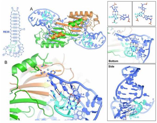
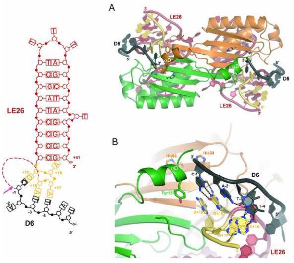
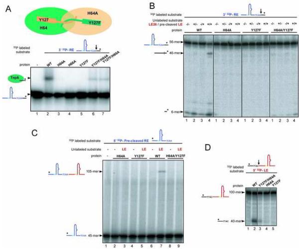
The smallest known DNA transposases are those from the IS200/IS605 family. Here we show how the interplay of protein and DNA activates TnpA, the Helicobacter pylori IS608 transposase, for catalysis. First, transposon end binding causes a conformational change that aligns catalytically important protein residues within the active site. Subsequent precise cleavage at the left and right ends, the steps that liberate the transposon from its donor site, does not involve a site-specific DNA-binding domain. Rather, cleavage site recognition occurs by complementary base pairing with a TnpA-bound subterminal transposon DNA segment. Thus, the enzyme active site is constructed from elements of both protein and DNA, reminiscent of the interdependence of protein and RNA in the ribosome. Our structural results explain why the transposon ends are asymmetric and how the transposon selects a target site for integration, and they allow us to propose a molecular model for the entire transposition reaction.


Figures

Figure 1
Figure 1. IS608 Transposition
(A) Alignment (adapted from Kersulyte et al. [2002]) of the termini and flanking sequences of IS608 elements from Helicobacter pylori. The strain is indicated on the left; all the work reported here was done with IS608 from PeCan2A (top line). Flanking DNA is in black, bases of the left end (LE) are shown in red and orange, and bases of the right end (RE) are shown in shades of blue. Boxed sequences underlined with inverted arrows delineate the Imperfect Palindromes (IP) at each end. The bases are numbered such that, at each end, the cleavage site is between base−1 and base+1 where bases 5′ of the cleavage site are negative, and those 3′ are positive. (B) Model of single-stranded transposition from Guynet et al. (in press). Intermediates in the reaction are a circular transposon junction and a precisely sealed donor backbone.
Figure 2
Figure 2. Conformational Change Induced in TnpA by a Four Nucleotide Extension of the Imperfect Palindrome
(A) Active site of TnpA bound to the RE22 hairpin (shown in blue). The two monomers of the TnpA dimer are colored green and orange. In this inactive conformation, the nucleophilic Y127 of one monomer is hydrogen-bonded to Ser110 of the other. (B) The same region of TnpA when bound to LE26 (shown in red and yellow) in which the IP is extended by 4 nt in the 5′ direction. Base A+18 has displaced Y127. Note that in the hairpin regions, LE and RE differ only by one T at the tip. The conformational change also necessarily shifts the residues that follow helix αD; we are only able to trace these in one of the monomers in the TnpA/LE26 structure.
Figure 3
Figure 3. The IS608 Right End is Directed into the TnpA Active Site by Internal DNA Interactions, not by Protein-DNA Recognition
(A) Overall view of theTnpA/RE35 structure. (B) Close-up of the active site showing the base pairs between the four bases at the RE of IS608 (TCAA) and bases 5′ of IPRE (GAAT) from base G−35 to T−32, in light blue). The grey sphere is bound Mn2+. (C) Two base triplets (top) are central to the RE35 conformation (shown in two orthogonal views in the middle and bottom panels).
Figure 4
Figure 4. Ternary Complex of TnpA Bound to LE26 and the LE Donor Flank Echoes Right End Recognition
(A) Overall view of the TnpA/LE26/D6 structure. On the left is a schematic of the oligonucleotides used where the dotted line indicates the break between LE26 and the donor flank, D6 (in black). The red arrow indicates the LE cleavage site. (B) Close-up of the active site showing that although the mode of interaction resembles the TnpA/RE35 structure, the specific bases differ.
Figure 5
Figure 5. Transposon End Cleavage Can Be Re-Directed
(A) SDS-PAGE gel showing the results of cleavage assays. Upon cleavage of an 8-mer that spans the LE cleavage site, TnpA (green oval) becomes covalently attached to four nt and can be resolved from unmodified TnpA. TnpA cleaves LE and RE cleavage substrates (cs) only when the appropriate 5′ extension is added to IP sequences. No covalent complex is formed with the Y127F active site mutant. (B) Modification of cleavage specificity. Shown are the results of LE cleavage assays using LE26 and LEcs, and two variants.
Figure 6
Figure 6. Mixed Mutant Dimers Suggest Transposon End Cleavage is in Trans and Resolution is in Cis
(A) Results of RE cleavage assays using mixed active site point mutants. The radiolabeled oligonucleotide is detected. (B) RE cleavage and donor backbone formation by mixed active site mutants was assessed by mixing a RE cleavage substrate (56-mer) with a LE flank (40-mer). The product of RE cleavage is a 6-mer, and the 46-mer is the sealed donor backbone that forms between the LE flank and the RE cleavage product. (C) Transposon junction formation by mixed active site mutants was assessed by mixing a LE cleavage substrate (100-mer which is cleaved to yield a 60-mer transposon LE) with a precleaved transposon RE (45-mer). The transposon junction is formed by the WT protein (lane 7) but not by mixed active site mutants (lane 9). (D) LE cleavage is catalyzed by mixed active site mutants (lane 3), but not by the single point mutants (lanes 4 and 5).
Figure 7
Figure 7. Model for IS608 Transposition
Upon LE and RE binding (A), Y127 of each monomer cleaves the transposon ends and becomes covalently attached to the 5′ side of the gap (B). Movement of αD helices from trans to cis and resolution of the phosphotyrosine intermediates results in a transposon junction and a sealed donor backbone (C). For integration (D), the donor backbone is replaced by target DNA containing TTAC which is recognized by an element of LE DNA. Cleavage (E), movement of the αD helices, and resolution of the phosphotyrosine intermediates (F) results in transposon insertion into target DNA immediately 3′ of TTAC.

References

    1. Belfort M, Derbyshire V, Parker MM, Cousineau B, Lambowitz AM. In: Mobile Introns: Pathways and Proteins in Mobile DNA II. Craig NL, et al., editors. ASM Press; Washington, D.C.: 2002.
    1. Brünger AT, Adams PD, Clore GM, Delano WL, Gros P, Grossekunstleve RW, Jiang JS, Kuszewski J, Nilges M, Pannu NS, et al. Crystallography and Nmr System - a New Software Suite For Macromolecular Structure Determination. Acta Crystallogr. D. 1998;54:905–921. - PubMed
    1. Curcio MJ, Derbyshire KM. The outs and ins of transposition: From Mu to kangaroo. Nature Rev. Mol. Cell Biol. 2003;4:1–13. - PubMed
    1. Debets-Ossenkopp YJ, Pot RGJ, van Westerloo DJ, Goodwin A, Vandenbroucke-Grauls CMJE, Berg DE, Hoffman PS, Kusters JG. Insertion of mini-IS605 and deletion of adjacent sequences in the nitroreductase (rdxA) gene cause metronidazole resistance in Helicobacter pylor NCTC11637. Antimicrob. Agents Chemother. 1999;43:2657–2662. - PMC - PubMed
    1. DeLano WL. The PyMol Molecular Graphics System. 2002. at www.pymol.org.

Publication types

MeSH terms

LinkOut - more resources