. 2008 Jan 25;132(2):247-58.
doi: 10.1016/j.cell.2007.12.016.

Rumi is a CAP10 domain glycosyltransferase that modifies Notch and is required for Notch signaling

Affiliations

Rumi is a CAP10 domain glycosyltransferase that modifies Notch and is required for Notch signaling

Melih Acar et al. Cell. .

Abstract

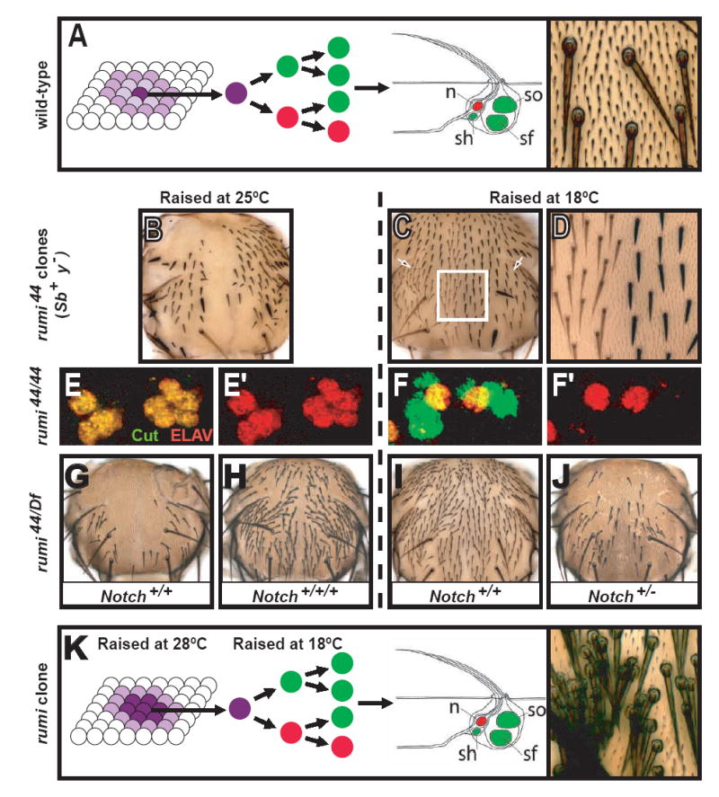
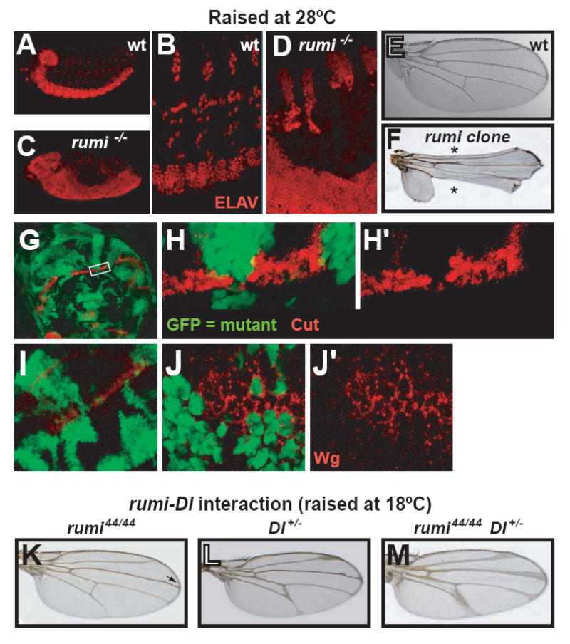
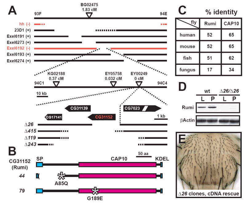
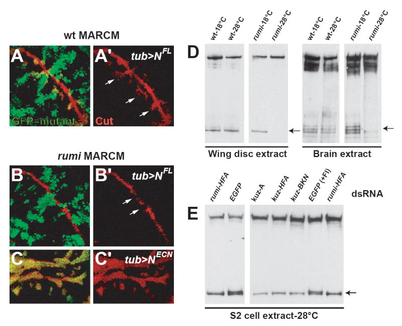
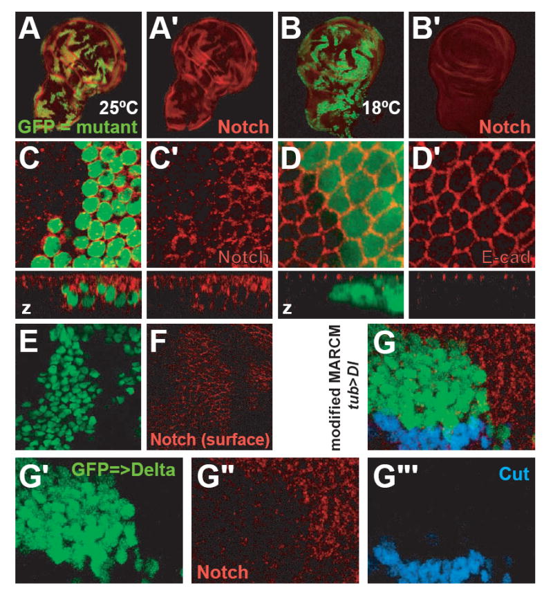
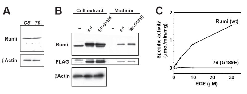
Notch signaling is broadly used to regulate cell-fate decisions. We have identified a gene, rumi, with a temperature-sensitive Notch phenotype. At 28 degrees C-30 degrees C, rumi clones exhibit a full-blown loss of Notch signaling in all tissues tested. However, at 18 degrees C only a mild Notch phenotype is evident. In vivo analyses reveal that the target of Rumi is the extracellular domain of Notch. Notch accumulates intracellularly and at the cell membrane of rumi cells but fails to be properly cleaved, despite normal binding to Delta. Rumi is an endoplasmic reticulum-retained protein with a highly conserved CAP10 domain. Our studies show that Rumi is a protein O-glucosyltransferase, capable of adding glucose to serine residues in Notch EGF repeats with the consensus C1-X-S-X-P-C2 sequence. These data indicate that by O-glucosylating Notch in the ER, Rumi regulates its folding and/or trafficking and allows signaling at the cell membrane.


Figures

Figure 1
Figure 1. rumi mutations cause a ts Notch phenotype
(A) Formation of an external sensory (es) organ from a single sensory organ precursor (SOP) cell is shown. Once the SOP is specified, it undergoes a series of asymmetric cell divisions to form the sensory organ structure. Only a single neuron forms in each sensory organ (red). The right picture shows wild-type microchaetae located on the thorax. n: neuron, so: socket cell, sh: sheath cell, sf: shaft cell. Bristle schematic adapted from (Lai and Orgogozo, 2004). (B) Bristle development is impaired in rumi clones at 25°C. Heterozygote bristles are marked with Sb and y+ markers. Note that not all bristles are lost at this temperature. (C and D) Bristles form in a denser pattern in rumi clones raised at 18°C. Non-Sb and yellow bristles mark rumi clones (compare the regions marked by the two arrows in (C)). (D) is a close-up of the white square. (E-F’) Cell fate specification is impaired in the adult microchaetae lineages of rumi mutants raised at 25°C, as evidenced by presence of multiple ELAV+ cells (red) per cluster (E and E’). Note that when raised at 18°C, only one of the Cut+ cells (green) per cluster expresses ELAV (F and F’). (G-J) Genetic interaction between Notch and rumi. (G) Hemizygous rumi44 flies that eclose at 25°C lose most of their microchaetae. (H) Adding an extra copy of Notch rescues many of the lost bristles (Compare G and H). (I) At 18°C, rumi mutant flies do not exhibit a significant bristle loss. (J) Flies heterozygous for Notch and hemizygous for rumi44, however, lose many bristles at 18°C (Compare I and J). (K) Tufts of bristles form in rumi mutant clones in flies that are kept at 28°C during SOP specification and at 18°C during asymmetric cell divisions, indicating an important role for rumi during lateral inhibition.
Figure 2
Figure 2. Loss of rumi causes loss of Notch signaling in various contexts
(A-D) rumi embryos laid by homozygous rumi flies show a neurogenic phenotype at 28°C (compare A and B with C and D). Neurons are marked with ELAV (red). (E-F) Wing margin development is impaired in rumi clones, causing Notching of the wings (asterisks in F). Panel (F) shows the wing of an adult who was subjected to the temperature-shift experiments described in Figure 1K. (G-J’) Expression of the Notch downstream targets Cut (red in G-H’) and Wg (red in I-J’) is lost in rumi clones at the dorso-ventral boundary of the third instar wing imaginal disc at 28°C in a cell-autonomous manner. GFP (green) marks rumi mutant cells. (K-M) Genetic interaction between Delta and rumi. All flies were raised at 18°C. (K) Wings of the homozygous rumi flies exhibit a mild Delta phenotype (arrow). (L) A wing of a fly which is heterozygous mutant for Delta. (M) There is a synergistic increase in wing vein expansion in flies that are homozygous mutant for rumi and heterozygous for Delta, indicating that rumi and Delta genetically interact.
Figure 3
Figure 3. rumi corresponds to CG31152
(A) The mapping strategy for rumi. The genomic region containing the rumi locus is shown. The broken lines depict deficiencies used to map rumi. hh and Exel6192 deletions (red lines) failed to complement rumi alleles. The genetic distance between the P-elements used for fine mapping and rumi is shown in centiMorgans (cM). Imprecise excision of EY00249 generated the additional alleles that remove rumi (CG31152) alone or in combination with CG31139, the only other fly gene capable of encoding a CAP10 domain. (B) rumi encodes a protein with a CAP10 domain, a KDEL ER-retention signal located at the C-terminus and a signal peptide (SP) located at the N terminus. Molecular lesions in alleles 44 and 79 are shown. Asterisks denote missense mutations. (C) Rumi protein, especially the CAP10 domain is highly conserved across different species. (D) Western blotting on the protein extracts from wild-type (wt) and rumiΔ26 larvae (L) and pupae (P) with a polyclonal α-Rumi antibody indicates that rumiΔ26 is a protein-null allele. (E) A UAS-rumi transgene can rescue the bristles lost in rumi clones. The bristles rescued by the transgene are yellow.
Figure 4
Figure 4. rumi is required in the signal-receiving cell upstream of the S3 cleavage
(A and A’) Ectopic expression of the full length Notch (NFL) induces aberrant Cut (red) expression in MARCM clones of a wild-type chromosome that are close to the dorso-ventral boundary (arrows in A’). GFP (green) marks the clones that ectopically express NFL in (A) and (B). (B and B’) Ectopic expression of NFL in MARCM rumi clones does not induce Notch signaling. Note that in rumi mutant cells Cut (red) expression is lost in the prospective wing margin despite NFL overexpression (arrows in B’). (C and C’) Ectopic expression of NECN induces Cut (red) expression in rumi cells. GFP marks MARCM rumi clones that ectopically express NECN, suggesting that the Rumi function is required upstream of the S3 cleavage of Notch. (D) Western blots showing that Notch processing is altered in the absence of Rumi function at the restrictive temperature. Anti-NICD antibody was used in all blots. The top bands in each blot correspond to full length Notch, which is around 300 kDa. In wing disc extracts one predominant band that corresponds to Notch cleavage product is visible in both wild-type (wt) and in rumΔ26/Δ26 (rumi) larvae kept at 18°C. This band is not detected in extracts of rumi larvae kept at 28°C for 10 hours prior to dissection (left arrow). In brain extracts four ~120 kDa fragments are visible in wt larvae and in rumi larvae that were kept at 18°C. The upper two bands however are lost in rumi larvae kept at 28°C (right arrow). (E) RNAi-mediated knockdown of Rumi and Kuz results in similar Notch processing defects in S2 cells. Western blotting with anti-NICD on protein extracts from S2 cells raised at 28°C is used to determine the pattern of Notch cleavage. Control cells (EGFP dsRNA) show two cleavage products (arrow). Addition of a Furin inhibitor (FI) does not alter this pattern. However, treatment of S2 cells with dsRNA against Rumi or Kuz results in the loss of the upper cleavage product, strongly suggesting that the Kuz-mediated S2 cleavage of Notch is affected by the loss of Rumi at the restrictive temperature. Note that three different kuz dsRNAs produce the same results.
Figure 5
Figure 5. High levels of Notch accumulate inside and at the membrane of rumi mutant cells at the restrictive temperature
(A-C’) rumi clones marked by GFP (green in A, B and C) show an accumulation of Notch (red) when raised at 25°C (A, A’, C, C’) but not when raised at 18°C (B, B’). The accumulation is evident with antibodies against both NECD (A, A’) and NICD (C, C’). A-B’ show third instar wing imaginal discs; C and C’ show a close-up from a pupa 12 hours after puparium formation (APF). Note the cell-autonomous increase in Notch levels (C, C’). Also, optical z sectioning shows that in mutant cells Notch is not mainly in the apical regions anymore (z in C, C’). (D and D’) E-Cadherin (E-cad, red) localization and levels do not change in rumi cells at 25°C. (E and F) Notch protein levels at the cell membrane increase in rumi cells. Shown are two different sections of the wing disc of a third instar larva with MARCM rumi clones (green) raised at the restrictive temperature, and fixed and stained with the α-NECD (red) antibody in the absence of detergent. (F) is close to the apical surface, and (E) is 700 nm basal to (F). Note the accumulation of Notch at apical regions (F) overlaying the clones marked by the nuclear GFP in (E). Absence of specific α-NECD staining in (E) indicates that the antibody has not entered the cell because of the lack of detergent. (G-G’’’) Ectopic over-expression of Delta in wild-type cells does not induce Notch signaling in adjacent cells that are mutant for rumi, despite the accumulation of Notch in these cells. Shown is an example of a modified MARCM clone located in the dorsal part of the wing pouch away from the wing margin (not shown), in which GFP (green) marks rumi+/+ cells that overexpress Delta, and Notch accumulation (red) marks rumi-/- cells. Note that Delta from green cells is able to induce Cut expression (blue) in rumi+/- cells but not in rumi-/- cells.
Figure 6
Figure 6. Rumi is a protein O-glucosyltransferase
(A) The unglycosylated form of 561CQINIDDCQSQPCR574 is present in the Rumi knockdown sample, but not in the control sample. The MS data from both samples were searched for the doubly charged form of the peptide, m/z 898.4 (see Figure S9A for MS and MS/MS spectra at 32.0 min). The ion can be clearly seen eluting at 32.0 minutes in the Rumi knockdown sample (-Rumi), but it cannot be detected above the noise in the control sample (+Rumi). (B) The O-glucosylated form of 561CQINIDDCQSQPCR574 is more abundant in the control sample than in the Rumi knockdown sample. The MS data from both samples was searched for the doubly charged form of the glycopeptide, m/z 978.7 (see Figure S9B for MS and MS/MS spectra at 30.8 min). The ion can be seen in both samples at 30.8 minutes, but more is present in the control sample (+Rumi) than in the Rumi knockdown sample (-Rumi). (C) Both cell extracts and culture media of Rumi-overexpressing S2 cells showed an O-glucosyltransferase activity in vitro. Inset: Coomassie staining after 10% SDS-PAGE of the equivalent amounts of the beads. Line indicates 50 kDa size marker. 1: cell extract from control cells, 2: cell extract from Rumi-overexpressing cells, 3: media from control cells, 4: media from Rumi-overexpressing cells. (D) O-glucosyltransferase activity was dependent on the amount of the purified Rumi protein in vitro. (E) O-glucosyltransferase activity of the purified Rumi protein was dependent on the concentration of factor VII EGF as acceptor substrate. (F) O-glucosyltransferase activity of the purified Rumi protein was dependent on the concentration of UDP-glucose as donor substrate. All O-glucosyltransferase assays were performed in duplicate. Error bars represent the range of duplicates.
Figure 7
Figure 7. O-glucosylation mediated by Rumi is required for Notch signaling
(A) Western blots showing the relative levels of Rumi and β-Actin proteins in wild-type (CS) and homozygous rumi79 (79) larval protein extracts. Late third instar larvae were used to prepare protein extracts for each genotype. Note that the G189E mutation in rumi79 larvae does not affect the level of the Rumi protein. (B) The G189E mutation does not affect the expression level of Rumi in S2 cells. Western blots showing the expression and secretion of Rumi-FLAG (RF) and Rumi-FLAG with the G189E mutation (RF-G189E) in S2 cells. RF and RF-G189E were expressed in S2 cells using pUAST and pAc-Gal4 vectors. Note that the RF and RF-G189E proteins expressed in S2 cells run slightly slower than the endogenous Rumi protein due to the presence of the FLAG tag (see anti-Rumi blot). Endogenous Rumi is not expressed at levels that lead to its secretion in the medium. Some of the RF and RF-G189E proteins are secreted into the medium when expressed in S2 cells. (C) The G189E mutation causes a complete loss of O-glucosyltransferase activity. Both wild-type and G189E Rumi proteins were overexpressed in S2 cells and purified from culture media by using the FLAG epitope as described in Experimental Procedures. The purified proteins were assayed for protein O-glucosyltransferase activity using increasing concentrations of the acceptor substrate, factor VII EGF repeat (wild-type Rumi, diamonds; G189E mutant, open squares). All assays were performed using 100 μM of UDP-glucose in duplicate. Error bars represent the range of duplicates.

Comment in

  • A notch sweeter.
    Irvine KD. Irvine KD. Cell. 2008 Jan 25;132(2):177-9. doi: 10.1016/j.cell.2008.01.005. Cell. 2008. PMID: 18243091

Similar articles

Cited by

References

    1. Artavanis-Tsakonas S, Rand MD, Lake RJ. Notch signaling: cell fate control and signal integration in development. Science. 1999;284:770–776. - PubMed
    1. Bellen HJ, Levis RW, Liao G, He Y, Carlson JW, Tsang G, Evans-Holm M, Hiesinger PR, Schulze KL, Rubin GM, et al. The BDGP gene disruption project: single transposon insertions associated with 40% of Drosophila genes. Genetics. 2004;167:761–781. - PMC - PubMed
    1. Bolos V, Grego-Bessa J, de la Pompa JL. Notch signaling in development and cancer. Endocrine reviews. 2007;28:339–363. - PubMed
    1. Brou C, Logeat F, Gupta N, Bessia C, LeBail O, Doedens JR, Cumano A, Roux P, Black RA, Israel A. A novel proteolytic cleavage involved in Notch signaling: the role of the disintegrin-metalloprotease TACE. Molecular cell. 2000;5:207–216. - PubMed
    1. Bruckner K, Perez L, Clausen H, Cohen S. Glycosyltransferase activity of Fringe modulates Notch-Delta interactions. Nature. 2000;406:411–415. - PubMed

Publication types

MeSH terms