. 2008 Jan 25;132(2):259-72.
doi: 10.1016/j.cell.2007.12.030.

Molecular and structural basis of cytokine receptor pleiotropy in the interleukin-4/13 system

Affiliations

Molecular and structural basis of cytokine receptor pleiotropy in the interleukin-4/13 system

Sherry L LaPorte et al. Cell. .

Abstract

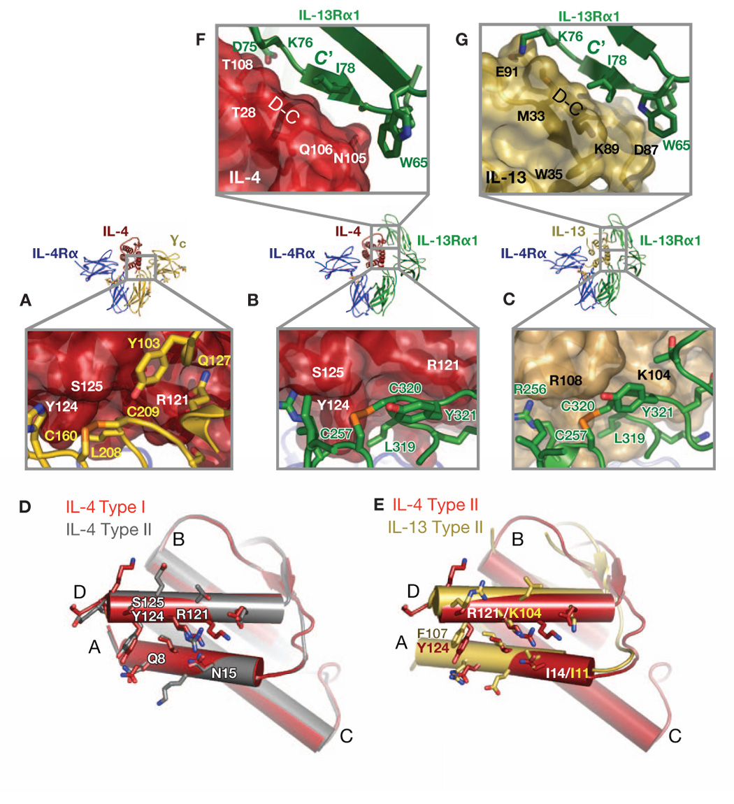
Interleukin-4 and Interleukin-13 are cytokines critical to the development of T cell-mediated humoral immune responses, which are associated with allergy and asthma, and exert their actions through three different combinations of shared receptors. Here we present the crystal structures of the complete set of type I (IL-4R alpha/gamma(c)/IL-4) and type II (IL-4R alpha/IL-13R alpha1/IL-4, IL-4R alpha/IL-13R alpha1/IL-13) ternary signaling complexes. The type I complex reveals a structural basis for gamma(c)'s ability to recognize six different gamma(c)-cytokines. The two type II complexes utilize an unusual top-mounted Ig-like domain on IL-13R alpha1 for a novel mode of cytokine engagement that contributes to a reversal in the IL-4 versus IL-13 ternary complex assembly sequences, which are mediated through substantially different recognition chemistries. We also show that the type II receptor heterodimer signals with different potencies in response to IL-4 versus IL-13 and suggest that the extracellular cytokine-receptor interactions are modulating intracellular membrane-proximal signaling events.


Figures

Figure 1
Figure 1. Structures of the Type I IL-4, Type II IL-4, and Type II IL-13 ternary complexes
(A) The Type I complex between IL-4Rα (blue), IL-4 (red) and γc (gold). (B) The Type II IL-4 complex between IL-4Rα (blue), IL-4 (red) and IL-13Rα1 (green). (C) The Type II IL-13 complex with IL-4Rα (blue), IL-13 (yellow-orange) and IL-13Rα1 (green). The complexes are shown from the side with a cartoon of a membrane underneath (left), and as viewed from the “top” (right). Glycan moieties on Asn residues are shown in ball-and-stick representation. All figures were generated with PyMol (DeLano, The PyMOL Molecular Graphics System, 2002).
Figure 2
Figure 2. Structural similarity between IL-2Rα and IL-13Rα1 cytokine engagement
(A) The quaternary complex of IL-2 (pink), IL-2Rα (cyan), IL-2Rβ (blue) and γc (gold). IL-2Rα is bound to the top surface of IL-2. (B) Ternary complex of IL-13 (yellow-orange), IL-4Rα (blue), and IL-13Rα1 (green). (C) As viewed from the top, the overlapping cytokine binding sites by IL-2Rα and IL-13Rα1 D1 domains are apparent. Semi-transparent ovals have been drawn around the D1 domain of IL-2Rα and IL-13Rα1 so that regions of overlap are clear.
Figure 3
Figure 3. Contrasting assembly sequences, binding thermodynamics and stabilities of the three ternary complexes
Each row illustrates the reaction in the order in which the binary and ternary complexes assemble (color scheme is maintained from Figure 1). Inset tables list the thermodynamic parameters calculated based upon the isothermal titration calorimetry (ITC) binding isotherm shown below each reaction diagram. Data are also summarized in Table S3 (A) Type I complex: Reaction diagram for the interaction of IL-4Rα and IL-4 followed by recruitment of the second receptor, γc. (B) Type II IL-4 complex: the interaction of IL-4Rα and IL-4 followed by the recruitment of the second receptor, IL-13Rα1. In both (A) and (B), the extremely high affinity of IL-4/IL-4Rα (Kd < 1nM) is an estimate due to the steepness of the ITC trace. (C) Type II IL-13 complex: the interaction of IL-13Rα1 and IL-13 followed by the recruitment of the second receptor, IL-4Rα.
Figure 4
Figure 4. The interactions of IL-4Rα with IL-4 and IL-13
For clarity the overall complexes are shown above blowups of the site I IL-4 (A) and IL-13 (B) interfaces, with color scheme retained from Figure 1. Zoomed up region of the site I interfaces focuses on the IL-4Rα EF loop interactions with cytokine A and C helices (labeled). Key interacting residues are shown in stick representation, and the “mimicked” Arg and Glu hotspot residues found in both IL-4 and IL-13 are thicker sticks and colored cyan, with polar contacts drawn as dashed green lines. (C) “Open book” view of binding epitopes presented by IL-4 and IL-13 to IL-4Rα. The resulting IL-4 and IL-13 cytokine (shown as cylinders) positions after their respective IL-4Rα were superimposed shows the relative structural overlap of the cytokine site I binding sites. (D) Structure-based sequence alignment between the interacting portions of helices A and C of IL-4 and IL-13 with IL-4Rα, where contact positions are in red boxes, common hydrophobic core residues are highlighted in gray and “hot spot” residues are highlighted in cyan to coincide with panels (A), (B) and (C).
Figure 5
Figure 5. The interactions of IL-4 and IL-13 with γc and IL-13Rα1
For clarity the overall complexes are shown in between blowups of the site IIa (underneath) and III (above) interfaces using the same color scheme as previous figures. Panels (A), (B) and (C) show zoomed views of the site IIa interfaces of the IL-4 Type I, IL-4 Type II, and IL-13 Type II complexes, respectively, where the receptor residues are projected on semi-transparent cytokine surfaces. IL-4(red) and IL-13(yellow-orange) residues underneath the surfaces are labeled in white and black, respectively, while γc (gold) and IL-13 Rα1(green) residues are labeled in yellow and green, respectively. (D) and (E) “Open book” views of the site IIa binding sites. Panel (D) shows the resulting structural positions of IL-4 on IL-4 after superposition of IL-4Rα from the Type I and Type II complexes, and panel (E) shows the resulting structural positions of IL-4 on IL-13 after superposition of IL-4Rα from the two different Type II complexes. All interacting residues in the site IIa interfaces are shown as sticks. Panels (F) and (G) illustrate the site III interfaces in IL-4 and IL-13 Type II complexes, respectively, using a similar coloring and labeling scheme as above. The anti-parallel beta sheet between the IL-13Rα1 c’ strand the cytokine C–D loop is labeled.
Figure 6
Figure 6. Basis of cross-reactive cytokine recognition by γc
(A) Structural alignment of the IL-2 quaternary (IL-2Rα not shown), and IL-4 Type I ternary complexes after superposition on γc (gold). The IL-2 complex is blue, and the IL-4 complex is green. (B) The conserved apolar ‘canyon’ on the cytokine surfaces (highlighted in blue) accommodates the protruding γc binding loops (gold and silver residues). (C) “Open book” views of the overlap of IL-2 versus IL-4 site IIa binding sites after superposition of γc. (D) Structure-based sequence alignment of all known γc cytokines for helices A and D based on γc contact positions by IL-2 and IL-4. Residue numbers are included and contact residues from IL-2 and IL-4 are marked with red boxes. Hydrophobic residues are shaded in gray and correspond predominantly with buried core residues.
Figure 7
Figure 7. Comparison of signaling activated by IL-4 and IL-13
(A) A549 express IL-4Rα and IL-13Rα1, but not IL-13Rα2 or γc. Expression of receptor subunits on A549 was analyzed by FACS as described in Methods. The heavy black histograms indicate staining with specific antibody and the dotted histograms indicate staining by the isotype-matched control. (B) A549 cells and Ramos cells were washed and treated with various concentrations of IL-4 or IL-13 as indicated for 30 minutes. Cell lysates were prepared and immunoprecipitated with anti-STAT6. Western blots were probed with anti-phosphotyrosine antibody. The blots were stripped and reprobed with anti-STAT6. STAT3 activation is analyzed in Figure S6. (C) A549 cells and Ramos cells were treated with various concentrations of IL-4 or IL-13 for various times; STAT6 phosphorylation was analyzed as described in (B). Data is quantitated in graphical format in Figure S7.

Comment in

References

    1. Aman MJ, Tayebi N, Obiri NI, Puri RK, Modi WS, Leonard WJ. cDNA cloning and characterization of the human interleukin 13 receptor alpha chain. J Biol Chem. 1996;271:29265–29270. - PubMed
    1. Andrews AL, Holloway JW, Holgate ST, Davies DE. IL-4 receptor alpha is an important modulator of IL-4 and IL-13 receptor binding: implications for the development of therapeutic targets. J Immunol. 2006;176:7456–7461. - PubMed
    1. Andrews AL, Holloway JW, Puddicombe SM, Holgate ST, Davies DE. Kinetic analysis of the interleukin-13 receptor complex. J Biol Chem. 2002;277:46073–46078. - PubMed
    1. Arima K, Sato K, Tanaka G, Kanaji S, Terada T, Honjo E, Kuroki R, Matsuo Y, Izuhara K. Characterization of the interaction between interleukin-13 and interleukin-13 receptors. J Biol Chem. 2005;280:24915–24922. - PubMed
    1. Barnes PJ. Cytokine modulators as novel therapies for asthma. Annu Rev Pharmacol Toxicol. 2002;42:81–98. - PubMed

Publication types

MeSH terms