. 2008 Jan 25;132(2):286-98.
doi: 10.1016/j.cell.2007.11.048.

Rab and Arl GTPase family members cooperate in the localization of the golgin GCC185

Affiliations

Rab and Arl GTPase family members cooperate in the localization of the golgin GCC185

Alondra Schweizer Burguete et al. Cell. .

Abstract

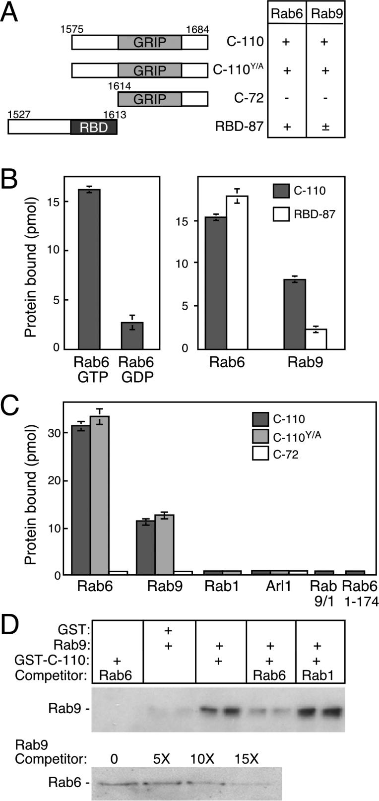
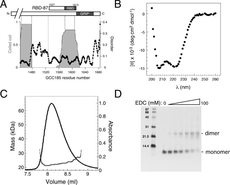
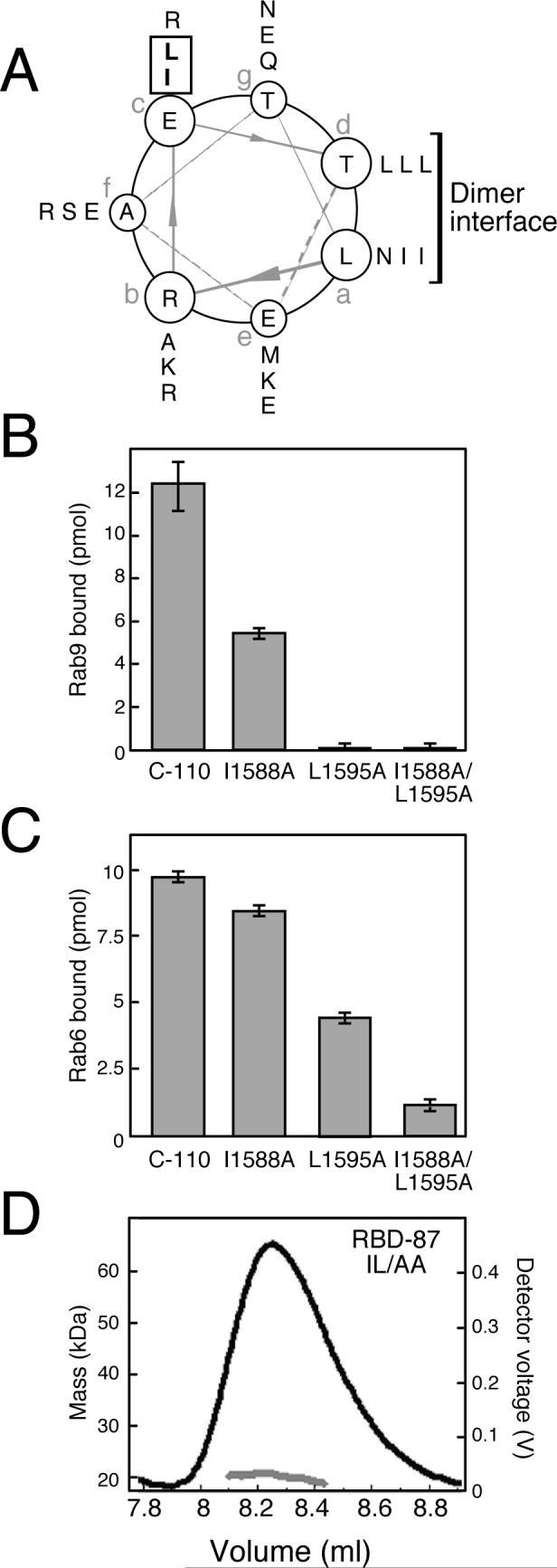
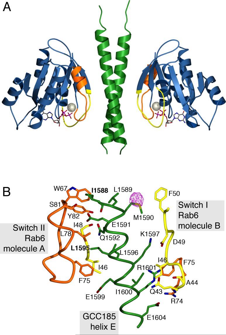
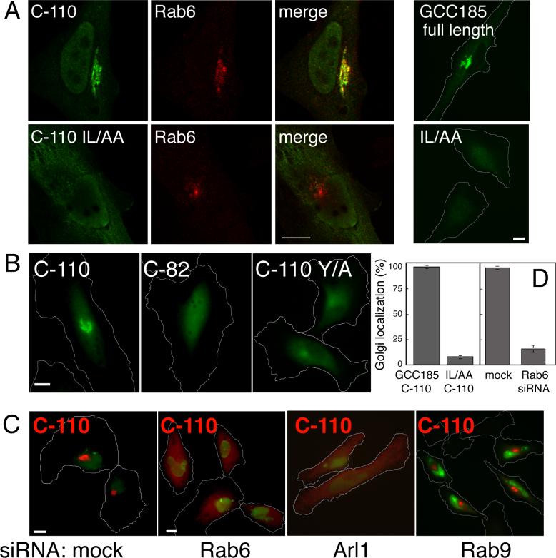
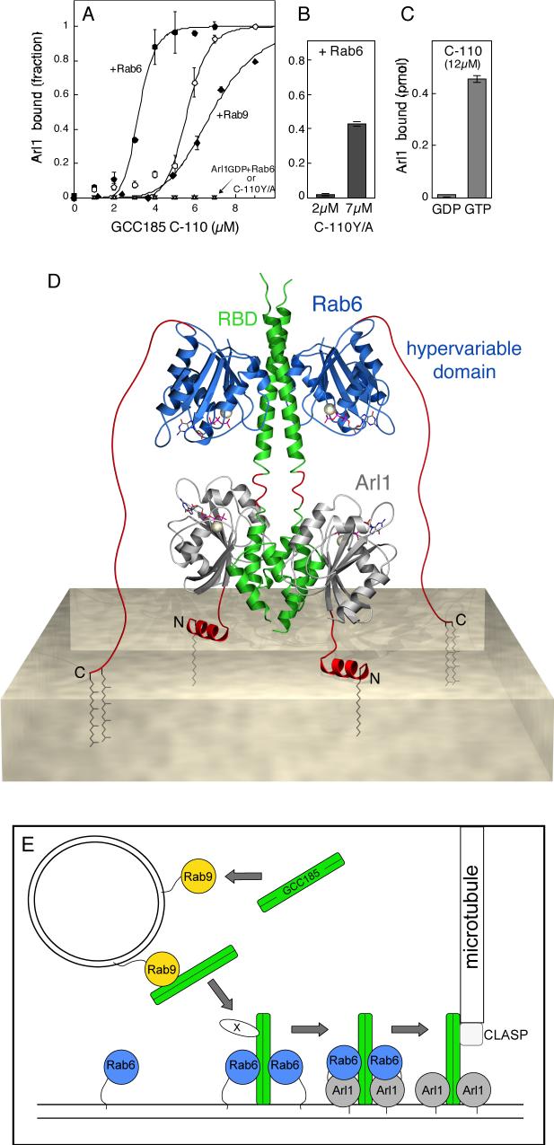
GCC185 is a large coiled-coil protein at the trans Golgi network that is required for receipt of transport vesicles inbound from late endosomes and for anchoring noncentrosomal microtubules that emanate from the Golgi. Here, we demonstrate that recruitment of GCC185 to the Golgi is mediated by two Golgi-localized small GTPases of the Rab and Arl families. GCC185 binds Rab6, and mutation of residues needed for Rab binding abolishes Golgi localization. The crystal structure of Rab6 bound to the GCC185 Rab-binding domain reveals that Rab6 recognizes a two-fold symmetric surface on a coiled coil immediately adjacent to a C-terminal GRIP domain. Unexpectedly, Rab6 binding promotes association of Arl1 with the GRIP domain. We present a structure-derived model for dual GTPase membrane attachment that highlights the potential ability of Rab GTPases to reach binding partners at a significant distance from the membrane via their unstructured and membrane-anchored, hypervariable domains.


Figures

Figure 1
Figure 1
Identification of a GCC185 Rab-binding domain. (A) Constructs used to map Rab-GCC185 interactions; numbers represent amino acid residues. The GRIP domain and a Rab binding domain (RBD) are shown. At right: summary of binding to Rab6 or Rab9. (B) GCC185 preferentially binds Rab6-GTP via residues upstream of the GRIP domain. Reactions contained 50 pmol His-Rab6 (left) or 1.2 nmol His-Rab6, or Rab9-His (right) using 35S-GTPγS or 3H-GDP-preloaded GTPase and either GST-C110 or GST-RBD-87 (2.8 μM). (C) The GRIP domain is not sufficient for Rab binding. GST-C-110, C-110Y/A, or C-72 (2 μM) was incubated with 35S-GTPγS-preloaded GTPases (∼500 pmol) (as in B. except Rab1 and Rab9 were untagged and or ArlQ71L-His was used). Untagged Rab9/1 and His-Rab6 1−174 were employed. (D) Rab6 specifically competes with Rab9 for GCC185 binding. GST-C-110:Rab9 complexes (pair of lanes in the center) were incubated for 3 min with ten fold excess competitor. Rab9-His was detected by immunoblot using a monoclonal anti-Rab9 antibody that did not cross react with Rab6 (see pair of lanes at far left). Lower panel, same as upper panel using indicated amounts of competitor Rab9. Data are mean ± SD.
Figure 2
Figure 2
The GCC185 Rab binding domain is a helical dimer. (A) Structure prediction of the GCC185 C-terminus. Coiled coil (shaded gray; cutoff=0.8) and disordered regions (black line and dots; cutoff=0.12) in GCC185 residues 1444−1684 were predicted with Paircoil and the DisEMBL™ programs, respectively. At top: bar diagram of the GCC185 C-terminus. (B) Circular dichroism spectrum of untagged RBD-87 (mean residue ellipticity). (C) Untagged RBD-87 forms a dimer in solution. Black line, A280. The mass at different elution volumes was calculated from multiple angle static light scattering data (dots). The polydispersity of the peak was 1.024. The mass of RBD-87 (92aa) monomer is 10.3 kDa. (D) Analysis of RBD-87 by crosslinking. Untagged protein (5 μM) was reacted at 20°C for 2 hr with 0−100 mM EDC (1-ethyl-3-[3-dimethylaminopropyl] carbodiimide hydrochloride).
Figure 3
Figure 3
Accessible hydrophobic residues in the predicted coiled coil are critical for Rab binding. (A) Helical wheel projection of a coiled coil predicted for GCC185 residues 1579−1606. Residues in registers “a-f” were predicted by the Paircoil program. Residues at positions “a” and “d” lie in the dimer interface. Boxed residues are candidates for binding interactions with Rab GTPases. (B, C) Effect of alanine substitutions on Rab binding. Reactions contained wild type or mutant GST-C-110 (B; 3 μM, C; 2 μM) and 35S-GTPγS-preloaded GTPases (B; 170 pmol Rab9-His, C; 190 pmol His-Rab6). Data are mean ± SD. (D) Mass determination of untagged RBD-87 I1588A/L1595A by multiple angle static light scattering. The gel filtration elution profile of the protein (black line) and molecular mass (grey line) are shown. Polydispersity of the peak was 1.001.
Figure 4
Figure 4
Structure of the Rab6-GCC185 complex. A. Ribbon representation of the GCC185 Rab binding domain dimer (green) and Rab6 (blue) bound to GTP (stick model) and magnesium (sphere). Switch I and II regions of Rab6 (Chattopadhyay et al., 2000) are colored yellow and orange respectively. B. View of the Rab6-GCC185 binding interface. A single GCC185 helix (E) out of the two-fold symmetric coiled coil is shown for clarity. Each helix contacts switch regions from two opposed Rab6 molecules A and B. Rab6 switch I and II (including W67), are colored yellow and orange, respectively. Protein backbone (α-carbon trace) and side chains involved in polar and hydrophobic interactions are shown. Carbonyl oxygens are shown for A44, I48 and I1588 and C-Cα bonds have been added to simplify the figure. An anomalous difference Fourier density map of the selenomethionine substituted crystal (pink, contoured at 6σ) is shown for GCC185.
Figure 5
Figure 5
GCC185 Golgi targeting requires the Rab binding domain and Rab6 and Arl1 GTPases. (A) Confocal micrograph of HeLa cells expressing myc-tagged wild type or I1588A/L1595A C-110, 19 h post transfection. c-myc epitope (green) and Rab6 (red) are shown. A confocal section is shown; image step size was 15.2 nm. Far right panels were analyzed as in B. (B) Conventional fluorescence micrograph of HeLa cells transfected with myc-C-110, myc-C-82 (20 h post transfection) or myc-C-110 Y1618A (29 h post transfection). Cells were stained as in A. (C) Conventional immunofluorescence micrograph of HeLa cells transfected with 6-FAM-conjugated RNA oligonucleotides (green, mock) or these plus siRNAs targeting Rab6, Arl1 or Rab9 as indicated. Post depletion, and 17 h prior to fixation, cells were transfected with myc-GCC185 C-110 (red). siRNA-siRNA-transfected cells are indicated by nuclear green fluorescence from 6-FAM conjugated RNA oligonucleotides.myc-C- (D). Quantification of panels A and part of C. Perinuclear myc-fluorescence above background that overlapped with Rab6 staining was scored as Golgi localization. Standard deviations are from two experiments, 100 cells counted per bar shown. For quantitation of C, perinuclear fluorescence above background was scored in mock (n=159 and 113) and Rab6 siRNA (n=314 and 210) treated cells in duplicate experiments. Bars, 10 μm.
Figure 6
Figure 6
Dual GTPase binding to GCC185. A. Rab6 stimulates Arl1 GTPase binding to GCC185. His-Rab6 or untagged Rab9 were preloaded with cold GTPγS; Arl1Δ17 was loaded with [35S] GTPγS or 3H-GDP. GST-C-110 (wild type or Y/A) was then incubated with Rab6- or Rab9-GTPγS (3.5μM) for 30 min at room temperature. Arl1Δ17 (1.2μM) bearing radioactive nucleotide was added to preformed complexes and samples were rotated for 1 h at room temperature in a total volume of 250μl. Data in (A) are fitted to an equation reflecting cooperative binding. Maximal binding was 2.2% of input Arl1 protein. Wild type C-110 binding to: Open circles, Arl1 alone; filled circles, Arl1 + Rab6; closed diamonds, Arl1 + Rab9; small triangles, Arl1GDP + Rab6; C-110 Y/A binding to: inverted triangles, Arl1 alone. B. Rab6 (3.5μM) was incubated with GST-C-110 Y/A at the indicated concentrations; Arl1 was then added as in A. Shown is the fraction Arl1 protein bound. C. Nucleotide dependence of Arl1 binding to GST-C-110. Reactions contained 22.8 pmol Arl1 protein. A-C, data are mean ± SD. D. Model of Rab6 and Arl1 bound to the GCC185 Rab binding domain (RBD) and GRIP domains, respectively. GCC185 (green) bound to Rab6 (blue) and Arl1 (grey). Modeled regions (red) include the Rab6 hypervariable domain (extended), the Arl1 N-terminus at the membrane and the junction between the GRIP and Rab binding domains of GCC185. Lipid anchors (brown) and the cytosolic leaflet of the membrane bilayer are shown. See text for details. E. Model for tether transfer from a vesicle to the Golgi membrane. Dimeric, cytosolic GCC185 is first recruited onto a Rab9-bearing vesicle. GCC185 on the vesicle is proposed to interact with a Golgi-bound GCC185 via a hypothetical linking protein (X) that may represent CLASP. Initial docking would permit SNARE pairing (not shown); independently, Rab6 displaces Rab9 from GCC185 and then promotes Arl1 binding. Soluble GCC185 may also be recruited to the Golgi by a Rab9 independent process. Proteins in E are not drawn to scale. At far right: GCC185-bound CLASP that nucleates microtubule polymerization at the Golgi.

Similar articles

Cited by

References

    1. Aivazian D, Serrano RL, Pfeffer S. TIP47 is a key effector for Rab9 localization. J. Cell Biol. 2006;173:917–926. - PMC - PubMed
    1. Barbero P, Bittova L, Pfeffer SR. Visualization of Rab9-mediated vesicle transport from endosomes to the trans-Golgi in living cells. J. Cell Biol. 2002;156:511–8. - PMC - PubMed
    1. Barr FA. A novel Rab6-interacting domain defines a family of Golgi-targeted coiled-coil proteins. Curr. Biol. 1999;9:381–384. - PubMed
    1. Bergbrede T, Pylypenko O, Rak A, Alexandrov A. Structure of the extremely slow GTPase Rab6A in the GTP bound form at 1.8 resolution. J. Struct. Biol. 2005;152:235–238. - PubMed
    1. Berger B, et al. Predicting coiled coils by use of pairwise residue correlations. Proc. Natl. Acad. Sci. 1995;92:8259–8263. - PMC - PubMed

Publication types

MeSH terms

Associated data