. 2008 Jan 25;132(2):299-310.
doi: 10.1016/j.cell.2007.11.047.

NFATc1 balances quiescence and proliferation of skin stem cells

Affiliations

NFATc1 balances quiescence and proliferation of skin stem cells

Valerie Horsley et al. Cell. .

Abstract

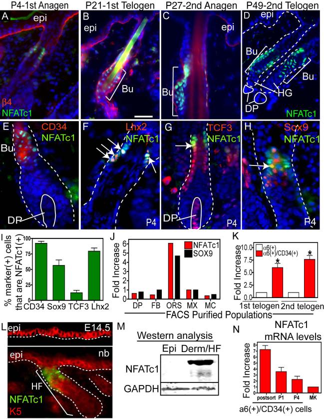
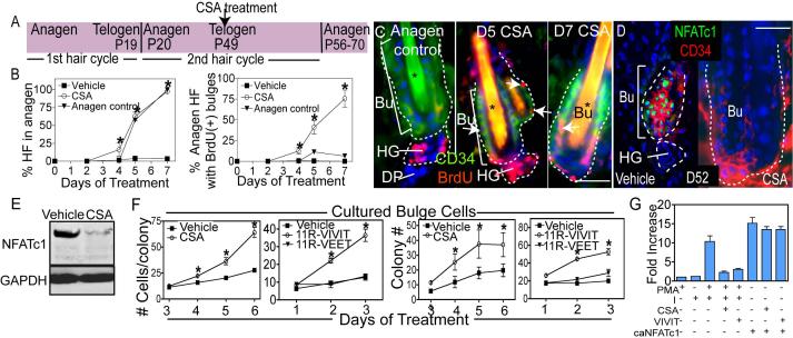
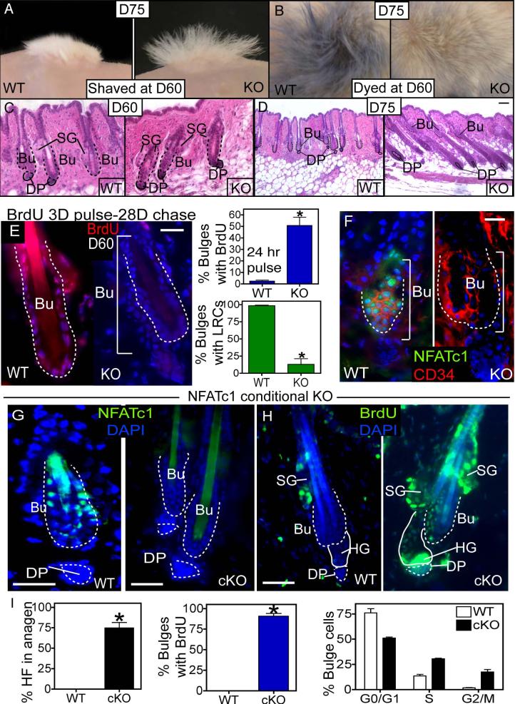
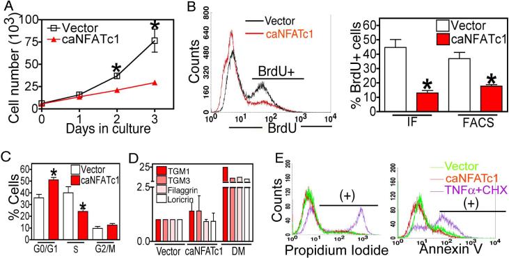
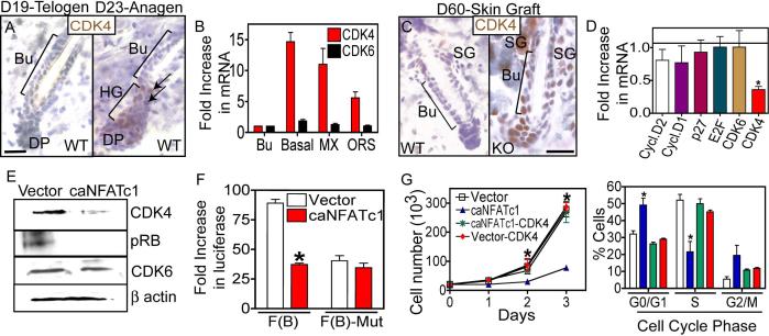
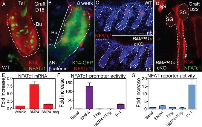
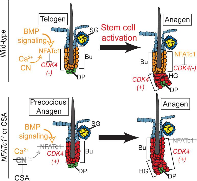
Quiescent adult stem cells reside in specialized niches where they become activated to proliferate and differentiate during tissue homeostasis and injury. How stem cell quiescence is governed is poorly understood. We report here that NFATc1 is preferentially expressed by hair follicle stem cells in their niche, where its expression is activated by BMP signaling upstream and it acts downstream to transcriptionally repress CDK4 and maintain stem cell quiescence. As stem cells become activated during hair growth, NFATc1 is downregulated, relieving CDK4 repression and activating proliferation. When calcineurin/NFATc1 signaling is suppressed, pharmacologically or via complete or conditional NFATc1 gene ablation, stem cells are activated prematurely, resulting in precocious follicular growth. Our findings may explain why patients receiving cyclosporine A for immunosuppressive therapy display excessive hair growth, and unveil a functional role for calcium-NFATc1-CDK4 circuitry in governing stem cell quiescence.


Figures

Figure 1
Figure 1. NFATc1: a marker of hair follicle stem cells
(A-D) NFATc1 is expressed the upper HF during anagen at P4 and P27 and at the base of the follicle during telogen (P21 and P49). β4 integrin marks the dermo-epidermal interface. (C-H) Immunohistochemistry showing NFATc1 colocalization with bulge cell markers, CD34, Lhx2, TCF3 and Sox9. Arrows denote examples of co-expression. (I) Quantification of the % of cells with bulge cell markers that colocalize with NFATc1. Data are mean ± SEM. N=50−176 cells. (J-K) Real-time PCR analysis of NFATc1 and Sox9 mRNA in FACS isolated populations at P4 (J) and NFATc1 mRNA in the α6/CD34-positive bulge compartment during the 1st (P19) and 2nd (P49) telogen. (K). Data are mean ± SEM. N=2 (J) and N=3 (K) FACS isolated populations. Data are mean ± SEM. Asterisks indicate significance, p<0.05. (L) Expression of NFATc1 in the keratin 5 (K5)(+) epidermis at E14.5 or newborn (nb). (M) Isolated P4 epidermis and the dermis (containing HFs) were subjected to western analysis. (N) Real Time PCR analysis of NFATc1 mRNA from FACS isolated α6(+)/CD34(+) bulge cells, after passage (P1, P4) and in epidermal keratinocytes (MK). Abbreviations: DP, dermal papillae; Bu, bulge; ORS, outer root sheath, epi, epidermis, derm, dermis HG, hair germ; FB, fibroblasts; MX, matrix, MC, melanocytes. Dapi staining (blue) shows nuclear localization. Scale Bars, 30 μm.
Figure 2
Figure 2. Impairing calcineurin/NFAT signaling results in hair growth and stem cell proliferation
(A) Schematic illustrating the experimental design of CSA experiments. (B-C) Quantification of anagen induction and BrdU incorporation in follicle stem cells of the bulge (Bu) with cyclosporine A (CSA) treatment. The anagen control used was the first hair cycle (P18-P25). Data are the mean ± SEM for 50−100 follicles for 3 individual mice for each timepoint. Asterisks in (C) indicate hair shaft autofluorescence. (D-E) Immunohistochemistry and western analysis of NFATc1 expression following 3d treatment with vehicle or CSA. (F) Colony formation and cell number of bulge cells following treatment with vehicle, CSA, 11R-VIVIT, or 11R-VEET. N= 3 experiments with independent sorted populations. (G) NFAT reporter activity in response to calcium ionophore (I), phorbol myristate acetate (PMA), caNFATc1, CSA or 11R-VIVIT. Data are mean ± SEM. N=3−5 individual experiments for each treatment. Asterisks indicate significance, p<0.05. Scale bars, 30μm. Dapi staining (blue) shows nuclear localization. Abbreviations: HF, hair follicle; Bu, bulge; HG, hair germ; DP, dermal papillae.
Figure 3
Figure 3. Full and conditional NFATc1 deletion activates precocious follicular growth and enhanced stem cell activity
(A-B) When shaved or dyed blue during telogen (D60), the KO follicles regrow white hair (A and B) and shed their dyed hairs (B) by D75. (C-D) Histological analysis of the WT and KO HFs at D60 (C) and D75 (D). (E) Immunostaining of WT and KO follicles after BrdU label retaining experiments (BrdU pulse at D24−27 post-graft followed by 28 days of chase). Quantification of bulge cell proliferation (24h BrdU pulse) and label retention at D60 in WT and NFATc1-null follicles. Data are mean ± SEM. N=3 mice for each genotype. (F) Immunostaining for NFATc1 and CD34 in WT and NFATc1 null follicles. (G) Immunolocalization of NFATc1 in WT and NFATc1 cKO mice at P56. (H) BrdU immunolabeling after a 48 hour pulse in WT and NFATc1 cKO follicles at P56. Data are mean ± SEM. N=6 mice for WT and N=5 mice for NFATc1 cKO. (I) Quantification of BrdU incorporation and cell cycle analysis of a6(+)/CD34(+) bulge cells from WT and NFATc1 cKO mice. Data are mean ± SEM. Asterisks indicate significance, p<0.05. Scale Bars, 30μm. Dapi staining (blue) shows nuclear localization. Abbreviations: Bu, bulge; SG, sebaceous gland; DP, dermal papillae; HG, hair germ.
Figure 4
Figure 4. NFATc1 inhibits cellular proliferation by blocking G1/S phase progression
(A) Retroviral expression of constitutively active NFATc1 (caNFATc1) represses cell proliferation in epidermal MKs. N= 3 individual experiments for each timepoint. (B-C) Quantification of BrdU incorporation as analyzed by FACS or immunostaining (IF) (B) or combined with DNA content (C) in control or caNFATc1 infected MKs. Representative FACS histogram plot of BrdU immunostaining is shown (left). Data are mean ± SEM. N= 3−4 individual experiments. (D) Real time PCR analysis of the epidermal terminal differentiation markers transglutaminase (TGM) 1 and 3, filaggrin, or loricrin in control, caNFATc1 infected MKs, or cells in high calcium media (Diff. Media). Data are mean ± SEM. N=3 individual experiments. (E) FACS analysis of propidium iodide (dead cells) and Annexin V (apoptotic cells) in control, caNFATc1 expressing cells, or cells treated with TNFα (100 ng/ml) and cycloheximide (CHX) (5ug/ml). Data are mean ± SEM. Asterisks indicate significance, p<0.05.
Figure 5
Figure 5. NFATc1 acts by repressing CDK4 expression to regulate G1/S progression in hair follicle stem cells
(A) Anti-CDK4 immunohistochemistry during the telogen (D19) to anagen (D23) transition. Hematoxylin counterstain. (B) Real Time PCR analysis of CDK4 and CDK6 mRNA expression in bulge (Bu), all basal (α6+), ORS and matrix (MX) cell populations. Data are mean ± SEM. N=3 for each sample. (C) Immunostaining of CDK4 in WT and NFATc1-null follicles. Bu, bulge. (D) Expression of cell cycle regulatory genes in control or caNFATc1 expressing cells. Data are mean ± SEM. N=3 individual experiments. (E) Western analysis for CDK4, phosphorylated retinoblastoma protein (Rb), and CDK6 in control and caNFATc1 expressing keratinocytes (MKs). (F) Reporter assays using the CDK4 promoter with NFAT sites (F(B)) or mutated NFAT sites (F(B)-mut) in conjuction with caNFATc1 in MKs. Data are mean ± SEM. N=4 individual experiments. (G-H) Expression of CDK4 rescues MK growth and S phase progression of cells expressing caNFATc1. N=3 individual experiments. Data are mean ± SEM. Asterisks indicate significance, p<0.05. Scale Bar, 30 μm.
Figure 6
Figure 6. Expression of NFATc1 in mutant mouse models with altered hair follicle stem cell biology
(A-D) K14 expression (immunolocalization and K14-GFP) and NFATc1 immunolocalization are shown for grafted skin from WT (A), heterozygous stabilized β-catenin (ΔN) (B) and BMPR1a conditional knockout (cKO) mice (C and D). (E) Real Time PCR of NFATc1 mRNA levels upon MK treatment with BMP4 and/or the BMP inhibitor, noggin. Data are mean ± SEM. N=3 individual experiments. (F) The activity of a 3.6-kb NFATc1 promoter construct following treatment of MKs with BMP4 and/or noggin or PMA (P) and ionomycin (I). Data are mean ± SEM. N=3 individual experiments. (G) NFAT reporter activity upon BMP4, noggin or P/I treatment of MKs. Data are mean ± SEM. N=3 individual experiments. Dapi staining (blue) shows nuclear localization. Bu, bulge, CN, calcineurin, DP, dermal papillae, HG, hair germ, epi, epidermis.
Figure 7
Figure 7. Model describing the role of NFATc1 signaling in bulge stem cell quiescence
Calcium/calcineurin (CN) activity and BMP signaling are required to maintain NFATc1 expression and activity, which transcriptionally represses CDK4 gene expression. Upon activation of hair growth, BMP signaling is inhibited, leading to a loss in NFATc1 expression, and relief of CDK4 repression. When CN signaling is blocked by CSA or loss of NFATc1 expression, CDK4 is expressed and precocious activation of the stem cells occurs within the bulge.

References

    1. Abbott KL, Friday BB, Thaloor D, Murphy TJ, Pavlath GK. Activation and cellular localization of the cyclosporine A-sensitive transcription factor NF-AT in skeletal muscle cells. Mol Biol Cell. 1998;9:2905–2916. - PMC - PubMed
    1. Al-Daraji WI, Grant KR, Ryan K, Saxton A, Reynolds NJ. Localization of calcineurin/NFAT in human skin and psoriasis and inhibition of calcineurin/NFAT activation in human keratinocytes by cyclosporin A. J Invest Dermatol. 2002;118:779–788. - PubMed
    1. Andl T, Ahn K, Kairo A, Chu EY, Wine-Lee L, Reddy ST, Croft NJ, Cebra-Thomas JA, Metzger D, Chambon P, et al. Epithelial Bmpr1a regulates differentiation and proliferation in postnatal hair follicles and is essential for tooth development. Development. 2004;131:2257–2268. - PubMed
    1. Andl T, Reddy ST, Gaddapara T, Millar SE. WNT signals are required for the initiation of hair follicle development. Dev Cell. 2002;2:643–653. - PubMed
    1. Aramburu J, Garcia-Cozar F, Raghavan A, Okamura H, Rao A, Hogan PG. Selective inhibition of NFAT activation by a peptide spanning the calcineurin targeting site of NFAT. Mol Cell. 1998;1:627–637. - PubMed

Publication types

MeSH terms