. 2008 Feb 1;29(2):191-206.
doi: 10.1016/j.molcel.2007.11.026.

Functional anatomy of phospholipid binding and regulation of phosphoinositide homeostasis by proteins of the sec14 superfamily

Affiliations

Functional anatomy of phospholipid binding and regulation of phosphoinositide homeostasis by proteins of the sec14 superfamily

Gabriel Schaaf et al. Mol Cell. .

Abstract

Sec14, the major yeast phosphatidylinositol (PtdIns)/phosphatidylcholine (PtdCho) transfer protein, regulates essential interfaces between lipid metabolism and membrane trafficking from the trans-Golgi network (TGN). How Sec14 does so remains unclear. We report that Sec14 binds PtdIns and PtdCho at distinct (but overlapping) sites, and both PtdIns- and PtdCho-binding activities are essential Sec14 activities. We further show both activities must reside within the same molecule to reconstitute a functional Sec14 and for effective Sec14-mediated regulation of phosphoinositide homeostasis in vivo. This regulation is uncoupled from PtdIns-transfer activity and argues for an interfacial presentation mode for Sec14-mediated potentiation of PtdIns kinases. Such a regulatory role for Sec14 is a primary counter to action of the Kes1 sterol-binding protein that antagonizes PtdIns 4-OH kinase activity in vivo. Collectively, these findings outline functional mechanisms for the Sec14 superfamily and reveal additional layers of complexity for regulating phosphoinositide homeostasis in eukaryotes.


Figures

Figure 1.
Figure 1.. General Properties of a PL-Bound Sfh1 Structure
(A) Superposition of two different orientations of the open Sec14 (light orange) on the Sfh1::PtdEtn complex (gray). The dashed line represents the distance between the α carbons on F231 in Sec14 in the ,open conformation- and F233 in Sfh1::PtdEtn complex located at the C-terminal end of the A9/T3 helix (A10/T4 helix in Sec14). (B and C) Ribbon diagram of the Sfh1 component of the Sfh1::PtdEtn complex (α helices in green, 310 helices in orange, and β strands in yellow).
Figure 2.
Figure 2.. Structures of Sfh1::PtdEtn and Sfh1::PtdCho Complexes
(A–D) 2Fo − Fc annealed omit electron density (contoured at 1σ) for the bound PtdEtn (A) and bound PtdCho (C). Residues within 4.2 Å of the bound PLs are shown as white spheres. Residues that coordinate the headgroup moieties are shown as sticks with H bonds represented as dashed lines. (B) PtdEtn and (D) PtdCho headgroup interactions with Sfh1. (E) Depiction of the hydrophilic patch formed by the side chains of Y109, Q111, Y124, and E126 (as indicated) and immobilized H2O (red spheres) on the floor of the hydrophobic Sfh1 PL binding pocket. Bound PtdCho is also shown, and the sn-2 acyl chain C5 position is highlighted.
Figure 3.
Figure 3.. Structure of the Sfh1::PtdIns Complexes
(A) Structure of the Sfh1::PtdIns complex with PtdIns rendered in cyan as sticks and transparent spheres. (B) 2Fo − Fc annealed omit electron density (contoured at 1σ) for bound 18:0/18:1 PtdIns. Residues within 4.2 Å of bound PtdIns are shown as white spheres. Residues that coordinate the PtdIns moiety are shown as sticks with H bonds represented as dashed lines. (C) Stereoview of critical PtdIns headgroup interactions with Sfh1. (D) Position of both PtdCho and PtdIns within the fold. (E) 2Fo − Fc annealed omit electron density (contoured at 1σ) for bound 18:0/18:1-PtdIns and 16:0/18:1-PtdCho derived from the mixed crystal.
Figure 4.
Figure 4.. Biochemical Validation of Structure-Based Mutant Sec14 Derivatives
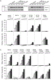
(A) Cytosolic fractions assayed in (A) were evaluated for Sec14 content by immunoblotting with an anti-Sec14 monoclonal antibody. Kes1 was evaluated as a control for proper normalization. (B) PtdCho and PtdIns-transfer assay data are presented. Cytosols prepared from the sec14Δ strain CTY303 harboring the indicated YEp(sec14) plasmids were assayed. PtdIns-transfer assays used 0.12, 0.40, 1.20, and 4.0 mg cytosol. PtdCho-transfer assays employed 0.06, 0.20, 0.60, and 2.0 mg cytosol. YEp(URA3) and YEp(SEC14) derivatives were vector (negative) and positive controls. Average values and standard deviations are given (n = 3). (C) Purified recombinant Sec14 proteins were assayed in a step series of 5-fold mass increases (0.004, 0.2, 1, 5, and 25 μg) for PtdCho- and PtdIns-transfer activity. Average values and standard deviations are given (n = 4).
Figure 5.
Figure 5.. Functional Characterization of Structure-Based Mutant Sec14 Derivatives
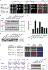
(A) Left and middle panels, isogenic sec14–1ts and sec14–1ts spo14Δ yeast strains (identified at top) transformed with YCp plasmids carrying the designated sec14 alleles (indicated at left) were spotted in 10-fold dilution series onto YPD agar plates and incubated at the restrictive temperature of 37°C. Rescue of growth defect reports functionality. YCp(URA3) and YEp(SEC14) derivatives served as vector (negative) and positive controls, respectively, in all experiments described in this Figure. Right panel, an ade2 ade3 sec14Δ/YEp(SEC14, LEU2, ADE3) yeast strain (strain CTY558; see Experimental Procedures) was transformed with the indicated YCp(sec14, URA3) or high-copy YEp(sec14, URA3) plasmids and dilution spotted onto YPD agar where all nutritional selection is removed. Functionality of mutant sec14 product is manifested as appearance of white segregant colonies that acquire leucine and histidine auxotrophies, signifying loss of parental YEp(SEC14, LEU2, ADE3). Retention of parental plasmid (i.e., nonfunctionality of the mutant sec14) is reported by red colony color (Phillips et al., 1999). (B) Immunoblots of lysates prepared from sec14–1ts yeast carrying the designated YCp(sec14) plasmids at 30°C (upper panel) and after 37°C challenge for 2 hr (lower panel). Equal amounts of total protein were loaded, and Sec14 and Kes1 (normalization control) were visualized with monoclonal antibodies specific for each (molecular mass standards at left). (C) Invertase secretion indices are shown for isogenic sec14–1ts strains transformed with low-copy YCp plasmids carrying the designated sec14 alleles after a 2 hr shift to 37°C. Average values and standard deviations are given (n = 3). (D) The indicated sec14–1ts/YCp(sec14) strains were shifted to 37°C for 2 hr, and radiolabeled with [35S] amino acids for 30 min followed by a 10 min chase. Vacuolar mCPY and TGN p2 CPY forms are identified at right. (E) The ade2 ade3 sec14Δ/YEp(SEC14, LEU2, ADE3) yeast strain CTY558 was cotransformed with YCp(myc-sec14 KanMX) and YCp(myc-sec14 URA3) plasmids under conditions that select for parental YEp(SEC14,LEU2,ADE3). Transformants were dilution spotted onto YPD agar (relieves nutritional selection for SEC14 plasmid, left panel), YPD agar supplemented with G418 (selects for myc-sec14 KanMX, middle panel), or selective media as used for the primary transformation (demands presence of all three plasmids, right panel). Functionality or nonfunctionality of combined expression of myc-sec14 products from KanMX or the URA3 low-copy plasmid is scored by loss or retention of parental SEC14 plasmid, respectively. YEp(SEC14) loss events are manifested by appearance of white segregants (left and middle panels). YCp(URA3) and YCp(KanMX) plasmids served as mock controls. (F) Nucleotide sequence of cDNA generated from yeast carrying both YCp(myc-sec14R65A,T236D KanMX) and YCp(myc-sec14S173I,T175I URA3) confirmed expression of mutant genes (lower panel). YCp(myc-SEC14 KanMX) and YCp(URA3) transformants were positive controls (upper panel). Base pair and amino acid substitutions are indicated (bottom and top, respectively). Nucleotide sequences were determined with myc tag-specific forward and sec14-specific reverse primers that do not amplify parental SEC14 cDNA. (G) Lysates prepared from CTY558 transformants carrying designated YCp(myc-sec14 KanMX) plasmids cultured at 30°C (in presence of G418) were probed for Sec14 antigen by immunoblotting. Loading was normalized by total protein. Kes1 was a normalization control. The myc-tagged sec14 products and the parental plasmid SEC14 product are identified.
Figure 6.
Figure 6.. Sec14 PL Binding Activities and PIP Homeostasis
(A) PIP analyses. Derivatives of the sec14Δ cki1 strain CTY303 carrying designated YCp plasmids were radiolabeled for 18 hr at 30°C with 20 μCi/ml [3H]-Ins. Deacylated PIPs were quantified (see Experimental Procedures). PtdIns-3-P, PtdIns-4-P, and PtdIns(4,5)P2 are as indicated. Average values and standard deviations are given (n = 4). YCp(URA3) and YCp(SEC14) derivatives served as negative and positive controls, respectively. (B) Stt4 activity as a function of mutant sec14 proteins. A sec14–1ts sac1–26 yeast mutant (CTY100) and isogenic derivatives expressing the indicated Sec14 proteins were radiolabeled to steady state with [3H]-Ins and shifted to 37°C for 2 hr. PtdIns-4-P and other Ins-PLs were extracted, resolved by thin layer chromatography, and identified by cochromatography with radiolabeled standards, and Stt4-dependent PtdIns-4-P accumulation was quantified as described (Phillips et al., 1999). The URA3 and SEC14 derivatives represented negative and positive controls, respectively. Average values and standard deviations are given (n = 4). (C) In vitro PtdIns kinase stimulation. The indicated recombinant Sec14 proteins were assayed in a step series of 3-fold mass increases (0.03, 0.09, 0.81, and 2.43 μg) in reactions containing 2.5 μg PtdIns 4-OH kinase, PtdIns:PtdCho liposomes, and [γ32P]-ATP. Data are plotted as percentage of total achievable PtdIns-4-P signal as determined in mixed micelle assays where Triton X-100 (0.4%) was incorporated into the system (see Experimental Procedures). Average values and standard deviations are given (n = 3). (D) In situ phosphorylation of Sec14-bound PtdIns. Sec14 and mutant derivatives were loaded with PtdIns and repurified from PtdIns liposomes on cobalt affinity resin followed by extensive washing and dialysis. For the in situ kinase reaction, 1.5 μg Sec14 was used and processed as described for in vitro kinase assays that employed liposomes. Average values and standard deviations are given (n = 3). (E) PIP analyses in sec14 kes1 mutants. Isogenic derivatives of the SEC14 (CTY182), sec14–1ts (CTY1–1A), sec14–1ts kes1 (CTY159), and sec14–1ts kes1 spo14Δ (CTY1098) were radiolabeled for 18 hr at 30°C with 20 μCi/ml [3H]-Ins and then shifted to the restrictive temperature of 37°C for 2 hr. Deacylated PIPs were quantified, and PtdIns-3-P, PtdIns-4-P, and PtdIns(4,5)P2 are as indicated (n = 5). SEC14 and sec14–1ts strains were positive and negative controls, respectively. The sec14–1ts kes1 and sec14–1ts kes1 spo14Δ mutants exhibited elevated PtdIns-4-P relative to sec14–1ts controls. Average values and standard deviations are given (n=5; p < 0.01 and p < 0.02, respectively).
Figure 7.
Figure 7.. Sec14 Function in PIP Homeostasis in the Yeast TGN
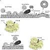
A two-ligand Sec14 priming model for PtdIns presentation to PtdIns kinases is shown. (A) Heterotypic exchange results in PtdCho entering the Sec14 pocket and ejecting bound PtdIns through an unobstructed portal (bottom panel). Egress occurs headgroup-first, and the exposed intermediate (◆) is highly susceptible to modification by Pik1 PtdIns 4-OH kinase in the TGN. This potentiation of Pik1 activity overcomes the net inhibitory effects of Kes1 and likely couples to the Ypt31/32 pathway for vesicle biogenesis. (B) Alternatively, slow egress of bound PtdCho interferes with entry of an invading PtdIns and delays it as a partially incorporated and exposed intermediate (◆) that is vulnerable to Pik1 modification. Completion of PtdCho unloading generates an apo intermediate that reloads with PtdIns or PtdCho (left panel). This mechanism suggests invading PtdIns and the leaving PtdCho collide in spatially overlapping entry/exit portals (right panel).

Comment in

  • The lipid trade.
    Prinz WA. Prinz WA. Nat Rev Mol Cell Biol. 2014 Feb;15(2):79. doi: 10.1038/nrm3740. Epub 2014 Jan 17. Nat Rev Mol Cell Biol. 2014. PMID: 24434885 Free PMC article. No abstract available.

Similar articles

Cited by

References

    1. Al-Aidroos K, and Bussey H (1978). Chromosomal mutants of Saccharomyces cerevisiae affecting the cell wall binding site for killer factor. Can. J. Microbiol 24, 228–237. - PubMed
    1. Ammerer G, Hunter CP, Rothman JH, Saari GC, Valls LA, and Stevens TH (1986). PEP4 gene of Saccharomyces cerevisiae encodes proteinase A, a vacuolar enzyme required for processing of vacuolar precursors. Mol. Cell. Biol 6, 2490–2499. - PMC - PubMed
    1. Aravind L, and Koonin EV (1999). Gleaning non-trivial structural, functional and evolutionary information about proteins by iterative database searches.J. Mol. Biol 287, 1023–1040. - PubMed
    1. Bankaitis VA, Malehorn DE, Emr SD, and Greene R (1989). The Saccharomyces cerevisiae SEC14 gene encodes a cytosolic factor that is required for transport of secretory proteins from the yeast Golgi complex. J. Cell Biol 108, 1271–1281. - PMC - PubMed
    1. Bankaitis VA, Aitken JR, Cleves AE, and Dowhan W (1990). An essential role for a PL transfer protein in yeast Golgi function. Nature 347, 561–562. - PubMed

Publication types

MeSH terms