. 2008 Feb 26;26(9):1195-203.
doi: 10.1016/j.vaccine.2007.12.032. Epub 2008 Jan 14.

An HSV-1 gD mutant virus as an entry-impaired live virus vaccine

Affiliations

An HSV-1 gD mutant virus as an entry-impaired live virus vaccine

Sita Awasthi et al. Vaccine. .

Abstract

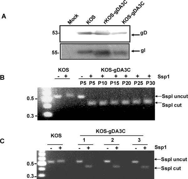
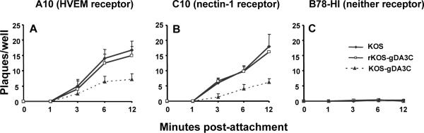
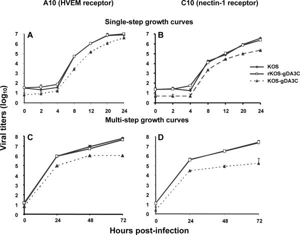
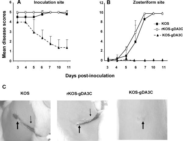
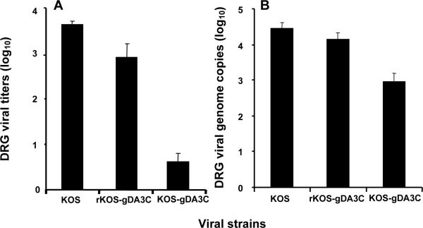
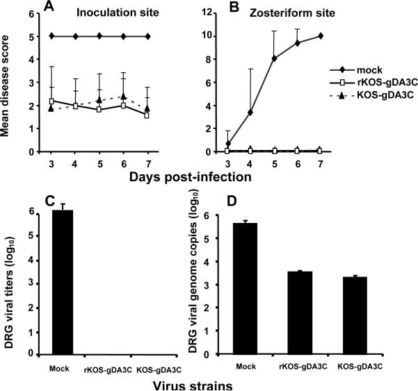
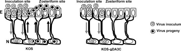
HSV-1 glycoprotein D (gD) interacts with HVEM and nectin-1 cell receptors to initiate virus entry. We prepared an HSV-1 strain with mutations in the gD gene at amino acid residues 3 and 38 by changing alanine to cysteine and tyrosine to cysteine, respectively (A3C/Y38C). These mutations were constructed with the intent of evaluating infection in vivo when virus enters by HVEM but not nectin-1 receptors and were based on prior reports demonstrating that purified gDA3C/Y38C protein binds to HVEM but not to nectin-1. While preparing a high-titered purified virus pool, the cysteine mutation at position 38 reverted to tyrosine, which occurred on two separate occasions. The resultant HSV-1 strain, KOS-gDA3C, had a single amino acid mutation at residue 3 and exhibited reduced entry into both HVEM and nectin-1 expressing cells. When tested in the murine flank model, the mutant virus was markedly attenuated for virulence and caused only mild disease, while the parental and rescued viruses produced much more severe disease. Thirty days after KOS-gDA3C infection, mice were challenged with a lethal dose of HSV-1 and were highly resistant to disease. The KOS-gDA3C mutation was stable during 30 passages in vitro and was present in each of 3 isolates obtained from infected mice. Therefore, this gD mutant virus impaired in entry may represent a novel candidate for an attenuated live HSV-1 vaccine.


Figures

Figure 1
Figure 1. Characterization and stability of KOS-gDA3C virus
A. Western blot to detect gD (Us6) and gI (Us7) in infected cell extracts. Vero cells were mock infected or infected with KOS, rKOS-gDA3C, or KOS-gDA3C and probed for gD or gI. The position of molecular weight markers is indicated on the left. B. Stability of the KOS-gDA3C virus in vitro. Agarose gel showing Ssp1 digestion of PCR-amplified gD gene fragments of KOS or KOS-gDA3C repeated every fifth passage from passage 5 (P5) to passage 30 (P30). C. Stability of the KOS-gDA3C mutant virus in vivo. A PCR-amplified gD fragment from three different isolates obtained from the DRG of KOS-gDA3C infected mice was cut with Ssp1 or left uncut. A 100 base pair DNA ladder indicating 0.5 and 0.3 kb markers is shown on the left in (B) and (C).
Figure 2
Figure 2. Entry of KOS, rKOS-gDA3C and KOS-gDA3C virus
Entry was measured into (A) A10, (B) C10, or (C) B78-H1 cells. Results are the mean ± SE of three separate infections each done in triplicate. The AUC for KOS-gDA3C is significantly less than KOS or r-KOS-gDA3C in (A) or (B) (P < 0.001).
Figure 3
Figure 3. Growth curves
Single-step (A, B) and multi-step (C, D) growth curves of KOS, rKOS-gDA3C and KOS-gDA3C were performed in A10 (A, C) or C10 (B, D) cells. Results are the mean ± SE of three separate infections. The AUC for KOS-gDA3C is significantly less than KOS or rKOS-gDA3C in (C) and (D) (P < 0.01 and < 0.001, respectively), while differences among the viruses are not significant in (A) and (B).
Figure 4
Figure 4. Disease in the murine flank model
Inoculation (A) and zosteriform (B) site disease scores in mice inoculated with 5×10⁵ PFU of KOS, rKOS-gDA3C or KOS-gDA3C. Thirty mice are in the KOS and KOS-gDA3C groups, and 10 mice are in the rKOS-gDA3C group. Error bars represent SE. The AUC for KOS-gDA3C is significantly less than for the other two viruses at the inoculation (P < .001) and zosteriform (P < .001) sites. C. Photographs of mice flanks taken 10 days post-infection. The mouse infected with KOS or rKOS-gDA3C has extensive inoculation (thick arrow) and zosteriform site disease (thin arrow), while the mouse inoculated with KOS-gDA3C has only minimal disease at the inoculation site and no zosteriform site disease.
Figure 5
Figure 5. Virus titers and genome copy numbers in DRG
DRG from mice infected with KOS, rKOS-gDA3C or KOS-gDA3C were assayed for virus titers (A) or viral genome copy number (B). Five animals are in each group. Results represent the mean ± SE. Comparing viral titers or genome copy number for KOS-gDA3C with rKOS-gDA3C or KOS, P < .001, while differences between KOS and rKOS-gDA3C are not significant, P = 0.07.
Figure 6
Figure 6. Prior infection with KOS-gDA3C protects against WT HSV-1 challenge
Mice were mock immunized or infected with 5×10⁵ PFU rKOS-gDA3C or KOS-gDA3C. Thirty days later, mice were challenged on the opposite flank with 106 PFU of WT HSV-1 strain NS. Ten mice were evaluated in each group. Results represent mean disease scores ± SE at the inoculation (A) and zosteriform (B) sites from days 3-7 post-infection (comparing rKOS-gDA3C or KOS-gDA3C with mock-infected mice, P < 0.001). DRG viral titers (C) and genome copy number (D) were measured 5 days post-challenge with NS. Five mice were assayed in each group. Results represent the mean ± SE. The rKOS-gDA3C and KOS-gDA3C values in figures (C) and (D) are both significantly less than mock immunized (P < 0.001).
Figure 7
Figure 7. Model for KOS-gDA3C infection in mice
KOS infects epithelial cells (E) and produces disease at the inoculation site. The virus spreads to neurons (N) in the DRG, replicates and spreads to adjacent neurons and then travels back to epithelial cells in the skin to cause zosteriform disease. KOS-gDA3C is impaired in entry and infects fewer epithelial cells, which results in fewer neurons becoming infected in the DRG. The defect in entry also reduces infection of adjacent neurons in the DRG and results in reduced zosteriform disease.

References

    1. Smith JS, Robinson NJ. Age specific prevalence of infection with herpes simplex virus type 2 and 1: a global review. Journal of Infectious Diseases. 2002;186:S3–28. - PubMed
    1. Xu Y, Sternberg MR, Kottiri BJ, McQuillan GM, Lee FK, Nehmias AJ, Barman SM, Markowitz LE. Trends in herpes simplex type 1 and type 2 seroprevalence in the United States. JAMA. 2006;296:964–73. - PubMed
    1. Whitley Rj, Lakeman F. Herpes virus infections of central nervous system: therapeutic and diagnostic considerations. Clinical Infectious Diseases. 1995;20:414–20. - PubMed
    1. Raschilas FWM, Delatour F, Chaffaut C, De Broucker T, Chevret S, Lebon P, Canton P, Rozenberg F. Outcome of and prognostic factors for herpes simplex encephalitis in adult patients: results of a multicenter study. Clinical Infectious Diseases. 2002;35:254–60. - PubMed
    1. Liesegang TJ. Epidemology of ocular herpes simplex. Natural history in Rochester, Minn, 1950 through 1982. Archives of Ophthalmology. 1889;107:171–85. - PubMed

Publication types

MeSH terms