

Neurotrophins and electrical stimulation for protection and repair of spiral ganglion neurons following sensorineural hearing loss
- PMID: 18243608
- PMCID: PMC2630855
- DOI: 10.1016/j.heares.2007.12.005
Neurotrophins and electrical stimulation for protection and repair of spiral ganglion neurons following sensorineural hearing loss
Abstract
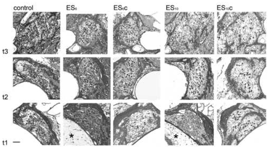
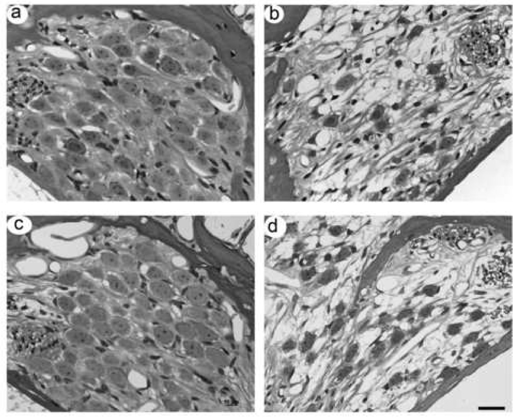
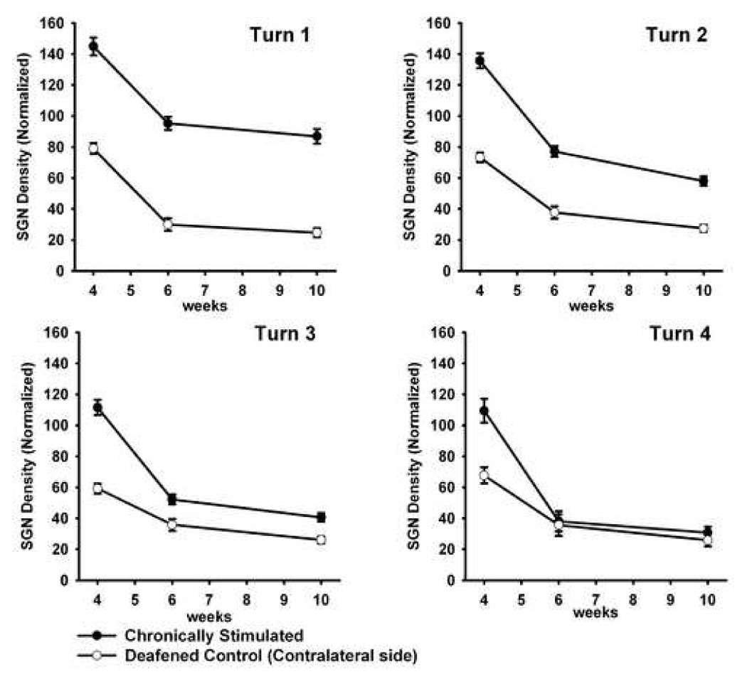
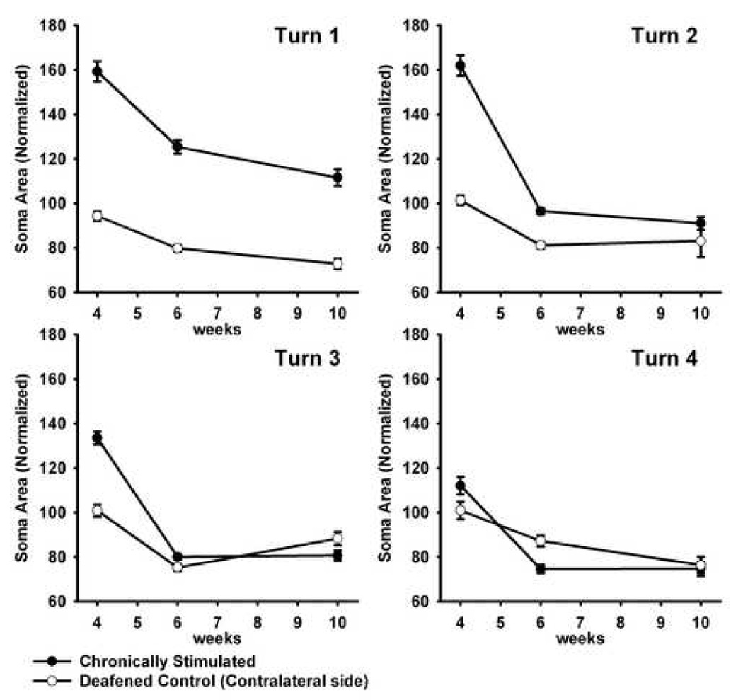
Exogenous neurotrophins (NTs) have been shown to rescue spiral ganglion neurons (SGNs) from degeneration following a sensorineural hearing loss (SNHL). Furthermore, chronic electrical stimulation (ES) has been shown to retard SGN degeneration in some studies but not others. Since there is evidence of even greater SGN rescue when NT administration is combined with ES, we examined whether chronic ES can maintain SGN survival long after cessation of NT delivery. Young adult guinea pigs were profoundly deafened using ototoxic drugs; five days later they were unilaterally implanted with an electrode array and drug delivery system. Brain derived neurotrophic factor (BDNF) was continuously delivered to the scala tympani over a four week period while the animal simultaneously received ES via bipolar electrodes in the basal turn (i.e., turn 1) scala tympani. One cohort (n=5) received ES for six weeks (i.e., including a two week period after the cessation of BDNF delivery; ES(6)); a second cohort (n=5) received ES for 10 weeks (i.e., a six week period following cessation of BDNF delivery; ES(10)). The cochleae were harvested for histology and SGN density determined for each cochlear turn for comparison with normal hearing controls (n=4). The withdrawal of BDNF resulted in a rapid loss of SGNs in turns 2-4 of the deafened/BDNF-treated cochleae; this was significant as early as two weeks following removal of the NT when compared with normal controls (p<0.05). Importantly, there was not a significant reduction in SGNs in turn 1 (i.e., adjacent to the electrode array) two and six weeks after NT removal, as compared with normal controls. This result suggests that chronic ES can prevent the rapid loss of SGNs that occurs after the withdrawal of exogenous NTs. Implications for the clinical delivery of NTs are discussed.
Figures
References
-
- Alam SA, Robinson BK, Huang J, Green SH. Prosurvival and proapoptotic intracellular signaling in rat spiral ganglion neurons in vivo after the loss of hair cells. J Comp Neurol. 2007;503:832–852. - PubMed
-
- Andrew J. Honours Thesis. Melbourne: University of Melbourne; 2003. Rehabilitation of the deafened auditory nerve with Schwann cell transplantation.
-
- Araki S, Kawano A, Seldon L, Shepherd RK, Funasaka S, Clark GM. Effects of chronic electrical stimulation on spiral ganglion neuron survival and size in deafened kittens. Laryngoscope. 1998;108:687–695. - PubMed
MeSH terms
Substances
Grants and funding
LinkOut - more resources
Full Text Sources
Other Literature Sources
Research Materials
