

Review
doi: 10.1016/j.smim.2007.12.004.
Epub 2008 Feb 19.
Ectopic lymphoid tissues and local immunity
Affiliations
- PMID: 18243731
- PMCID: PMC2276727
- DOI: 10.1016/j.smim.2007.12.004
Item in Clipboard
Review
Ectopic lymphoid tissues and local immunity
Semin Immunol.
2008 Feb.
Abstract
Ectopic or tertiary lymphoid tissues develop at sites of inflammation or infection in peripheral, non-lymphoid organs. These tissues are architecturally similar to conventional secondary lymphoid organs, with separated B and T cell areas, specialized populations of dendritic cells, well-differentiated stromal cells and high endothelial venules. Ectopic lymphoid tissues are often associated with the local pathology that results from chronic infection or chronic inflammation. However, there are also examples in which ectopic lymphoid tissues appear to contribute to local protective immune responses. Here we review how ectopic lymphoid structures develop and function in the context of local immunity and pathology.
Figures
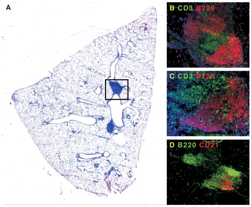
Mice were infected with murine γ-herpesvirus-68 and allowed to clear the lytic phase of the infection. A. Although the majority of the inflammatory response in the lung has resolved, areas of pulmonary lymphoid tissue remain next to major airways. B. The boxed area in panel A contains B220+ B cell areas (red), and CD3+ T cell areas (green). C. A magnification of the B cell areas shows infiltrating T cells. D. In a serial section of panel B, it is clear that some of the B cell follicles in iBALT (green) have CD21+ FDCs (red).

A. This panel shows an H&E stained section of a lung biopsy from a patient with bronchioalvaolar carcinoma. Numerous ectopic follicles (blue) can be observed. B and C. Some of the B cells in ectopic follicles express Proliferating Cell Nuclear Antigen (PCNA), suggesting that they are activated B cells that could contribute to germinal centers.

Lungs from naïve mice (A) or mice that had recovered from influenza infection (B) were probed with antibodies to B cells (red) or lymphatic vessels (green). C. CCL21 (red) is expressed on vascular endothelium surrounding B cell follicles (green) in iBALT. D. CXCL13 (red) is expressed in a reticular pattern in the B cell follicle (green) of iBALT.
References
-
- Goodnow CC. Chance encounters and organized rondezvous. Immunol Rev. 1997;156:5–10. - PubMed
-
- Cyster JG. Chemokines and cell migration in secondary lymphoid organs. Science. 1999;286(5447):2098–102. - PubMed
-
- Mowat AM, Viney JL. The anatomical basis of intestinal immunity. Immunol Rev. 1997;156:145–166. - PubMed
-
- Gretz JE, Anderson AO, Shaw S. Cords, channels, corridors and conduits: critical architectural elements facilitating cell interactions in the lymph node cortex. Immunol Rev. 1997;156:11–24. - PubMed
Publication types
MeSH terms
Grants and funding
LinkOut - more resources
Full Text Sources
Other Literature Sources
Medical
