

Review
doi: 10.1016/j.mib.2007.12.002.
Epub 2008 Feb 20.
A novel pathway for exotoxin delivery by an intracellular pathogen
Affiliations
- PMID: 18243772
- PMCID: PMC2323430
- DOI: 10.1016/j.mib.2007.12.002
Item in Clipboard
Review
A novel pathway for exotoxin delivery by an intracellular pathogen
Curr Opin Microbiol.
2008 Feb.
Abstract
Fundamental to the biology of many bacterial pathogens are bacterial proteins with the capacity to modulate host cellular functions. These bacterial proteins are delivered to the host's molecular targets by a great diversity of mechanisms of varying complexity. The different delivery mechanisms are adapted to the specific biology of the pathogen. Here we focus our attention on a recently described delivery pathway adapted to the biology of an intracellular pathogen, in which an exotoxin is delivered from an intracellular location to its molecular target through autocrine and paracrine pathways.
Figures

In the simplest strategy, represented by the large clostridial cytotoxins, all the necessary information for toxin delivery and enzymatic activity is encoded in a single polypeptide (A). Another strategy, represented by the AB toxins, utilizes the B subunit to deliver the enzymatically active A subunit (B). The most complex strategy, represented by the type III and type IV secretions systems, involves multi protein machines to deliver multiple effectors (C) (see text for details).

Immunofluorescence staining of cells infected with a strain of S. Typhi that encodes a 3x-FLAG epitope-tagged CdtB. Cells were infected and stained at different times post-infection (as indicated) with a mouse monoclonal antibody specific for the FLAG epitope (green), and a rabbit antibody specific for S. Typhi (red), and DAPI (blue). Scale bar, 5 µm.
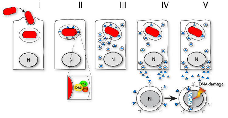
After internalization (I) S. Typhi reaches a compartment where expression of cdtB, pltA, and pltB can take place (II). The CdtB/PltA/PltB complex is secreted from the bacteria into the lumen of the S. Typhi containing vacuole and it is recognized and packaged into transport carriers (III). The complex is then transported to the plasma membrane and secreted to the extracellular medium (IV) from where it can target susceptible non-infected cells (e. g. cells of the immune system) and induce DNA damage (V). Infected cells that do not express a receptor for the toxin would be resistant to the toxin and provide a safe haven for the bacteria (IV and V) (adapted from[13])
References
-
- Schirmer J, Aktories K. Large clostridial cytotoxins: cellular biology of Rho/Ras-glucosylating toxins. Biochim Biophys Acta. 2004;1673:66–74. - PubMed
-
- Gurcel L, Abrami L, Girardin S, Tschopp J, van der Goot FG. Caspase-1 activation of lipid metabolic pathways in response to bacterial pore-forming toxins promotes cell survival. Cell. 2006;126:1135–1145. - PubMed
-
- Fan E, Merritt EA, Verlinde CL, Hol WG. AB(5) toxins: structures and inhibitor design. Curr Opin Struct Biol. 2000;10:680–686. - PubMed
Publication types
MeSH terms
Substances
Grants and funding
LinkOut - more resources
Full Text Sources
Other Literature Sources
