. 2008 Apr 4;283(14):9089-100.
doi: 10.1074/jbc.M710012200. Epub 2008 Feb 1.

Mitochondrial import and accumulation of alpha-synuclein impair complex I in human dopaminergic neuronal cultures and Parkinson disease brain

Affiliations

Mitochondrial import and accumulation of alpha-synuclein impair complex I in human dopaminergic neuronal cultures and Parkinson disease brain

Latha Devi et al. J Biol Chem. .

Abstract

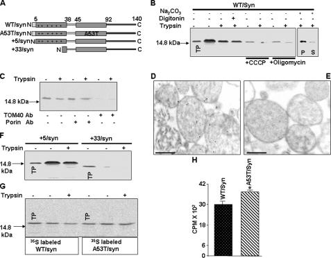
Alpha-synuclein, a protein implicated in the pathogenesis of Parkinson disease (PD), is thought to affect mitochondrial functions, although the mechanisms of its action remain unclear. In this study we show that the N-terminal 32 amino acids of human alpha-synuclein contain cryptic mitochondrial targeting signal, which is important for mitochondrial targeting of alpha-synuclein. Mitochondrial imported alpha-synuclein is predominantly associated with the inner membrane. Accumulation of wild-type alpha-synuclein in the mitochondria of human dopaminergic neurons caused reduced mitochondrial complex I activity and increased production of reactive oxygen species. However, these defects occurred at an early time point in dopaminergic neurons expressing familial alpha-synuclein with A53T mutation as compared with wild-type alpha-synuclein. Importantly, alpha-synuclein that lacks mitochondrial targeting signal failed to target to the mitochondria and showed no detectable effect on complex I function. The PD relevance of these results was investigated using mitochondria of substantia nigra, striatum, and cerebellum of postmortem late-onset PD and normal human brains. Results showed the constitutive presence of approximately 14-kDa alpha-synuclein in the mitochondria of all three brain regions of normal subjects. Mitochondria of PD-vulnerable substantia nigra and striatum but not cerebellum from PD subjects showed significant accumulation of alpha-synuclein and decreased complex I activity. Analysis of mitochondria from PD brain and alpha-synuclein expressing dopaminergic neuronal cultures using blue native gel electrophoresis and immunocapture technique showed the association of alpha-synuclein with complex I. These results provide evidence that mitochondrial accumulated alpha-synuclein may interact with complex I and interfere with its functions.


Figures

FIGURE 1.
FIGURE 1.
Characterization of mitochondrial targeting properties of α-synuclein by mitochondrial import assay. A, description of wild-type (WT), familial, and N-terminal deletion constructs of α-synuclein. B, in vitro mitochondrial import of 35S-labeled WT/synuclein, in isolated rat liver mitochondria as described under “Materials and Methods” using 200 μg of trypsin/ml of reaction (+). Mitochondria were preincubated with or without added inhibitors (50 μm carbonyl cyanide-m-lorophenylhydrazone (CCCP) or 50 μm oligomycin) at 25 °C for 10 min before initiating the in vitro transport of WT/synuclein. C, effects of antibodies to porin and TOM40 on mitochondrial import of α-synuclein. Mitochondria were preincubated with antibodies (5 μg/ml) to porin and TOM40 at room temperature for 30 min prior to the initiation of import. Immunoelectron microscopy of mitochondrial imported unlabeledα-synuclein using rabbit polyclonal antibodies toα-synuclein (D) and the preabsorbed α-synuclein antibody (E). Sections were stained with secondary antibody conjugated to 10 nm gold. Bar = 500 nm. F, in vitro mitochondrial import of +5/synuclein and +33/synuclein. G and H, effects of A53T mutation on mitochondrial import of α-synuclein (G, autoradiogram and H, quantitation of trypsin-protected α-synuclein bands). Values are mean ± S.D. from three separate experiments. *, p < 0.05 as compared with WT/synuclein (Student's t test). 250 μg of mitochondrial protein were used for electrophoresis. TP = translation product (20% of total input), S = supernatant, P = pellet.
FIGURE 2.
FIGURE 2.
Mitochondrial targeting of WT/α-synuclein and +33/α-synuclein in DAN neuronal cultures. DAN cells were transfected with WT/synuclein-FLAG (A) and +33/synuclein-FLAG (D). Cytosol (cyto.) and mitochondrial (mito.) fractions (100 μg) were probed with FLAG antibodies. B and C show the levels of loading controls: TOM20 and actin, respectively, for A. E and F show the levels of loading controls: TOM20 and actin, respectively, for D. G, quantitation of WT/synuclein and +33/synuclein in the mitochondrial and cytosolic fractions by ELISA at different time points of transfection. Values are mean ± S.D. from three separate experiments. *, p < 0.05 compared with 20-h transfection. H, following the transfection, the whole cell lysates (homogenate) from DAN cells were tested for the levels of α-synuclein at different time points using quantitative ELISA. Values are mean ± S.D. from three separate experiments. No significant variation was observed (one-way analysis of variance with Tukey-Kramer post test).
FIGURE 3.
FIGURE 3.
Mitochondrial localization of WT/α-synuclein and +33/α-synuclein by immunofluorescence microscopy. DAN cells were transfected with WT/synuclein-FLAG (A–C) or +33/synuclein-FLAG (D–F). Cells (A and B, and D and E) were double immunostained with antibodies to FLAG and TOM20, respectively. C and F are the merged image of A and B, and D and E, respectively. Bar = 10 mm.
FIGURE 4.
FIGURE 4.
Mitochondrial complex activities in DAN cells expressing various α-synuclein constructs. A, Western immunoblot analysis of mitochondrial (100 μg) localization of WT/synuclein-FLAG and A53T/synuclein-FLAG with time using antibodies to FLAG. B, levels of TOM20. Mitochondrial complex I activity (C) and the production of ROS (D) were estimated at various time points following the expression of WT/synuclein-FLAG, A53T/α-synuclein-FLAG, and +33/synuclein-FLAG cDNA constructs. *, p < 0.05 compared with vector control. **, p < 0.05 compared with vector control, and +33/α-synuclein-FLAG constructs. #, p < 0.05 compared with WT/synuclein-FLAG (one-way analysis of variance with Tukey-Kramer post test). Western immunoblot analysis of endogenous α-synuclein (E), actin (F), and TOM20 (G) in total homogenate (100 μg) and mitochondria (100 μg) following 48-h transfection of α-synuclein-specific siRNA in DAN cells. Mitochondrial NADH cytochrome c reductase (H) and complex I (I) in control and α-synuclein-specific siRNA transfected DAN cells. *, p < 0.05 (Student t test).
FIGURE 5.
FIGURE 5.
Subcellular purity and mitochondrial localization of α-synuclein in human PD and normal brains. Western immunoblot analysis of marker proteins for different subcellular fractions from substantia nigra (100 μg of protein each) was carried out using antibodies to p97, TOM20, actin (A), and Sec 61 and TGN 38 (B). C, amounts of α-synuclein present in the mitochondrial fractions from substantia nigra of normal and PD subjects as quantitated by ELISA. Values are mean ± S.D. from three separate experiments. Western immunoblot analysis of mitochondria (50 μg) using antibodies to α-synuclein of substantia nigra (SN, D), striatum (ST, G), and cerebellum (CE, J) and. F, I, and L are Western immunoblot analysis of TOM20 levels (as a loading control) in D, G, and J blots, respectively. E, H, and K represent the quantification of α-synuclein immunoreactivity in Western blots D, G, and J, respectively. Values are mean ± S.D. from three separate experiments. *, p < 0.05 compared with corresponding normal levels (one-way analysis of variance with Tukey-Kramer post test). NS = normal subjects, N = nucleus, M = mitochondria, MC = microsomes, and C = cytosol.
FIGURE 6.
FIGURE 6.
Biochemical and immunoelectron microscopy characterization of mitochondrial α-synuclein in human PD and normal brains. Western blot analysis of 50 μg of mitochondrial fraction from substantia nigra of PD #1 for α-synuclein (A), mitochondrial (mt)HSP70 (B), CcO (C), and TOM20 (D) following the treatment with trypsin, digitonin, and sodium carbonate. Sup = supernatant, Dig = digitonin. E, Western blot analysis of 50 μg of cytosol from substantia nigra of PD #1 treated with or without trypsin (30 μg/mg of protein) using antibodies to α-synuclein. Immunoelectron microscopy analysis of α-synuclein in the mitochondria of substantia nigra of PD subject (PDS #1) (F) and normal subject (NS #11) (G) using anti rabbit antibodies toα-synuclein. The sections were stained with secondary antibody conjugated to 20 nm (E) and 10 nm (F), respectively. M = mitochondrion, V = vesicle, C = cytosol. Bar = 100 nm. Arrows indicate association of gold particle to the outer membrane of mitochondria.
FIGURE 7.
FIGURE 7.
Correlation between mitochondrial α-synuclein and complex I activity in PD subjects. Distribution of complex I activities in the mitochondria of substantia nigra (A) and cerebellum (B) from PD and control subjects. Scatter plots of mitochondrial α-synuclein levels versus complex I activity in substantia nigra of PD subjects (C) and scatter plots of mitochondrial α-synuclein levels versus complex I activity in cerebellum (D). Regression analysis was carried out using Origin 7.5 software.
FIGURE 8.
FIGURE 8.
Interaction of α-synuclein with mitochondrial complex I. A, effect of in vitro incubation of mitoplast from substantia nigra of control subject #9 with dilution buffers (a) and varying concentrations of α-synuclein (b). Effect of α-synuclein (1.5 μg/mg of mitochondrial protein) on the activities of complex II (B), complex III (C), and complex IV (D). Mitochondria from striatum of a normal subject (NS) and a PD subject (PDS), as well as DAN cells expressing α-synuclein constructs were solubilized and run on blue native PAGE. Following the separation, complexes were transferred on polyvinylidene difluoride membranes and stained with antibodies to NDUFA9 (E) and α-synuclein (F). Co-immunoprecipitation of α-synuclein using complex I immunocapture kit (Mitosciences). Immunocaptured mitochondrial complexes I (4 μg) from striatum of PD and normal subjects as well as DAN cells expressing α-synuclein constructs were resolved on 14% SDS-PAGE and subjected to Western immunoblot using antibodies to α-synuclein (G) and NDUFA9 (H).

References

    1. Bennett, M. C. (2005) Pharmacol. Ther. 105 311-331 - PubMed
    1. Dawson, T. M., and Dawson, V. L. (2003) Science 302 819-822 - PubMed
    1. Shults, C. W. (2006) Proc. Natl. Acad. Sci. U. S. A. 103 1661-1668 - PMC - PubMed
    1. Abou-Sleiman, P. M., Muqit, M. M., McDonald, N. Q., Yang, Y. X., Gandhi, S., Healy, D. G., Harvey, K., Harvey, R. J., Deas, E., Bhatia, K., Quinn, N., Lees, A., Latchman, D. S., and Wood, N. W. (2006) Ann. Neurol. 60 414-419 - PubMed
    1. Fiskum, G., Starkov, A., Polster, B. M., and Chinopoulos, C. (2003) Ann. N. Y. Acad. Sci. 991 111-119 - PubMed

Publication types

MeSH terms