. 2008 Apr;74(7):2187-99.
doi: 10.1128/AEM.01214-07. Epub 2008 Feb 1.

Packaging of live Legionella pneumophila into pellets expelled by Tetrahymena spp. does not require bacterial replication and depends on a Dot/Icm-mediated survival mechanism

Affiliations

Packaging of live Legionella pneumophila into pellets expelled by Tetrahymena spp. does not require bacterial replication and depends on a Dot/Icm-mediated survival mechanism

Sharon G Berk et al. Appl Environ Microbiol. 2008 Apr.

Abstract

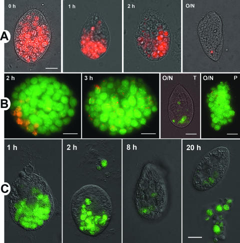
The freshwater ciliate Tetrahymena sp. efficiently ingested, but poorly digested, virulent strains of the gram-negative intracellular pathogen Legionella pneumophila. Ciliates expelled live legionellae packaged in free spherical pellets. The ingested legionellae showed no ultrastructural indicators of cell division either within intracellular food vacuoles or in the expelled pellets, while the number of CFU consistently decreased as a function of time postinoculation, suggesting a lack of L. pneumophila replication inside Tetrahymena. Pulse-chase feeding experiments with fluorescent L. pneumophila and Escherichia coli indicated that actively feeding ciliates maintain a rapid and steady turnover of food vacuoles, so that the intravacuolar residence of the ingested bacteria was as short as 1 to 2 h. L. pneumophila mutants with a defective Dot/Icm virulence system were efficiently digested by Tetrahymena sp. In contrast to pellets of virulent L. pneumophila, the pellets produced by ciliates feeding on dot mutants contained very few bacterial cells but abundant membrane whorls. The whorls became labeled with a specific antibody against L. pneumophila OmpS, indicating that they were outer membrane remnants of digested legionellae. Ciliates that fed on genetically complemented dot mutants produced numerous pellets containing live legionellae, establishing the importance of the Dot/Icm system to resist digestion. We thus concluded that production of pellets containing live virulent L. pneumophila depends on bacterial survival (mediated by the Dot/Icm system) and occurs in the absence of bacterial replication. Pellets of virulent L. pneumophila may contribute to the transmission of Legionnaires' disease, an issue currently under investigation.


Figures

FIG. 1.
FIG. 1.
Early kinetics of L. pneumophila ingestion and pellet production. (A to D) Images of green fluorescent Lp02 overlaid on their corresponding DIC images of Tetrahymena cells. Samples were taken at the indicated times after addition of the bacterial inoculum to show the progressive formation and accumulation of food vacuoles. The arrowhead in panel A points to the clearly seen vestibulum of the cytopharynx, at the end of which a food vacuole seems to be forming. The arrowhead in panel D points to a pellet being expelled. The size bar in panel A represents 10 μm and applies to all panels.
FIG. 2.
FIG. 2.
Examples of ultrastructural similarity between Tetrahymena sp. food vacuoles at different times postinoculation. Electron micrographs of single food vacuoles showing virtually identical features in ciliates fixed after 30 min (A), 4 h (B), 8 h (C), and 13 h (D) of feeding on L. pneumophila strain Lp1-SVir. The arrow in panel B points to a region with a marked warping of the vacuolar membrane (which follows the contour of the contained bacteria), and the arrowhead indicates a mitochondrion in tight apposition to the vacuolar membrane. The arrow in panel C points to a structurally degraded bacterial cell, and that in panel D points to the membranous material present in all food vacuoles. All size bars represent 0.5 μm.
FIG. 3.
FIG. 3.
Expelled free pellets show one of several morphologies. (A) Low-magnification electron micrograph showing a group of sectioned pellets depicting different ultrastructural features. Features: 1, pellets containing tightly packed Lp02 cells held together by an amorphous material and membrane fragments; 2, pellets with membrane fragments between bacteria and wrapped around the pellet's surface; 3, pellet containing a few bacteria and abundant vesicular and membranous material; 4, pellet with no obvious peripheral or interbacterial binding material. Bar represents 2 μm. (B to D) High-magnification electron micrographs showing ultrastructural detail of a tightly packaged pellet (B), a pellet wrapped in membrane fragments (C), and a pellet lacking any apparent binding material (D). Bars represent 1.0 μm.
FIG. 4.
FIG. 4.
Pulse-chase experiments suggest a steady and rapid turnover of food vacuoles in feeding ciliates. (A) Overlay images of red fluorescent E. coli DH5α and DIC images of Tetrahymena cells showing the chase phase of fluorescent E. coli with nonfluorescent E. coli at the times shown. Notice the polarized displacement of fluorescent vacuoles. The bar in the 0 h overlay represents 10 μm and applies to all images in panel A. (B) Overlay images of red fluorescent E. coli DH5α, chased by green fluorescent L. pneumophila Lp02. Only the chase phase is shown at the times indicated, where the O/N indicates an overnight incubation (∼16 h). T, Tetrahymena-associated fluorescence; P, pellet-associated fluorescence. DIC images of Tetrahymena cells or pellets were omitted (except for the O/N-T overlay) for visual clarity. Red fluorescent E. coli was not packaged into pellets, except for a few cells apparently copackaged with L. pneumophila (O/N-P). Size bars represent 10 μm. (C) Overlay images of green fluorescent L. pneumophila Lp02 and DIC images of Tetrahymena cells showing the chase phase of fluorescent L. pneumophila with nonfluorescent L. pneumophila at the times shown. The polarized displacement of vacuoles and the transfer of fluorescence to expelled pellets should be noted. The bar in the 20-h overlay represents 10 μm and applies to all images in panel C.
FIG. 5.
FIG. 5.
Tetrahymena efficiently digests E. coli cells. Transmission electron micrographs of intravacuolar E. coli JM109 showing signs of structural degradation (A) and a single dispersed pellet expelled by Tetrahymena feeding on E. coli DH5α showing no surviving bacterial cells and abundant membranous whorls (B). The size bars in panels A and B represent 500 nm.
FIG. 6.
FIG. 6.
The interaction of L. pneumophila with ciliates is associated with a loss in bacterial viability or culturability. Graphs of two independent feeding experiments, each sampled in triplicate, for strains Lp1-SVir (A) and Lp02 (B), show a decrease in total L. pneumophila CFU per milliliter of Tetrahymena culture. Control curves represent L. pneumophila alone suspended in Osterhout's solution. Means ± standard deviations (n = 3) for each experiment are shown. A group of pellets expelled during feeding experiments with L. pneumophila strain 33216 stained with the BacLight LIVE/DEAD kit, as observed in DIC (C) or confocal fluorescence microscopy to detect live green fluorescent bacteria (shown here in grayscale) (D).
FIG. 7.
FIG. 7.
Electron micrographs showing expelled pellets (A and B) or food vacuoles (C and D) produced by Tetrahymena cells feeding on dotA mutant JV309 (A and C) or dotB mutant JV303 (B and D). Pellet samples were fixed at 24 h postinoculation, whereas food vacuole samples were fixed at 4 h postinoculation. The arrows in panels C and D point at structurally degraded bacteria. Notice the abundance of membranous whorls in pellets and membranous and vesicular material in food vacuoles. All size bars represent 0.5 μm.
FIG. 8.
FIG. 8.
Electron micrographs of sections labeled with OmpS-specific polyclonal antibodies and a secondary antibody conjugated to 10-nm gold particles, confirming the bacterial origin of the abundant membranous material present in pellets and food vacuoles. (A) Pellet of dotA mutants. (B) Portion of a food vacuole containing some apparently intact dotB mutants and a degraded mutant (arrow). (C) Small pellet of virulent Lp02 cells. Notice the specific labeling of the membrane fragments and the outer membrane of structurally preserved bacterial cells. All specimens were fixed 24 h postinoculation. Size bars represent 0.5 μm.
FIG. 9.
FIG. 9.
The resistance to digestion and, consequently, production of numerous pellets containing live legionellae is Dot/Icm system dependent. (A to C) Electron micrographs of the pellets produced by ciliates feeding on the ΔdotB mutant JV918 (A), the genetically complemented ΔdotB mutant JV1170 (B), and the mock-complemented ΔdotB mutant JV1133 (C). (D to F) Low-magnification phase-contrast micrographs showing Tetrahymena sp. cells and pellets in a live culture fed with the ΔdotB mutant JV918 (D), the genetically complemented ΔdotB mutant JV1170 (E), and the mock-complemented ΔdotB mutant JV1133 (F). Only ciliates feeding on the genetically complemented ΔdotB mutant often acquired a round shape (arrowhead in panel E) and produced massive aggregative pellets (arrow in panel E) that contained numerous bacterial cells (B). Cytoplasmic inclusions that were not properly infiltrated with epoxy resin appear bubbled and enlarged (A and B). Ciliates feeding on Dot/Icm-defective L. pneumophila looked slender, swam very actively, and produced a few dispersed pellets (D and F). The size bars in panels A to C represent 0.5 μm. The length of the arrow in panel E represents 33 μm and applies to panels D and F.

References

    1. American Public Health Association, American Water Works Association, and Water Environment Federation. 1992. Plankton (10200) sample collection, p. 10-14. In A. E. Greenberg, L. S. Clesceri, and A. D. Eaton (ed.), Standard methods for the examination of water and wastewater, 18th ed. American Public Health Association, Washington, DC.
    1. Arroyo, R., J. Engbring, J. Nguyen, O. Musatovova, O. Lopez, C. Lauriano, and J. F. Alderete. 1995. Characterization of cDNAs encoding adhesin proteins involved in Trichomonas vaginalis cytoadherence. Arch. Med. Res. 26:361-369. - PubMed
    1. Barbaree, J. M., B. S. Fields, J. C. Feeley, G. W. Gorman, and W. T. Martin. 1986. Isolation of protozoa from water associated with a legionellosis outbreak and demonstration of intracellular multiplication of Legionella pneumophila. Appl. Environ. Microbiol. 51:422-424. - PMC - PubMed
    1. Berger, K. H., and R. R. Isberg. 1993. Two distinct defects in intracellular growth complemented by a single genetic locus in Legionella pneumophila. Mol. Microbiol. 7:7-19. - PubMed
    1. Berger, K. H., J. J. Merriam, and R. R. Isberg. 1994. Altered intracellular targeting properties associated with mutations in the Legionella pneumophila dotA gene. Mol. Microbiol. 14:809-822. - PubMed

Publication types

Substances

LinkOut - more resources