. 2008 Feb;178(2):883-901.
doi: 10.1534/genetics.107.082487. Epub 2008 Feb 1.

A Drosophila gain-of-function screen for candidate genes involved in steroid-dependent neuroendocrine cell remodeling

Affiliations

A Drosophila gain-of-function screen for candidate genes involved in steroid-dependent neuroendocrine cell remodeling

Tao Zhao et al. Genetics. 2008 Feb.

Abstract

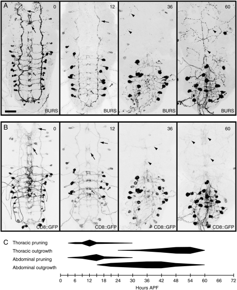
The normal functioning of neuroendocrine systems requires that many neuropeptidergic cells change, to alter transmitter identity and concentration, electrical properties, and cellular morphology in response to hormonal cues. During insect metamorphosis, a pulse of circulating steroids, ecdysteroids, governs the dramatic remodeling of larval neurons to serve adult-specific functions. To identify molecular mechanisms underlying metamorphic remodeling, we conducted a neuropeptidergic cell-targeted, gain-of-function genetic screen. We screened 6097 lines. Each line permitted Gal4-regulated transcription of flanking genes. A total of 58 lines, representing 51 loci, showed defects in neuropeptide-mediated developmental transitions (ecdysis or wing expansion) when crossed to the panneuropeptidergic Gal4 driver, 386Y-Gal4. In a secondary screen, we found 29 loci that produced wing expansion defects when crossed to a crustacean cardioactive peptide (CCAP)/bursicon neuron-specific Gal4 driver. At least 14 loci disrupted the formation or maintenance of adult-specific CCAP/bursicon cell projections during metamorphosis. These include components of the insulin and epidermal growth factor signaling pathways, an ecdysteroid-response gene, cabut, and an ubiquitin-specific protease gene, fat facets, with known functions in neuronal development. Several additional genes, including three micro-RNA loci and two factors related to signaling by Myb-like proto-oncogenes, have not previously been implicated in steroid signaling or neuronal remodeling.


Figures

F<sc>igure</sc> 1.—
Figure 1.—
Head eversion and wing expansion phenotypes produced by gene GOF in peptidergic neurons. (A) Oregon R (wild-type) adult female. (B) Examples of y, w; CCAP-Gal4/EY(2)04392 females with normal wings (normal), partially expanded wings (PEW), and unexpanded wings (UEW). The PEW and UEW phenotypes were scored as described (Luan et al. 2006). Arrows indicate folds due to incomplete wing expansion in the PEW animal. (C and D) Dorsal (C) and ventral (D) views of an Oregon R (wild-type) pupa and two y, w/w; 386Y-Gal4/EY(3)10546 pupae displaying weak and strong microcephalic (mc) phenotypes (cf. Hadorn and Gloor 1943; Hewes et al. 2000). Cryptocephalic pupae are very similar to the strong microcephalic pupae, except that the head structures are found entirely within the thorax (Hadorn and Gloor 1943; Chadfield and Sparrow 1985; Hewes et al. 2000; Park et al. 2003). The pronged bars in C indicate the anterior edges of the head and thorax and the posterior edge of the thorax. The solid and dashed lines in D indicate the posterior edges of the legs and wings, respectively. Asterisks, pupal abdomen lacking external bristles; b, posterior gas bubble due to failed anterior translocation of the gas bubble during head eversion; D, dorsal thorax dimpled; N, tanning incomplete; P, ptilinum permanently soft and partially extended; S, scutellum wrinkled and scutellar bristles crossed and directed toward anterior; T, darkened cuticle in shape of trident on dorsal thorax; X, nonglossy cuticle surface.
F<sc>igure</sc> 2.—
Figure 2.—
Scatter plot of target element insertion-site distances from the nearest promoter or exon. The distance for each target element to the 5′ end of the nearest promoter (black diamonds) or exon (magenta circles) was plotted on the y-axis. Negative values represent insertions located upstream of the respective promoter or exon splice acceptor site, and positive values refer to insertions that were located 3′ of these landmarks (while still 5′ of significant portions of the transcript). Only the first locus within 30 kb and in the same orientation as the direction of Gal4-directed transcription off of each EP, EY, or WH element (unidirectional) or XP element (bidirectional) was included, and the distances to putative LOF (antisense) transcripts (supplemental Table 3) are not shown. Insertions within the predicted coding sequence (CDS) of a transcript, and insertions located in introns located downstream of an exon containing CDS, are indicated with open symbols.
F<sc>igure</sc> 3.—
Figure 3.—
Staining patterns and morphologies of the CCAP/bursicon neurons. (A) Anti-bursicon (BURS) and (B) anti-CCAP (CCAP) immunostaining in wandering third-instar larval CNS (CCAP-Gal4/+). Anti-bursicon immunostaining was also observed in a cluster of neurites located over the corpora cardiaca (not shown). (C) mCD8∷GFP (CD8∷GFP) fluorescence in a wandering third-instar CNS (CCAP-Gal4, UAS-mCD8∷GFP/+). (D) Anti-bursicon immunostaining in a stage P14 pharate adult CNS (CCAP-Gal4/+). A midline protocerebral brain arbor (MPB) was also bursicon immunoreactive (not shown, see Figure 5C). Abbreviations: AA, abdominal arbor; formula image, eight neurons located toward the anterior of the abdominal ganglia; formula image, six neurons located in the posterior abdominal ganglia; DP, dorsal protocerebral neuron (weakly bursicon positive and CCAP negative); CTA, circum-neuropilar (ventral) thoracic arbor; DTA, dorsal thoracic arbor; LB(S, T1–3, A1–A7), lateral branch (leading from LLT to MA) in the subesophageal region and segments T1–T3 and A1–A7; LLT, lateral longitudinal tract; LSE(1–3), lateral subesophageal neurons; MA(S, T1–3, A1–A7), midline arbor in the indicated segments; MLT, midline longitudinal tract; MP, midline protocerebral neurons; MPA, midline protocerebral arbor; PA, protocerebral arbor; PAA, posterior abdominal arbor; SA, subesophageal arbor; TA, tritocerebral arbor; VA, ventral abdominal neuron. Bar, 50 μm.
F<sc>igure</sc> 4.—
Figure 4.—
Remodeling of the CCAP/bursicon neurons during metamorphosis. (A) Anti-bursicon immunostaining of the ventral nerve cord (VNC) at 0, 12, 36, and 60 hr after puparium formation (APF) (n = 6–19). Additional time points are shown in supplemental Figure 1. (B) mCD8∷GFP fluorescence in the same preparations as in A (CCAP-Gal4, UAS-mCD8∷GFP/+). (C) Time courses for pruning and outgrowth of the neuritic arborizations in the thoracic and abdominal ganglia. The relative extent of pruning and outgrowth (indicated by the relative height, along the y-axis, of the horizontal shapes) was quantified for each of the time points represented with tick marks on the x-axis. The images in A and B are representative of the changes at the selected times. Arrows, thoracic neurites that were pruned back by the next stage shown; arrowheads, new adult-specific thoracic neurites; double-feathered arrows, abdominal neurites that were pruned back by the next stage shown; double-feathered arrowheads, new adult-specific abdominal neurites. Bar, 50 μm.
F<sc>igure</sc> 5.—
Figure 5.—
GOF of Ptr produced neurite pathfinding defects in larval CCAP/bursicon neurons. In both larvae (A and B) and pharate adults (C), misexpression of Ptr (A′, B′, and C′) led to the loss of some somata and neurites (labels with dashed lines). Ectopic or mistargeted neurites were also observed (arrows). (A) mCD8∷GFP (UAS-mCD8∷GFP) expressed under the control of CCAP-Gal4 either with (A′) or without (A) EP(2)2003, which is inserted just 5′ of the Ptr gene (supplemental Table 3). CNS dissections were performed at the wandering third-instar stage (n = 8). (B) Anti-CCAP immunostaining in a CCAP-Gal4/EP(2)2003 wandering third-instar larval CNS (B′) and a CCAP-Gal4/+ control (B) (n = 5). In a second CCAP-Gal4/EP(2)2003 CNS (B′ inset), MLT was missing on one side. (C) Anti-bursicon immunostaining in the CNS from two CCAP-Gal4/EP(2)2003 P14-stage pharate adults (C′ and C″) and a CCAP-Gal4/+ control (C) (n = 7). The labels are defined in the Figure 3 legend. Arrowheads, thicker neurites with larger than normal varicosities; feathered arrows, small-diameter somata. Bars: A–C, 50 μm; B′ inset, 100 μm.
F<sc>igure</sc> 6.—
Figure 6.—
GOF of cbt prevented the outgrowth of adult-specific bursicon-immunoreactive neurites and the increase in diameter of the CCAP/bursicon cell somata during metamorphosis. (A–D) Anti-bursicon immunostaining in the CNS (A and C) and peripheral neurites (B and D). The staining was performed on tissue dissected at the wandering third-instar larval stage (A and B) and the P14 pharate adult stage (C and D) from CCAP-Gal4/EP(2)2237 animals (A′–D′) and CCAP-Gal4/+ controls (A–D) (n = 5–7). Note that the morphology of the CCAP/bursicon neurons was largely unaffected following cbt GOF in larvae. In contrast, the pharate adult morphology of the cells was severely disrupted. (E–H) Anti-GFP immunostaining in the CNS (E and F) and peripheral neurites (G and H) at 18 hr APF (E), 30 hr APF (G), or 54 hr APF (F and H). Membrane-associated mCD8∷GFP (UAS-mGFP) was expressed in CCAP-Gal4/+ (E–H) and CCAP-Gal4/EP(2)2237 (E′–H′) animals. In the CNS, cbt GOF caused the neurite projections to remain short, thick, and with few branches (F′), similar to the appearance of control animals 36 hr earlier (E). In the periphery, the efferents displayed some initial outgrowth, but they failed to form a complete adult arbor (H′). Asterisk, fine neuritic arbor; bicolor (solid/open) arrows, nonbranching efferent projections; solid arrows, traces of remaining neurites; solid arrowheads, smaller somata; solid double-feathered arrows, strongly immunoreactive axon terminals; solid double-feathered arrowheads, weakly immunoreactive axons; open arrows, adult-specific longitudinal axon tracts (forming or completed); open arrowheads, developing neurite branches; open double-feathered arrows, persistent larval projections after muscle detachment and before pruning is completed; open double-feathered arrowheads, adult efferents near points of exit from the CNS. Bars: A and C, 50 μm; B, D, G, and H, 100 μm; E and F, 10 μm.
F<sc>igure</sc> 7.—
Figure 7.—
GOF of klar resulted in specific loss of adult-specific bursicon-immunoreactive neurites and loss of six to eight CCAP/bursicon cell somata. (A and B) Anti-bursicon immunostaining in wandering third-instar (A) and P14 pharate adult (B) stage CNS from CCAP-Gal4/EY(3)00559 animals (A′ and B′) and CCAP-Gal4/+ controls (A and B) (n = 6–8). Most elements of the CCAP/bursicon cell projection pattern were retained in the larval CNS (A′), but there was a dramatic reduction of central neurites and BAG somata number in the pharate adult (B′). Arrows, traces of remaining neurites; arrowhead, atrophied soma. Bars, 50 μm.
F<sc>igure</sc> 8.—
Figure 8.—
GOF of the miR-310–miR-313 micro-RNA cluster during an early- to midmetamorphosis critical period prevented bursicon secretion in adults. (A) In situ hybridization (magenta) for miR-310 (antisense LNA probe) in a wandering third-instar larval CNS. Autofluorescence (green) was detected using an FITC/GFP filter set. EP(2)2587 was expressed under the control of CCAP-Gal4. Comparable staining was observed with LNA probes for miR-312 and miR-313 (not shown; miR-311 was not tested). (B) Anti-bursicon staining immediately after eclosion (0 hr AE) and 1 hr after eclosion (1 hr AE) in control flies (CCAP-Gal4/+) and flies with miR-310–miR-313 GOF [CCAP-Gal4/EP(2)2587]. (C) Quantification of bursicon levels for the treatments shown in B (n = 5–6). *P < 0.05; NS, not significant (P = 0.000224, one-way ANOVA; Tukey–Kramer multiple-comparison post hoc test). The differences in staining intensities were confirmed by blind scoring. (D) Percentage of adult flies with unexpanded wings (UEW) or partially expanded wings (PEW) following miR-310–miR-313 GOF at different developmental stages [n = 7–119 (41.0 ± 1.1) per point]. All animals had one copy each of 386Y-Gal4, EP(2)2587, and tubulinP-Gal80ts. The shift-up group (black squares, solid line) was collected as embryos at the permissive temperature (19°) and then shifted at the times shown on the x-axis to the restrictive temperature (30°). The shift-down group (open circles, dashed line) was collected as embryos at the restrictive temperature and then shifted at the times shown to the permissive temperature. The curves are fifth-order polynomials—only the center portions of the curves are shown. The gray bar indicates the critical window for the EP(2)2587 effect on wing expansion: It begins at the shift time that produced wing expansion defects in ∼50% of the shift-down animals, and it ends at the shift time that produced displayed wing expansion defects in ∼50% of the shift-up animals. Bars: A, 50 μm; B, 100 μm.

Similar articles

Cited by

References

    1. Abdelilah-Seyfried, S., Y. M. Chan, C. Zeng, N. J. Justice, S. Younger-Shepherd et al., 2000. A gain-of-function screen for genes that affect the development of the Drosophila adult external sensory organ. Genetics 155 733–752. - PMC - PubMed
    1. Allan, D. W., S. E. Pierre, I. Miguel-Aliaga and S. Thor, 2003. Specification of neuropeptide cell identity by the integration of retrograde BMP signaling and a combinatorial transcription factor code. Cell 113 73–86. - PubMed
    1. Arancio, O., and M. V. Chao, 2007. Neurotrophins, synaptic plasticity and dementia. Curr. Opin. Neurobiol. 17 325–330. - PubMed
    1. Aravin, A. A., M. Lagos-Quintana, A. Yalcin, M. Zavolan, D. Marks et al., 2003. The small RNA profile during Drosophila melanogaster development. Dev. Cell 5 337–350. - PubMed
    1. Bainbridge, S. P., and M. Bownes, 1981. Staging the metamorphosis of Drosophila melanogaster. J. Embryol. Exp. Morphol. 66 57–80. - PubMed

Publication types