. 2008 Feb;178(2):989-1002.
doi: 10.1534/genetics.107.082453. Epub 2008 Feb 1.

The conserved proteins CHE-12 and DYF-11 are required for sensory cilium function in Caenorhabditis elegans

Affiliations

The conserved proteins CHE-12 and DYF-11 are required for sensory cilium function in Caenorhabditis elegans

Taulant Bacaj et al. Genetics. 2008 Feb.

Abstract

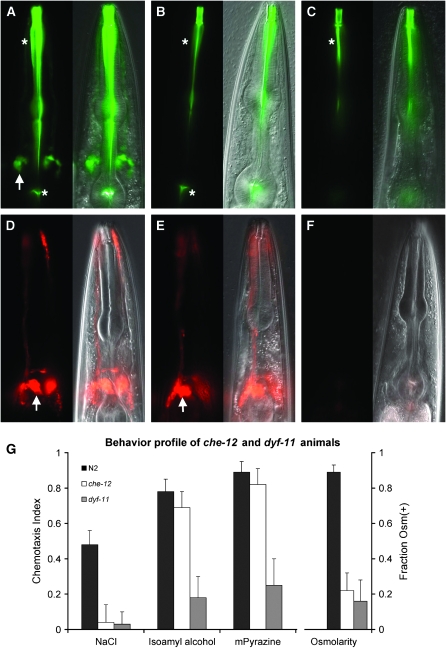
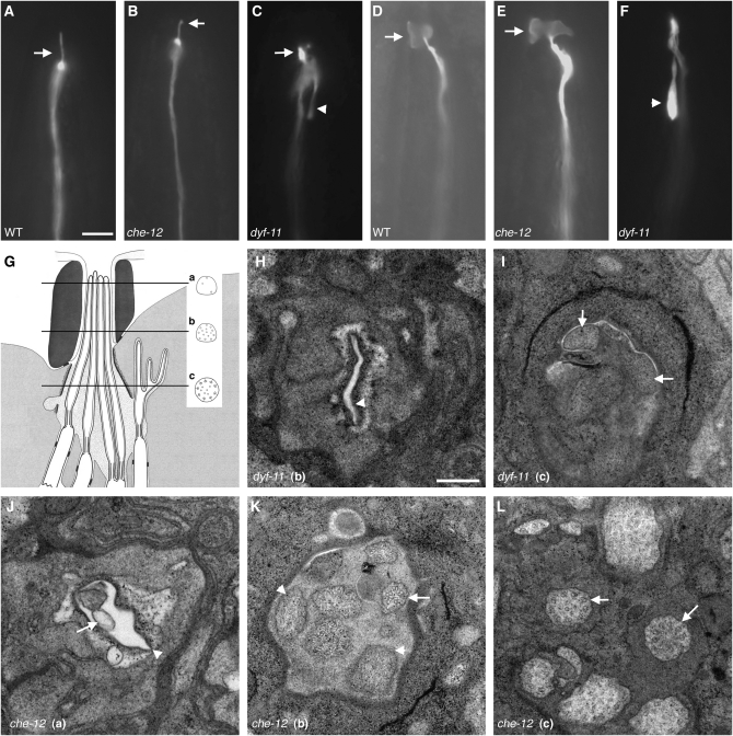
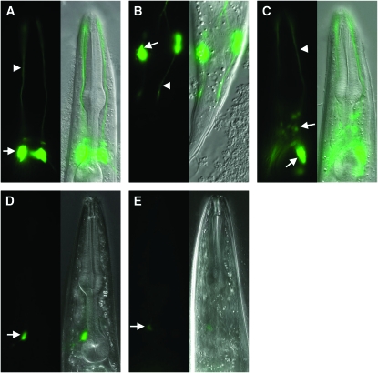
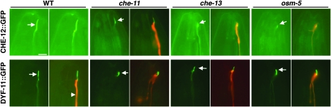
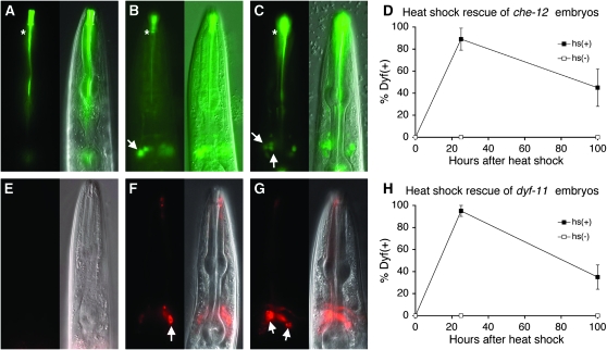
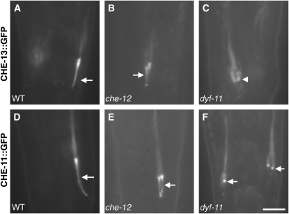
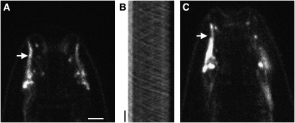
Sensory neuron cilia are evolutionarily conserved dendritic appendages that convert environmental stimuli into neuronal activity. Although several cilia components are known, the functions of many remain uncharacterized. Furthermore, the basis of morphological and functional differences between cilia remains largely unexplored. To understand the molecular basis of cilia morphogenesis and function, we studied the Caenorhabditis elegans mutants che-12 and dyf-11. These mutants fail to concentrate lipophilic dyes from their surroundings in sensory neurons and are chemotaxis defective. In che-12 mutants, sensory neuron cilia lack distal segments, while in dyf-11 animals, medial and distal segments are absent. CHE-12 and DYF-11 are conserved ciliary proteins that function cell-autonomously and are continuously required for maintenance of cilium morphology and function. CHE-12, composed primarily of HEAT repeats, may not be part of the intraflagellar transport (IFT) complex and is not required for the localization of some IFT components. DYF-11 undergoes IFT-like movement and may function at an early stage of IFT-B particle assembly. Intriguingly, while DYF-11 is expressed in all C. elegans ciliated neurons, CHE-12 expression is restricted to some amphid sensory neurons, suggesting a specific role in these neurons. Our results provide insight into general and neuron-specific aspects of cilium development and function.


Figures

F<sc>igure</sc> 1.—
Figure 1.—
Characterization of che-12 and dyf-11 mutants. (A) Uptake of FITC by amphid sensory neurons in a wild-type animal. Left, fluorescence image; right, fluorescence and DIC overlay of the same image. Arrow, cell bodies; asterisks, nonspecific pharyngeal staining. (B) A che-12(mn389) mutant showing FITC-uptake defect. (C) dyf-11(mn392) animals also fail to take up FITC. (D) DiI staining pattern of a wild-type animal. (E) che-12(mn389) animals can also take up DiI. (F) dyf-11(mn392) animals fail to take up DiI. (G) Behavioral defects of che-12(mn399) and dyf-11(mn392) mutants. Both mutants fail to chemotax to 0.2 m NaCl and are unable to respond to an osmotic barrier. dyf-11, but not che-12, animals show defects in odortaxis toward 1% isoamyl alcohol and 1% methyl pyrazine. In all figures anterior is up unless otherwise indicated.
F<sc>igure</sc> 2.—
Figure 2.—
che-12 and dyf-11 mutants have defects in cilium structure. (A) The ASER cilium (arrow) of a wild-type animal expressing gcy-5 pro∷GFP. (B) che-12(mn399) animals have a short ASER cilium. (C) The stunted ASER cilium (arrow) of a dyf-11 animal. A backward process is visible (arrowhead). (D) The AWC cilium (arrow) of a wild-type animal expressing str-2 pro∷GFP. (E) In che-12(mn399), the AWC cilium retains its winged morphology (arrow). (F) The AWC cilium fails to spread in dyf-11 animals. A backward process is present (arrowhead). Bar, A–F, 5 μm. (G) A cartoon of the amphid channel, adapted from Perkins et al. (1986). Three of the eight channel neurons are depicted. The locations of EM cross sections are shown. The normal cross-sectional profile of a cilium at each level is diagrammed. (H) EM cross section at level “b” of a dyf-11(mn392) animal. Note the empty cavity (arrowhead) where amphid cilia should be located. (I) EM cross section of a dyf-11(mn392) animal at level “c” showing the absence of doublet or any other microtubules in neuronal profiles (arrows). (J) EM cross section of a che-12(mn389) mutant at level “a”. The amphid cilia are short, and so only 2 of the 10 neuronal profiles are evident. Amphid opening (arrowhead). The beginning of a cilium (arrow). (K) EM cross section of the distal segment of a che-12(mn389) animal at level “b”. No singlet microtubules are seen in most cilia (arrowheads). A cilium containing doublet microtubules is shown (arrow). (L) Proximal EM cross section of a che-12(mn389) animal at level “c”. The cilia appear normal and contain doublet microtubules (arrows). Bar, H–L, 300 nm. In H–L, dorsal is up.
F<sc>igure</sc> 3.—
Figure 3.—
The genomic structures of che-12 and dyf-11. (A) che-12 was mapped to the indicated region of chromosome V. Rescue of the Dyf defect could be achieved by injection of the B0024 cosmid or the B0024.8 gene. (B) Genomic structure of B0024.8. The positions of the three alleles are shown. (C) dyf-11 was mapped to the indicated region of chromosome X. A single gene, C02H7.1, within cosmid C02H7, could rescue the Dyf defect. (D) Genomic structure of C02H7.1 and position of the mn392 allele.
F<sc>igure</sc> 4.—
Figure 4.—
CHE-12 and DYF-11 are expressed in ciliated neurons in a DAF-19-dependent manner. (A) CHE-12 is expressed in a subset of amphid neurons. Neuronal cell bodies (arrow); dendritic processes (arrowhead). (B) CHE-12 is also present in phasmid neurons. (C) Expression of DYF-11 is seen in most ciliated neurons including those of the amphid and labial sensilla (arrows). (D) Expression of che-12 pro∷GFP is greatly reduced in daf-16; daf-19 animals. Compare with A. In D and E, image exposure was at least twice as long as in A and C. (E) The expression of dyf-11 pro∷GFP also depends on the transcription factor DAF-19. Compare with C.
F<sc>igure</sc> 5.—
Figure 5.—
CHE-12 and DYF-11 localize to cilia. Left image of a pair, GFP alone. Right image, GFP and mCherry overlay. CHE-12 localizes to the cilium of ASER (arrows). The localization of CHE-12 is disrupted in che-13(1805) and osm-5(184) IFT-B mutants (note low intensity of GFP within cilium) and to a lesser extent in che-11(1810) IFT-A mutants. DYF-11 is normally localized within cilia of wild-type animals. This localization is not affected by the che-11, che-13, and osm-5 mutations. All transgenes are driven by the gcy-5 promoter, which is expressed specifically in ASER. Bar, 5 μm.
F<sc>igure</sc> 6.—
Figure 6.—
CHE-12 and DYF-11 act cell-autonomously and are required continuously for cilium morphology and function. (A) che-12(mn389) animals fail to take up FITC; asterisk indicates nonspecific staining. Fourth larval stage (L4) animals are depicted in all images. (B) Expression of CHE-12 in a che-12(mn389) mutant animal within only two amphid neurons, ASH and ASI, using the sra-6 promoter, rescues the Dyf defect only within these neurons (arrow). (C) A che-12(mn389) animal that is provided CHE-12 via heat shock as an adult is able to take up FITC in several amphid neurons (arrows). (D) che-12(mn389) embryos carrying extrachromosomal arrays containing the hsp-16.2 pro∷che-12 cDNA transgene were heat-shocked for 30 min at 34°. Dye filling was performed after 24 hr to determine initial rescue or after 100 hr. (E) dyf-11 animals cannot take up DiI. (F) Expression of dyf-11 in ASH and ASI using the sra-6 promoter enables only these two neurons to fill with dye (arrow). (G) Providing DYF-11 to adult animals via heat shock rescues their Dyf defect. Animals contain an hsp-16.2 pro∷dyf-11 cDNA transgene. (H) Same as D, except that dyf-11(mn392) mutants carrying an hsp-16.2 pro∷dyf-11 transgene were used.
F<sc>igure</sc> 7.—
Figure 7.—
Localization of IFT-A and IFT-B components in che-12 and dyf-11 mutants. (A) Localization of CHE-13∷GFP in a phasmid cilium (arrow) of a wild-type animal. (B) In che-12(mn399) animals, CHE-13∷GFP localizes properly. (C) CHE-13∷GFP fails to localize to the cilia of dyf-11(mn392) animals; the base of the cilium is indicated by an arrowhead. (D) CHE-11∷GFP localization in a wild-type phasmid cilium. (E) In che-12(mn399) animals, CHE-11∷GFP localizes appropriately. (F) CHE-11∷GFP can occasionally localize to the base of the stunted cilia of dyf-11 animals. Bar, 4 μm.
F<sc>igure</sc> 8.—
Figure 8.—
DYF-11 may associate with IFT particle B. (A) Localization of DYF-11∷GFP in the cilia (arrow) of a wild-type animal. (B) Kymograph of the cilia seen in A, showing anterograde and retrograde movement of DYF-11∷GFP. Position is displayed on the horizontal axis and time on the vertical axis. The transition zone of the cilium is on the left. Vertical bar, 5 sec. (C) DYF-11∷GFP can enter the distal segment of cilia in a bbs-8(nx77) mutant animal (arrow). Bar, A–C, 2.5 μm.
F<sc>igure</sc> 9.—
Figure 9.—
A hypothetical model showing the functions of CHE-12 and DYF-11 in sensory cilia. The IFT subcomplexes A and B, linked by the BBS-8 protein, are moved along the ciliary microtubules by the kinesin-2 and OSM-3 motors. DYF-11 might interact with microtubules as well as with the IFT-B protein complex, acting to promote association of CHE-13 and OSM-5 proteins with the complex. CHE-12 is transiently associated with the IFT particle to be transported into the cilium, where it is released and accumulates. Not all known IFT particle components are shown in this diagram.

References

    1. Altschul, S. F., W. Gish, W. Miller, E. W. Myers and D. J. Lipman, 1990. Basic local alignment search tool. J. Mol. Biol. 215 403–410. - PubMed
    1. Andrade, M. A., and P. Bork, 1995. HEAT repeats in the Huntington's disease protein. Nat. Genet. 11 115–116. - PubMed
    1. Ansley, S. J., J. L. Badano, O. E. Blacque, J. Hill, B. E. Hoskins et al., 2003. Basal body dysfunction is a likely cause of pleiotropic Bardet-Biedl syndrome. Nature 425 628–633. - PubMed
    1. Avidor-Reiss, T., A. M. Maer, E. Koundakjian, A. Polyanovsky, T. Keil et al., 2004. Decoding cilia function: defining specialized genes required for compartmentalized cilia biogenesis. Cell 117 527–539. - PubMed
    1. Bargmann, C. I., and H. R. Horvitz, 1991. Chemosensory neurons with overlapping functions direct chemotaxis to multiple chemicals in C. elegans. Neuron 7 729–742. - PubMed

Publication types

MeSH terms

LinkOut - more resources