. 2008 Mar;178(3):1209-20.
doi: 10.1534/genetics.107.080135. Epub 2008 Feb 1.

Catalytic-site mutations in the MYST family histone Acetyltransferase Esa1

Affiliations

Catalytic-site mutations in the MYST family histone Acetyltransferase Esa1

Peter V Decker et al. Genetics. 2008 Mar.

Abstract

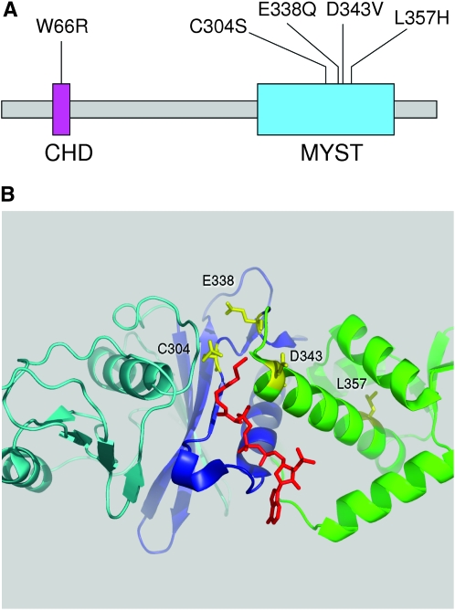
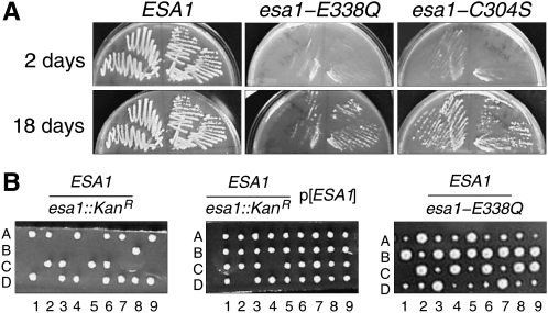
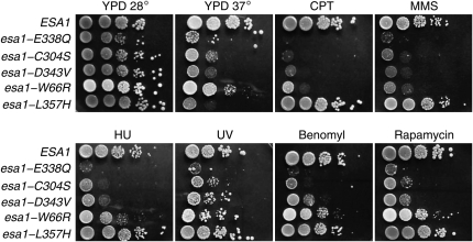
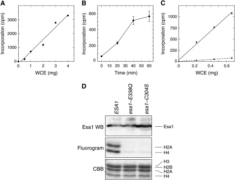
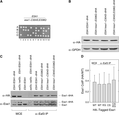
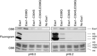
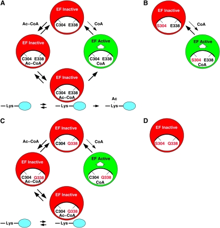
Esa1 is the only essential histone acetyltransferase (HAT) in budding yeast. It is the catalytic subunit of at least two multiprotein complexes, NuA4 and Piccolo NuA4 (picNuA4), and its essential function is believed to be its catalytic HAT activity. To examine the role of Esa1 in DNA damage repair, we isolated viable esa1 mutants with a range of hypersensitivities to the toposide camptothecin. Here we show that the sensitivity of these mutants to a variety of stresses is inversely proportional to their level of histone H4 acetylation, demonstrating the importance of Esa1 catalytic activity for resistance to genotoxic stress. Surprisingly, individual mutations in two residues directly involved in catalysis were not lethal even though the mutant enzymes appear catalytically inactive both in vivo and in vitro. However, the double-point mutant is lethal, demonstrating that the essential function of Esa1 relies on residues within the catalytic pocket but not catalysis. We propose that the essential function of Esa1 may be to bind acetyl-CoA or lysine substrates and positively regulate the activities of NuA4 and Piccolo NuA4.


Figures

F<sc>igure</sc> 1.—
Figure 1.—
Panel of esa1 mutants. (A) Mutants shown on the primary structure of Esa1. W66R lies in the chromodomain (CHD) while the others fall within the MYST family HAT domain. (B) Relevant residues (yellow) on the crystal structure of Esa1(160–435) in complex with coenzyme A (red).
F<sc>igure</sc> 2.—
Figure 2.—
Viability of esa1 catalytic-site mutants. (A) LEU2-marked plasmids bearing ESA1, esa1-E338Q, or esa1-C304S were transformed into a BY4743-derived esa1∷kan strain carrying an ESA1 URA3 plasmid. Resulting strains were then streaked onto 5-FOA to counterselect the ESA1 URA3 plasmid. (B) An esa1-E338Q allele was knocked into the genomic ESA1 locus in a W303 diploid strain, which was then sporulated and dissected. The esa1-E338Q allele supports viability (right, small colonies), while an esa1∷kan allele does not (left) unless covered with an ESA1 plasmid (middle).
F<sc>igure</sc> 3.—
Figure 3.—
Phenotypes of the esa1 panel. Serial dilutions of the esa1 panel were spotted on YPD-based plates containing no drug or 30 μg/ml camptothecin (CPT); 0.025% methylmethane sulfonate (MMS); 12.5 μg/ml benomyl; 0.1 m hydroxyurea; or 25 nm rapamycin. For the UV treatment, the YPD plate was irradiated with 0.015 J/cm2 of 254 nm UV light in a Stratalinker.
F<sc>igure</sc> 4.—
Figure 4.—
Characterization of esa1 mutants by Western blot. (A) The indicated strains were grown to mid-log phase and whole-cell extracts were prepared, separated by SDS–PAGE, and probed by Western blot for acetylated H4 (H4ac) and bulk histone H4 (H4). (B) The indicated strains were grown to mid-log phase, diluted, and then grown for 4 hr at either 28° or 37°. Whole-cell extracts were then prepared, separated by SDS–PAGE, and then probed by Western blot for Esa1 protein, histone H4 and H3 acetylation, and glucose-6-phosphate dehydrogenase (GPDH) as a loading control. The hhf1-ΔN allele encodes histone H4 missing N-terminal residues 1–27.
F<sc>igure</sc> 5.—
Figure 5.—
In vitro HAT activity of esa1 mutants in the context of their native complexes. (A) Increasing amounts of whole-cell extracts from ESA1 cells were immunoprecipitated with a constant 5 μl of anti-Esa1 antibody (Abcam). The immunoprecipitates were subjected to HAT assays by incubating them with [3H]-acetyl CoA and purified chicken core histone substrate for 60 min. The reactions were collected on filters, extensively washed, and measured by scintillation counting. Subsequent experiments used 5 μl of anti-Esa1 antibody per 2.0 mg of whole-cell extract protein. (B) Whole-cell extracts were prepared from ESA1 cells and assayed for HAT activity for increasing lengths of time. Each point represents the average and standard deviation of triplicate assays of 0.67 mg of starting whole-cell extract protein. Subsequent experiments were carried out for 40 min. (C) Whole-cell extracts were prepared from ESA1 (circles, solid line) and esa1-E338Q cells (squares, dashed line) and increasing amounts of protein were immunoprecipitated with anti-Esa1 antibody. Each point represents the average and standard deviation of triplicate assays carried out for 40 min. (D) Esa1-containing complexes were immunoprecipitated using an Esa1 antibody, and immune complexes were incubated with [3H]-acetyl CoA and chicken core histones. After reaction, SDS–PAGE loading buffer was added to 1× concentration and samples were boiled prior to SDS–PAGE. The bottom portion of the gel was then stained with Coomassie brilliant blue (CBB; bottom) and subjected to fluorography (middle) to determine incorporation of [3H]-acetyl groups into histones, while the top portion of the gel was transferred to nitrocellulose and probed for Esa1 protein with an Esa1 antibody (top).
F<sc>igure</sc> 6.—
Figure 6.—
Characterization of the esa1-C304S,E338Q allele. (A) The esa1-C304S,E338Q double-point mutant is inviable. A heterozygous ESA1/esa1-C304S,E338Q diploid strain was sporulated and the resulting tetrads were dissected. The growth of the four spore colonies (A–D) from 12 tetrads are shown. Tetrads gave predominantly a 2:2 pattern of viable:inviable spores, with all viable spores being Ura. In this example, two tetrads (7 and 10) produced four viable spore colonies, but in both cases all four spores were Ura and homozygous for the wild-type ESA1 allele presumably as a result of recombination. (B) Esa1-C304S,E338Q protein is expressed normally. Whole-cell extracts were prepared from four heterozygous diploid strains in which one ESA1 allele was wild type and encoded untagged protein, while the other allele was either wild type or mutant and expressed protein tagged with a C-terminal 6HA epitope. The extracts were then probed by Western blot with an anti-HA antibody to determine the level of expression of the different proteins. The anti-GPDH probing served to control for protein loading. (C) Esa1 proteins carrying catalytic-site mutations co-immunoprecipitate with the Eaf3 subunit of NuA4. Extracts were prepared from the heterozygous diploid strains indicated and immunoprecipitated with an anti-Eaf3 antibody. Aliquots of the whole-cell extracts (WCE) or immunoprecipitates (α-Eaf3 IP) were separated by PAGE. The gel was blotted and probed with anti-HA antibody to score the tagged Esa1 protein (α-HA) and with anti-Esa1 antibody (α-Esa1) to score both the tagged and the untagged proteins. (D) Relative recovery of tagged and untagged Esa1 proteins in anti-Eaf3 immunoprecipitates. The relative co-IP recoveries were determined for the data in C and two other independent experiments. Western blots probed with anti-Esa1 antibody were digitized and the intensities of the Esa1-6HA and Esa1 proteins present in the whole-cell extracts and recovered in the Eaf3 IPs were quantified for each mutant. The average ratio and standard deviation of HA-tagged to untagged protein is shown.
F<sc>igure</sc> 7.—
Figure 7.—
In vitro HAT activity of recombinant Esa1. The wild-type and catalytic-site mutants were N-terminally tagged with a His6 epitope tag and expressed in E. coli. After purification on Ni-NTA resin, relatively equal amounts (top, Esa1 CBB; note that the Esa1-E338Q samples have slightly more protein present) of Esa1 (or no Esa1 as a control) were combined with chicken core histones and [3H]-acetyl CoA at either pH 8.0 or pH 9.2 and incubated at room temperature for 30 min. To assay incorporation of [3H]-acetyl groups into the histones, we ran the reaction on SDS–PAGE, dried the gel, and subjected it to fluorography (middle). A Coomassie stain of the same region of the gel is shown to confirm equal loading of histones (bottom).
F<sc>igure</sc> 8.—
Figure 8.—
An Esa1 cofactor regulatory cycle. (A) The acetylation cycle for NuA4 complexes containing wild-type Esa1. NuA4 complexes inactive for essential functions are shown in red (“EF inactive”) and active complexes are shown in green (“EF active”). Here EF active and EF inactive refer to the essential functions of NuA4 complexes, such as transcriptional coactivation (Reid et al. 2000; Nourani et al. 2004; Durant and Pugh 2007) and not the catalytic activity of Esa1 itself. We propose that Esa1 positively activates NuA4 in the CoA-bound state but not the Ac-CoA-bound state. This cycle is facilitated by acetylation of lysine substrates in the histone tails, depicted in blue. Thus, the acetylation cycle of the wild-type enzyme produces both acetylated histones, or other protein substrates, and CoA-bound Esa1 to activate the essential functions of NuA4. (B) NuA4 complexes containing Esa1-C304S have an abbreviated regulatory cycle. The amino acid replacement S304, the Esa1 catalytic pocket, is indicated in red. We propose that Esa1-C304S is defective for binding Ac-CoA but retains binding of CoA on the basis of the results of acetylation reactions at pH 9.2 in vitro (Figure 7) and the crystallographic results of Yan et al. (2002). Thus, NuA4 complexes containing Esa1-C304S can be activated by CoA binding, conferring cell viability, but this is impaired relative to wild-type Esa1 in the absence of the full acetylation cycle. The binding of histone-tail lysines is unknown in this case. (C) NuA4 complexes containing Esa1-E338Q have a blocked regulatory cycle. The amino acid replacement Q338 is indicated in red. We propose that Esa1-E338Q retains the ability to bind both Ac-CoA and CoA but is unable to acetylate lysine substrates because the target ɛ-amino groups cannot be deprotonated in the absence of E338 (Yan et al. 2000, 2002; Berndsen et al. 2007a). Like Esa1-C304S, Esa1-E338Q is able to partially activate NuA4 complexes by binding CoA. However, this activity is impaired relative to Esa1-C304S since Ac-CoA now becomes a competitive inhibitor of CoA binding. Lysine substrates may bind, but they cannot be acetylated. (D) NuA4 complexes containing Esa1-C304S,E338Q. We propose that Esa1-C304S,E338Q is largely unable to bind either CoA or Ac-CoA, and therefore it fails to activate the essential functions of NuA4 complexes, leading to cell death. The binding status of histone-tail lysines is unknown.

Similar articles

Cited by

References

    1. Allard, S., R. T. Utley, J. Savard, A. Clarke, P. Grant et al., 1999. NuA4, an essential transcription adaptor/histone H4 acetyltransferase complex containing Esa1p and the ATM-related cofactor Tra1p. EMBO J. 18 5108–5119. - PMC - PubMed
    1. Amberg, D., D. Burke and J. Strathern, 2005. Methods in Yeast Genetics. Cold Spring Harbor Laboratory Press, Cold Spring Harbor, NY.
    1. Babiarz, J., J. Halley and J. Rine, 2006. Telomeric heterochromatin boundaries require NuA4-dependent acetylation of histone variant H2A.Z in Saccharomyces cerevisiae. Genes Dev. 20 700–710. - PMC - PubMed
    1. Berndsen, C. E., B. Albaugh, S. Tan and J. Denu, 2007. a Catalytic mechanism of a MYST family histone acetyltransferase. Biochemistry 46 623–629. - PMC - PubMed
    1. Berndsen, C. E., W. Selleck, S. J. McBryant, J. C. Hansen, S. Tan et al., 2007. b Nucleosome recognition by the Piccolo NuA4 histone acetyltransferase complex. Biochemistry 46 2091–2099. - PMC - PubMed

Publication types

MeSH terms

Substances