. 2008 Feb 12;105(6):1955-9.
doi: 10.1073/pnas.0709663105. Epub 2008 Feb 1.

Endothelial expression of the Notch ligand Jagged1 is required for vascular smooth muscle development

Affiliations

Endothelial expression of the Notch ligand Jagged1 is required for vascular smooth muscle development

Frances A High et al. Proc Natl Acad Sci U S A. .

Abstract

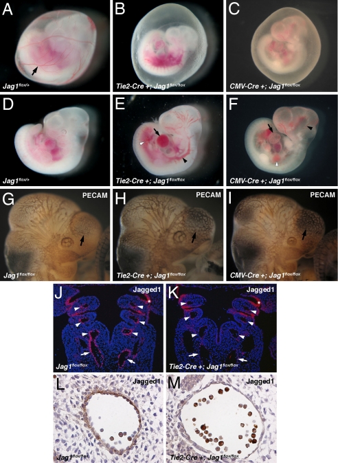
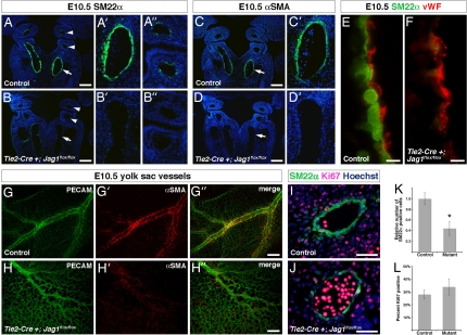
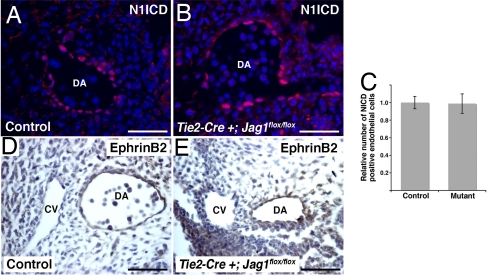
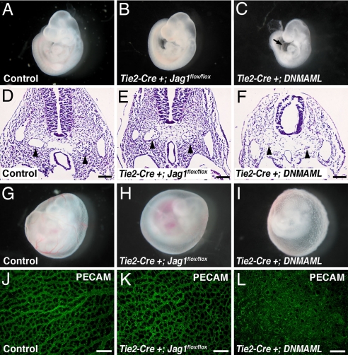
The Notch ligand Jagged1 (Jag1) is essential for vascular remodeling and has been linked to congenital heart disease in humans, but its precise role in various cell types of the cardiovascular system has not been extensively investigated. We show that endothelial-specific deletion of Jag1 results in embryonic lethality and cardiovascular defects, recapitulating the Jag1 null phenotype. These embryos show striking deficits in vascular smooth muscle, whereas endothelial Notch activation and arterial-venous differentiation appear normal. Endothelial Jag1 mutant embryos are phenotypically distinct from embryos in which Notch signaling is inhibited in endothelium. Together, these results imply that the primary role of endothelial Jag1 is to potentiate the development of neighboring vascular smooth muscle.


Conflict of interest statement

The authors declare no conflict of interest.

Figures

Fig. 1.
Fig. 1.
Endothelial-specific Jag1 mutants display cardiovascular defects. (A–C) E10.5 embryos with yolk sacs. Control yolk sacs (A) display prominent blood vessels (arrow) that are absent from both endothelial-specific (B) and global (C) Jag1 mutants. (D–F) E10.5 control (D), endothelial Jag1 mutant (E), and global Jag1 mutant (F) embryos demonstrating cardiovascular defects in mutants including pericardial effusions (arrows in E and F), dilated blood vessels (black arrowheads in E and F), and hemorrhage (white arrowheads in E and F). (G and H) Whole-mount PECAM immunostaining of E10.5 control (G), endothelial Jag1 mutant (H), and global Jag1 mutant (I) embryos. Mutant embryos show a less intricate vascular network over forebrain vesicles (arrows). (J and K) In situ hybridizations for Jag1 on frontal sections through E10.5 control (J) and mutant (K) embryos. Mutant embryos show loss of Jag1 expression in the dorsal aortae (arrows) and aortic arch arteries (arrowheads), but not pharyngeal endoderm (*). (L and M) Immunostaining for Jag1 on sections through the dorsal aorta of E10.5 control (L) and mutant (M) embryos, showing loss of endothelial Jagged1 protein in mutants. (Magnifications: A–F, ×40; G–K, ×100; L and M, ×400.)
Fig. 2.
Fig. 2.
Vascular smooth muscle development in endothelial-specific Jag1 mutants. (A–A″ and B–B″) Immunostaining for SM22α (green) on frontal sections through E10.5 control (A–A″) and mutant (B–B″) embryos. Control embryos show prominent SM22α expression in dorsal aorta (arrow in A, higher magnification in A′) and a few cells in the aortic arch arteries (arrowheads in A, higher magnification in A″). Mutant embryos show significant loss of SM22α in dorsal aorta (arrows in B and B′) and aortic arch arteries (arrowheads in B and B″). (C, C′, D, and D′) Immunostaining for αSMA (green) on frontal sections through E10.5 control (C and C′) and mutant (D and D′) embryos. Control embryos express αSMA in the dorsal aorta (arrows in C and C′), whereas mutant embryos lack αSMA expression (arrows in D and D′). (E and F) High-magnification images of the endothelial-smooth muscle interface in the dorsal aorta of E10.5 control (E) and mutant (F) embryos, immunostained for SM22α (green) and von Willebrand factor (vWF, red). (G–G″ and H–H″) E10.5 yolk sacs whole-mount immunostained for PECAM (green) and αSMA (red). Control yolk sacs (G–G″) show large blood vessels with prominent αSMA expression, whereas mutant yolk sacs (H–H″) show fewer large blood vessels that were abnormal in appearance and lacked αSMA expression. (I and J) Representative images of SM22α, Ki67 coimmunostaining on sections through the dorsal aorta of E10.5 control (I) and mutant (J) embryos. (K) Quantification of total number of SM22α-positive cells in the dorsal aortae of control and mutant embryos. (L) Quantification of percentage of SM22α-positive cells that coexpress Ki67 in the dorsal aortae of control and mutant embryos. Error bars indicate 1 SD. *, P < 0.001. (Scale bars: A–D′, 100 μm; E and F, 5 μm; G–H″, 200 μm; I and J, 50 μm.)
Fig. 3.
Fig. 3.
Endothelial Notch1 activation and EphrinB2 expression in endothelial-specific Jag1 mutants. (A and B) Immunostaining for Notch1 ICD (N1ICD, red) with Hoechst nuclear counterstaining (blue) on sections through the dorsal aorta (DA) of E10.5 control (A) and mutant (B) embryos. (C) Quantification of number of endothelial cells with positive nuclear staining for N1ICD in control and mutant embryos. Error bars indicate 1 SD. (D and E) Immunostaining for EphrinB2 (brown) showing expression in the dorsal aorta (DA) but not the cardinal vein (CV) of both control (D) and mutant (E) embryos. (Scale bars: 50 μm.)
Fig. 4.
Fig. 4.
Comparison of phenotypes of endothelial-specific Jag1 and endothelial-specific DNMAML mutants. (A–C) E9.5 embryos. Tie2-Cre+;Jag1flox/flox mutants (B) are grossly indistinguishable from controls (A), whereas Tie2-Cre+;DNMAML mutants (C) are developmentally delayed and show pericardial effusions (arrow in C). (D–F) Hematoxylin and eosin-stained sections through E9.5 embryos with dorsal aortae indicated by arrowheads. Dorsal aortae in Tie2-Cre+;Jag1flox/flox mutants are morphologically normal, whereas Tie2-Cre+;DNMAML mutant dorsal aortae are atretic. (G–I) E9.5 yolk sacs showing loss of large blood vessels in Tie2-Cre+;Jag1flox/flox mutants and abnormal rough texture in Tie2-Cre+;DNMAML mutants. (J–L) E9.5 yolk sacs whole-mount immunostained for PECAM (green). Tie2-Cre+;Jag1flox/flox yolk sac capillaries appear similar to controls, whereas Tie2-Cre+;DNMAML yolk sac vessels form a markedly abnormal, highly fused plexus. Images in A–C and G–I were photographed at ×40 magnification. (Scale bars: D–F, 100 μm; J–L, 200 μm.)

References

    1. Gale NW, Dominguez MG, Noguera I, Pan L, Hughes V, Valenzuela DM, Murphy AJ, Adams NC, Lin HC, Holash J, et al. Proc Natl Acad Sci USA. 2004;101:15949–15954. - PMC - PubMed
    1. Duarte A, Hirashima M, Benedito R, Trindade A, Diniz P, Bekman E, Costa L, Henrique D, Rossant J. Genes Dev. 2004;18:2474–2478. - PMC - PubMed
    1. Domenga V, Fardoux P, Lacombe P, Monet M, Maciazek J, Krebs LT, Klonjkowski B, Berrou E, Mericskay M, Li Z, et al. Genes Dev. 2004;18:2730–2735. - PMC - PubMed
    1. Krebs LT, Xue Y, Norton CR, Shutter JR, Maguire M, Sundberg JP, Gallahan D, Closson V, Kitajewski J, Callahan R, et al. Genes Dev. 2000;14:1343–1352. - PMC - PubMed
    1. Krebs LT, Shutter JR, Tanigaki K, Honjo T, Stark KL, Gridley T. Genes Dev. 2004;18:2469–2473. - PMC - PubMed

Publication types

MeSH terms