. 2008 Feb 1;68(3):946-55.
doi: 10.1158/0008-5472.CAN-07-3140.

Raf and MEK protein kinases are direct molecular targets for the chemopreventive effect of quercetin, a major flavonol in red wine

Affiliations

Raf and MEK protein kinases are direct molecular targets for the chemopreventive effect of quercetin, a major flavonol in red wine

Ki Won Lee et al. Cancer Res. .

Abstract

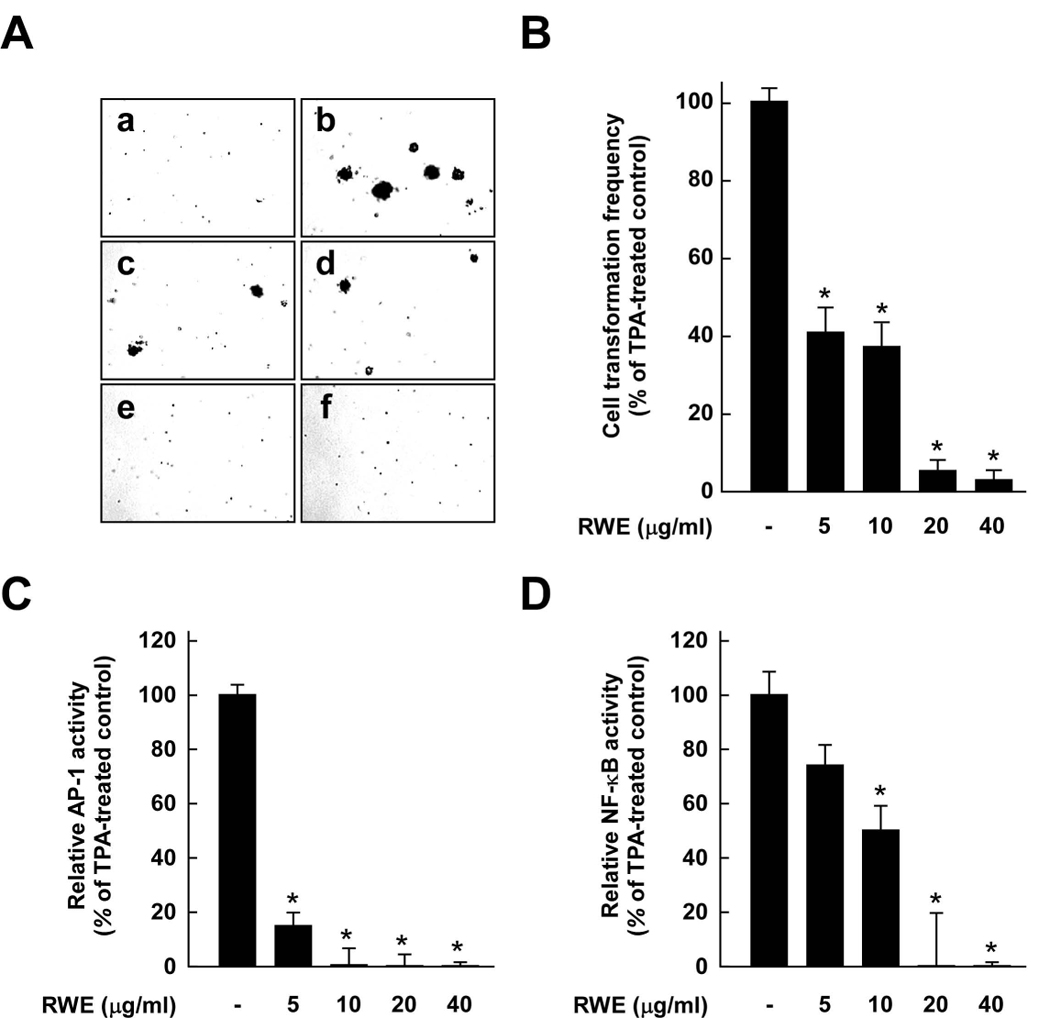
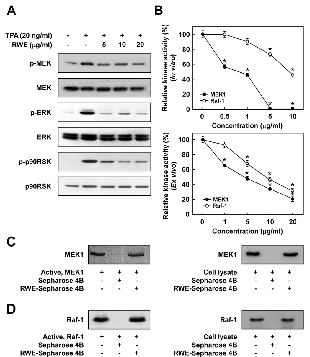
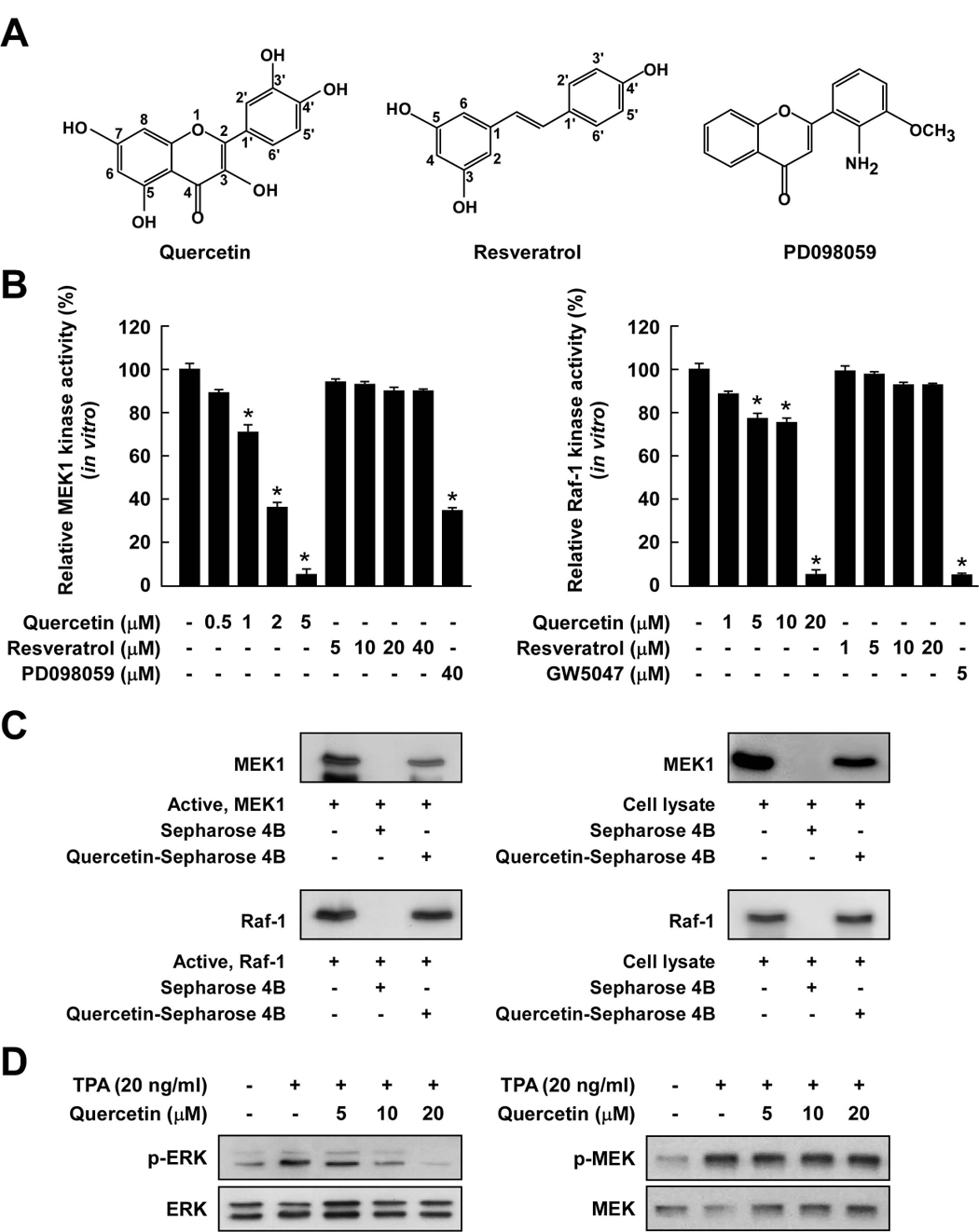
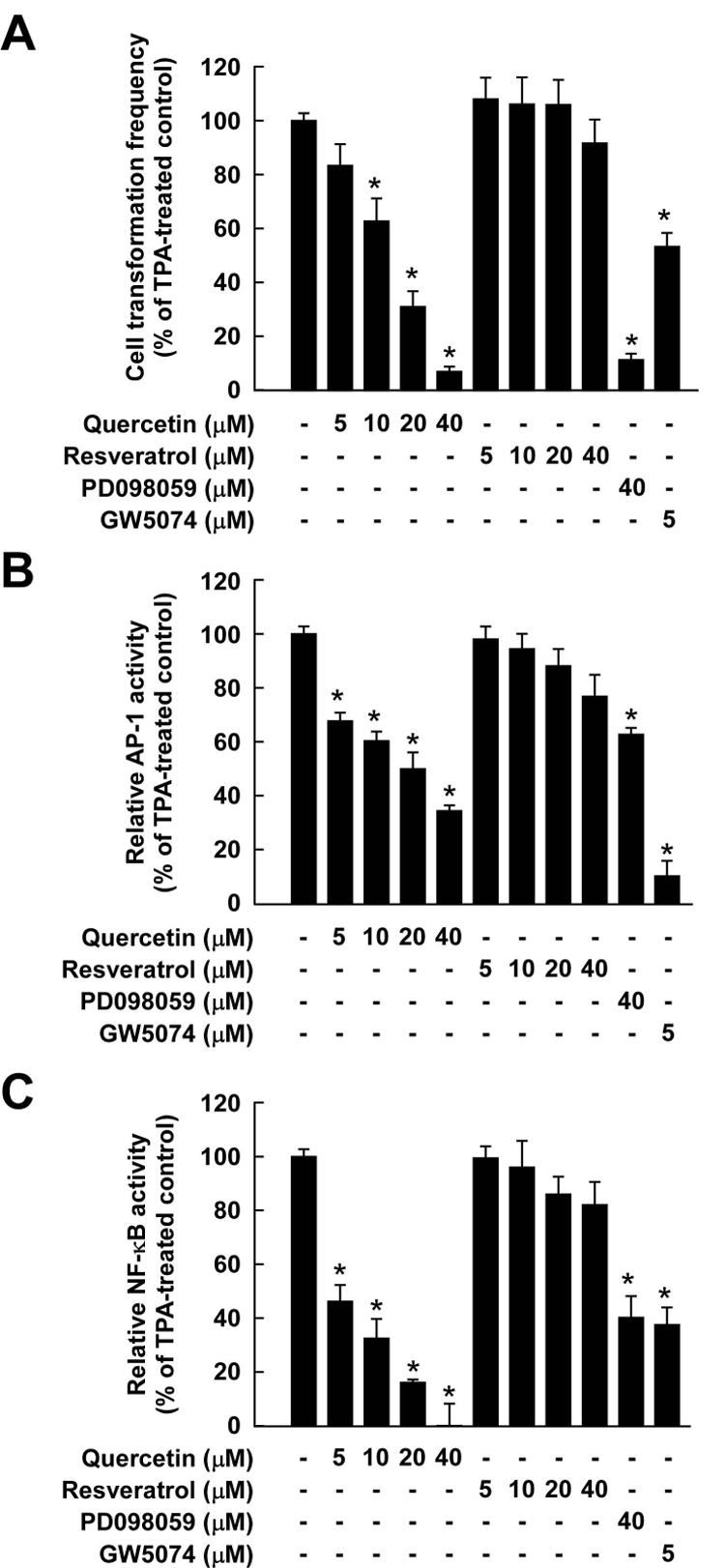
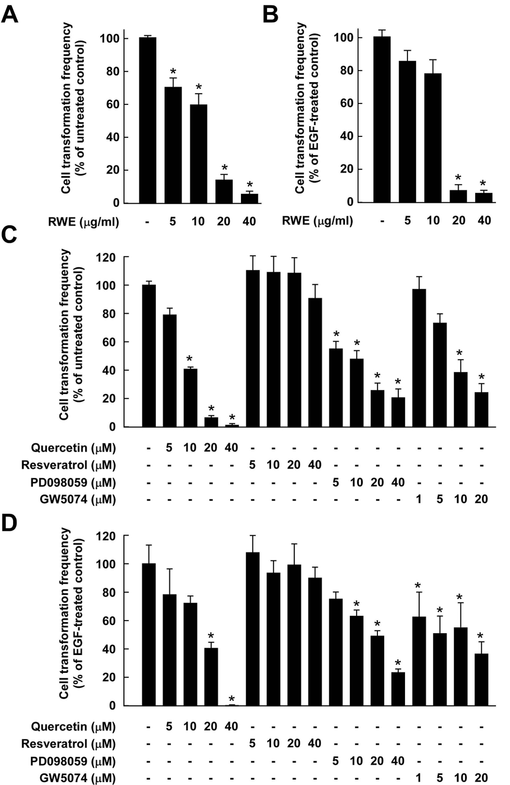
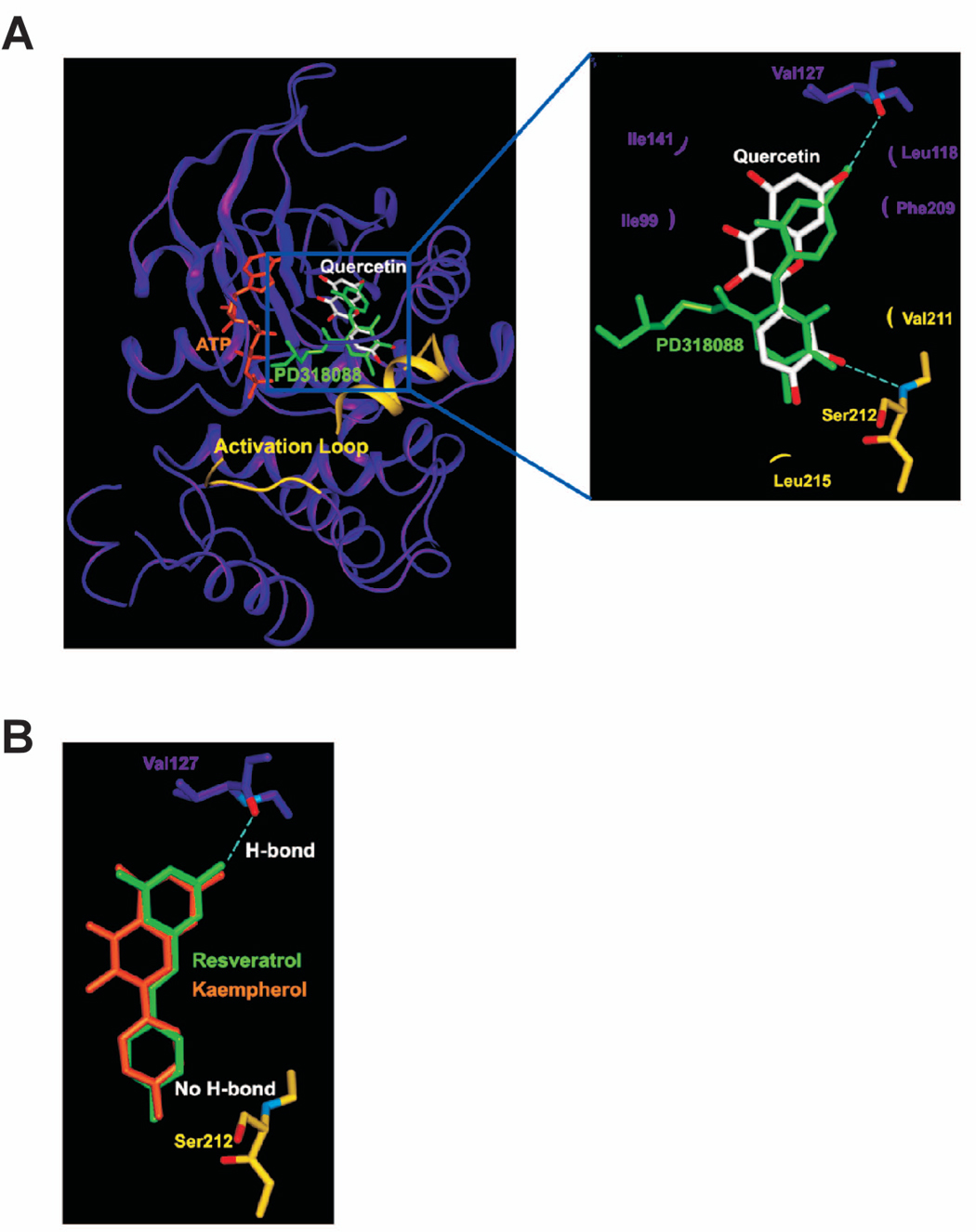
Considerable attention has focused on the health-promoting effects of red wine and its nonflavonoid polyphenol compound resveratrol. However, the underlying molecular mechanisms and molecular target(s) of red wine or other potentially active ingredients in red wine remain unknown. Here, we report that red wine extract (RWE) or the red wine flavonoid quercetin inhibited 12-O-tetradecanoylphorbol-13-acetate (TPA)-induced transformation of JB6 promotion-sensitive mouse skin epidermal (JB6 P+) cells. The activation of activator protein-1 and nuclear factor-kappaB induced by TPA was dose dependently inhibited by RWE or quercetin treatment. Western blot and kinase assay data revealed that RWE or quercetin inhibited mitogen-activated protein kinase/extracellular signal-regulated kinase (ERK) kinase (MEK) 1 and Raf1 kinase activities and subsequently attenuated TPA-induced phosphorylation of ERK/p90 ribosomal S6 kinase. Although either RWE or quercetin suppressed Raf1 kinase activity, they were more effective in inhibiting MEK1 activity. Importantly, quercetin exerted stronger inhibitory effects than PD098059, a well-known pharmacologic inhibitor of MEK. Resveratrol did not affect either MEK1 or Raf1 kinase activity. Pull-down assays revealed that RWE or quercetin (but not resveratrol) bound with either MEK1 or Raf1. RWE or quercetin also dose dependently suppressed JB6 P+ cell transformation induced by epidermal growth factor or H-Ras, both of which are involved in the activation of MEK/ERK signaling. Docking data suggested that quercetin, but not resveratrol, formed a hydrogen bond with the backbone amide group of Ser(212), which is the key interaction for stabilizing the inactive conformation of the activation loop of MEK1.


Figures

Figure 1
Figure 1
Effects of red wine extract (RWE) on TPA-induced neoplastic transformation, and AP-1 and NF-κB transactivation in JB6 P+ cells. A, RWE inhibited TPA-induced JB6 P+ cell transformation. JB6 P+ cells were treated as described in the Materials and Methods and colonies were counted 14 days later: untreated control (a), TPA alone (b), TPA and 5 µg/ml RWE (c), TPA and 10 µg/ml RWE (d), TPA and 20 µg/ml RWE (e), and TPA and 40 µg/ml RWE (f). B, Cell colonies were counted under a microscope with the aid of Image-Pro Plus software (v.4). The effects of RWE on neoplastic transformation of JB6 P+ cells are presented as the percent inhibition of cell transformation compared with cells treated only with TPA in soft agar. Data are presented as means ± S.D. of the percent inhibition as determined from three separate experiments. C and D, RWE inhibited TPA-induced AP-1 (C) or NF-κB (D) activation. The JB6 P+ cells, which were stably transfected with AP-1 or NF-κB luciferase reporter plasmids, were pretreated with RWE for 1 h at the indicated concentrations (5, 10, 20, or 40 µg/ml) followed by exposure to 20 ng/ml TPA for 24 h. The relative activity was measured by the luciferase assay as described in the Materials and Methods. Data are presented as means ± S.D. values of AP-1 and NF-κB luciferase activities calculated from three independent experiments. For B–D, the asterisk (*) indicates significant differences between groups treated with TPA and RWE together and the group treated with TPA alone (p < 0.05).
Figure 2
Figure 2
The Raf1/MEK/ERK/p90RSK signaling cascade is downregulated by RWE through the direct inhibition of both Raf1 and MEK1 activities. A, RWE inhibited TPA-induced phosphorylation of MEK, ERK, and p90RSK in JB6 P+ cells. JB6 P+ cells were treated with RWE (5, 10 or 20 µg/ml) for 1 h before being stimulated by 20 ng/ml TPA for an additional 15 min. The cells were lysed and the levels of phosphorylated and total MEK, ERK, and p90RSK proteins were determined by Western blot analysis as described in the Materials and Methods using specific antibodies against the respective phosphorylated and total proteins. Data are representative of three independent experiments that gave similar results. B, RWE inhibited MEK1 activity more than Raf1 activity both in vitro (upper panel) and ex vivo (lower panel). An in vitro MEK1 or Raf1 kinase assay was performed as described in the Materials and Methods, and the kinase activity is expressed as the percent inhibition relative to the activity of untreated MEK1 or Raf1 control. For the ex vivo MEK1 or Raf1 kinase assay, cells were pretreated with RWE at the indicated concentrations (1, 5, 10, or 20 µg/ml) for 1 h and then stimulated with 20 ng/ml TPA for 30 min. Cells were harvested, and immunoprecipitation and an ex vivo MEK1 or Raf1 kinase assay was performed. The kinase activity is expressed as the percent inhibition relative to cells treated with TPA only. The average 32P count was determined from three separate experiments, and the data are presented as means ± S.D. values. For in vitro kinase assays, the asterisk (*) indicates a significant decrease (p < 0.05) in kinase activity between the groups treated with active MEK1 (or Raf1) and RWE together and the group treated with active MEK1 (or Raf1) alone. For ex vivo kinase assays, the asterisk (*) indicates a significant decrease in kinase activity between cells treated with TPA and RWE together and the cells treated with TPA alone (p < 0.05). C, RWE specifically binds with MEK1 both in vitro and ex vivo. The MEK1–RWE binding in vitro was confirmed by immunoblotting using an antibody against MEK1 (left panel): lane 1 (input control), MEK1 protein standard; lane 2 (control), Sepharose 4B was used to pull down MEK1 as described in the Materials and Methods; and lane 3, RWE-Sepharose 4B affinity beads were used to pull down MEK1. The MEK1–RWE binding ex vivo was confirmed by immunoblotting using an antibody against MEK1 (right panel): lane 1 (input control), whole-cell lysates from JB6 P+ cells; lane 2 (control), a lysate of JB6 P+ cells precipitated with Sepharose 4B beads; and lane 3, whole-cell lysates from JB6 P+ cells precipitated by RWE-Sepharose 4B affinity beads. D, RWE specifically binds with Raf1 both in vitro and ex vivo. The Raf1–RWE binding in vitro was confirmed by immunoblotting using an antibody against Raf1 (left panel): lane 1 (input control), Raf1 protein standard; lane 2 (control), Sepharose 4B used to pull down Raf1 as described in the Materials and Methods; and lane 3, RWE-Sepharose 4B affinity beads were used to pull down Raf1. The Raf1–RWE binding ex vivo was confirmed by immunoblotting using an antibody against Raf1 (right panel): lane 1 (input control), whole-cell lysates from JB6 P+ cells; lane 2 (control), a lysate of JB6 P+ cells precipitated with Sepharose 4B beads; and lane 3, whole-cell lysates from JB6 P+ cells precipitated by RWE-Sepharose 4B affinity beads. Each experiment was performed two times and representative blots are shown.
Figure 3
Figure 3
Comparison of the inhibitory effects of quercetin or resveratrol against activation of Raf/MEK/ERK signaling cascades. A, Chemical structures of quercetin, resveratrol, and PD098059. B, Quercetin inhibited MEK1 activity more strongly than Raf1 activity, whereas resveratrol did not inhibit either kinase activity. An in vitro MEK1 (left panel) or Raf1 (right panel) kinase assay was performed as described in the Materials and Methods, and the kinase activity is expressed as the percent inhibition relative to the activity of untreated MEK1 or Raf1 control. The asterisk (*) indicates a significant decrease (p < 0.05) in kinase activity between the groups treated with active MEK1 or Raf1 and quercetin (or resveratrol or PD098059 or GW5074) together and the group treated with active MEK1 or Raf1 alone. C, Quercetin specifically binds with either MEK1 or Raf1. The in vitro MEK1 (or Raf1)–quercetin binding was confirmed by immunoblotting using an antibody against MEK1 (left-upper panel) or Raf1 (left-lower panel): lane 1 (input control), MEK1 or Raf1 protein standard; lane 2 (control), Sepharose 4B was used to pull down MEK1 or Raf1; and lane 3, MEK1 or Raf1 was pulled down using quercetin-Sepharose 4B affinity beads. The ex vivo MEK1 (or Raf1)–quercetin binding was confirmed by immunoblotting using an antibody against MEK1 (right-upper panel) or Raf1 (right-lower panel): lane 1 (input control), whole-cell lysates from JB6 P+ cells; lane 2 (control), a lysate of JB6 P+ cells precipitated with Sepharose 4B beads; and lane 3, whole-cell lysates from JB6 P+ cells precipitated by quercetin-Sepharose 4B affinity beads. Each experiment was performed two times and representative blots are shown. D, Quercetin inhibited TPA-induced ERK phosphorylation but had no effect on MEK phosphorylation in JB6 P+ cells. JB6 P+ cells were treated with quercetin at the indicated concentrations (5, 10, or 20 µM) for 1 h before being stimulated by 20 ng/ml TPA for an additional 15 min. The cells were lysed and the levels of phosphorylated and total ERK and MEK proteins were determined by Western blot analysis as described in the Materials and Methods using specific antibodies against the respective phosphorylated and total proteins. Data are representative of three independent experiments that gave similar results.
Figure 4
Figure 4
Comparison of inhibitory effects of quercetin or resveratrol against TPA-induced neoplastic transformation, and AP-1 and NF-κB transactivation in JB6 P+ cells. A, Quercetin was more effective than resveratrol at inhibiting TPA-induced JB6 P+ cell transformation. JB6 P+ cells were treated as described in the Materials and Methods, and colonies were counted 14 days later under a microscope with the aid of Image-Pro Plus software (v.4). The effects of quercetin or resveratrol on neoplastic transformation of JB6 P+ cells are presented as the percent inhibition of cell transformation compared with cells treated with only TPA in soft agar. Data are presented as means ± S.D. values of percent inhibition as determined from three separate experiments. The asterisk (*) indicates a significant difference (p < 0.05) between groups treated with TPA and quercetin (or resveratrol or PD098059 or GW5074) together and the group treated with TPA alone. B and C, TPA-induced activation of AP-1 (B) or NF-κB was inhibited more strongly by quercetin than by resveratrol. The JB6 P+ cells, which were stably transfected with AP-1 or NFκB luciferase reporter plasmids, were pretreated with quercetin (or resveratrol or PD098059 or GW5074) for 1 h followed by exposure to 20 ng/ml TPA for 24 h. The relative activity was measured by the luciferase assay as described in the Materials and Methods. Data are presented as means and S.D. values of AP-1 and NF-κB luciferase activities calculated from three independent experiments. The asterisks (*) indicate significant differences (p < 0.05) between groups treated with TPA and quercetin (or resveratrol or PD098059 or GW5074) together and the group treated with TPA alone (* indicates p < 0.05).
Figure 5
Figure 5
Effects of RWE, quercetin, or resveratrol on H-Ras- or EGF-induced cell transformation. Cell transformation was induced by H-Ras or EGF as described in the Materials and Methods and colonies were counted 14 days later under a microscope with the aid of Image-Pro Plus software (v.4). The effects of RWE, quercetin, or resveratrol on cell transformation of JB6 P+ cells are presented as the percent inhibition of cell transformation compared with H-Ras-or EGF-stimulated cells in soft agar. Data are presented as means ± S.D. values of the percent inhibition as determined from three independent experiments. A, RWE inhibited H-Ras-induced cell transformation of JB6 P+ cells. The asterisk (*) indicates a significant difference (p < 0.05) between groups treated with RWE and the untreated control group. B, RWE inhibited EGF-induced cell transformation of JB6 P+ cells. The asterisk (*) indicates a significant difference (p < 0.05) between groups treated with EGF and RWE together and the group treated with EGF alone. C, Quercetin (but not resveratrol) inhibited H-Ras-induced cell transformation of JB6 P+ cells. The asterisk (*) indicates a significant difference (p < 0.05) between groups treated with quercetin (or resveratrol or PD098059 or GW5074) and the untreated control group (p < 0.05). D, Quercetin (but not resveratrol) inhibited EGF-induced cell transformation of JB6 P+ cells. The asterisk (*) indicates a significant difference (p < 0.05) between groups treated with EGF and quercetin (or resveratrol or PD098059 or GW5074) together and the group treated with EGF alone.
Figure 6
Figure 6
Modeling study of the MEK1 binding of quercetin, resveratrol, or kaempherol. A. Hypothetical model of MEK1-quercetin complex. Quercetin (white color) binds to the pocket adjacent to the ATP (orange) binding site. PD318088 (green) is superimposed on the model structure of MEK1-quercetin complex for comparison. The partially disordered activation loop is colored yellow. The residues involved in the interactions with quercetin are indicated. The hydrogen bonds are depicted as dashed lines. B. Hypothetical model of MEK1 in complex with resveratrol (green) or kaempherol (orange). Although each of these compounds can retain the hydrogen bond with Val127 and the van der Waals interactions involved in the binding of quercetin to MEK1, neither compound can form a hydrogen bond with the activation loop of MEK1 due to the lack of a hydrogen bond acceptor at the 3′ position of their respective ring adjacent to the activation loop.

Similar articles

Cited by

References

    1. German JB, Walzem RL. The health benefits of wine. Annu Rev Nutr. 2000;20:561–593. - PubMed
    1. Damianaki A, Bakogeorgou E, Kampa M, et al. Potent inhibitory action of red wine polyphenols on human breast cancer cells. J Cell Biochem. 2000;78:429–441. - PubMed
    1. She QB, Bode AM, Ma WY, Chen NY, Dong Z. Resveratrol-induced activation of p53 and apoptosis is mediated by extracellular-signal-regulated protein kinases and p38 kinase. Cancer Res. 2001;61:1604–1610. - PubMed
    1. Kundu JK, Chun K-S, Kim SO, Surh Y-J. Resveratrol inhibits phorbol ester-induced cyclooxygenase-2 expression in mouse Skin : MAPKs and AP-1 as potential molecular targets. BioFactors. 2004;21:33–39. - PubMed
    1. Ribeiro de Lima MT, Waffo-Teguo P, Teissedre PL, et al. Determination of stilbenes (trans-astringin, cis- and trans-piceid, and cis- and trans-resveratrol) in Portuguese wines. J Agric Food Chem. 1999;47:2666–2670. - PubMed

Publication types

MeSH terms