

Activation of CCR9/CCL25 in cutaneous melanoma mediates preferential metastasis to the small intestine
- PMID: 18245522
- PMCID: PMC2760931
- DOI: 10.1158/1078-0432.CCR-07-2025
Activation of CCR9/CCL25 in cutaneous melanoma mediates preferential metastasis to the small intestine
Erratum in
- Clin Cancer Res. 2008 Mar 1;14(5):1598
- Clin Cancer Res. 2008 May 1;14(9):2892
Abstract
Purpose: Specific chemokines and their respective receptors have been implicated in distant tumor cell metastasis. Cutaneous melanoma has a distinct pattern of metastasis, preferentially targeting the submucosa of the small intestine. However, the underlying pathogenic mechanism remains unknown. Migration of CCR9(+) lymphocytes to the small intestine is known to occur in response to the chemoattractant effects of CCL25 (thymus-expressed chemokine). The integrin heterodimers alphabeta are also known to be important mediators of cellular adhesion. We hypothesize that the mechanism of small intestinal metastasis by melanoma is via the CCR9-CCL25 axis and specific integrins.
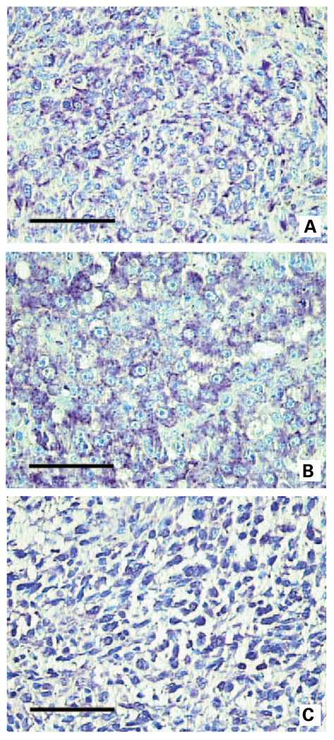
Experimental design: Quantitative reverse transcription-PCR, flow cytometry, and immunohistochemistry were used to assess melanoma tumors for CCR9 and CCL25. Integrin expression was assessed using flow cytometry. CCR9 expression by quantitative reverse transcription-PCR was assessed in primary (n = 23) and metastatic (n = 198) melanomas, and melanoma lines derived from small intestinal metastases (n = 23).
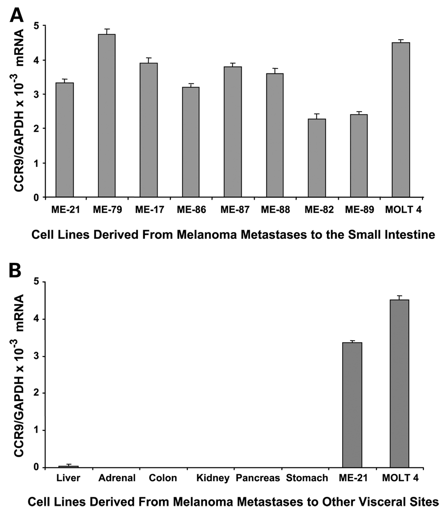
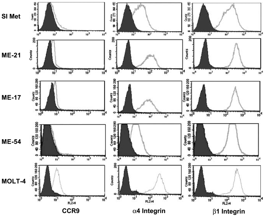
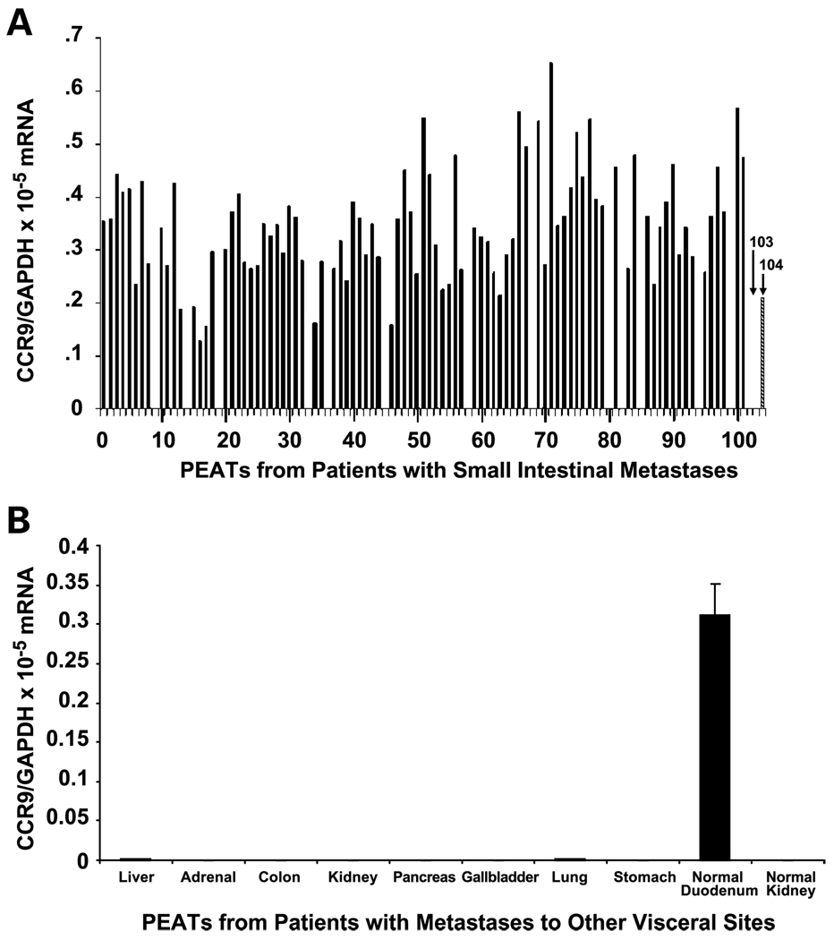
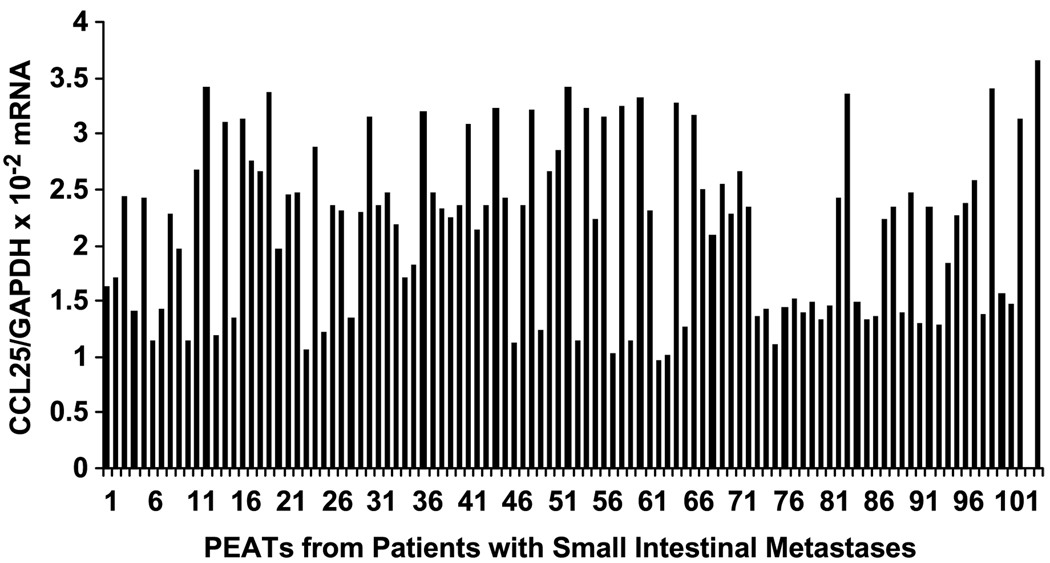
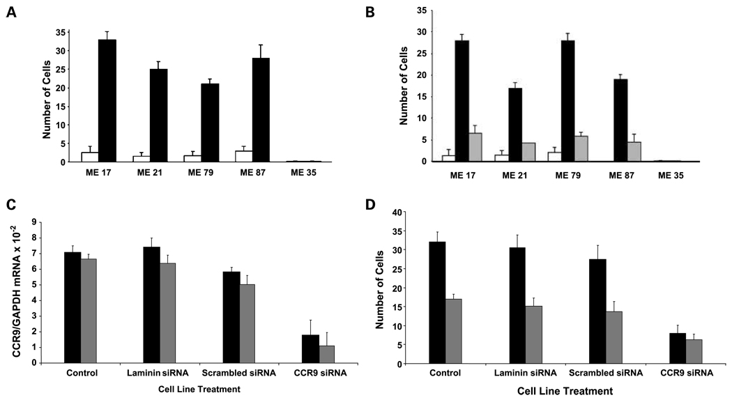
Results: We showed CCR9 expression in 88 of 102 paraffin-embedded metastatic melanomas from the small intestine, 8 of 8 melanoma lines derived from metastases in the small intestine, and 0 of 96 metastatic melanomas from other sites. In vitro migration and invasion studies done on CCR9(+) melanoma lines showed migration in response to CCL25 that was inhibited by anti-CCR9 antibody or by short interfering RNA CCR9. Flow cytometric analysis confirmed CCR9 expression by melanomas to the small intestine and showed concomitant alpha(4)beta(1) integrin expression.
Conclusions: Our findings show that functionally active CCR9 on melanoma cells facilitates metastasis to the small intestine. The CCR9-CCL25 axis may explain the high incidence of melanoma metastasis to this specific location.
Figures
Comment in
-
CCR9 homes metastatic melanoma cells to the small bowel.Clin Cancer Res. 2008 Feb 1;14(3):621-3. doi: 10.1158/1078-0432.CCR-07-2235. Clin Cancer Res. 2008. PMID: 18245518 No abstract available.
-
The CCR9-CCL25 axis mediates melanoma metastasis to the small intestine.Nat Clin Pract Oncol. 2008 Aug;5(8):440-1. doi: 10.1038/ncponc1174. Epub 2008 Jun 24. Nat Clin Pract Oncol. 2008. PMID: 18577981 No abstract available.
-
CCR9:CCL25 in melanoma metastatic to small intestine.Curr Oncol Rep. 2009 Sep;11(5):331-2. doi: 10.1007/s11912-009-0056-9. Curr Oncol Rep. 2009. PMID: 19679006 No abstract available.
References
-
- Agrawal S, Yao TJ, Coit DG. Surgery for melanoma metastatic to the gastrointestinal tract. Ann Surg Oncol. 1999;6:336–344. - PubMed
-
- Reintgen DS, Thompson W, Garbutt J, Seigler HF. Radiologic, endoscopic, and surgical considerations of melanoma metastatic to the gastrointestinal tract. Surgery. 1984;95:635–639. - PubMed
-
- Ashley SW, Wells SA., Jr Tumors of the small intestine. Semin Oncol. 1988;15:116–128. - PubMed
-
- Schuchter LM, Green R, Fraker D. Primary and metastatic diseases in malignant melanoma of the gastrointestinal tract. Curr Opin Oncol. 2000;12:181–185. - PubMed
-
- Ollila DW, Essner R, Wanek LA, Morton DL. Surgical resection for melanoma metastatic to the gastrointestinal tract. Arch Surg. 1996;131:975–979. 979–980. - PubMed
Publication types
MeSH terms
Substances
Grants and funding
LinkOut - more resources
Full Text Sources
Other Literature Sources
Medical
