

Proximal tibial osteochondromas in patients with fibrodysplasia ossificans progressiva
- PMID: 18245597
- PMCID: PMC3516450
- DOI: 10.2106/JBJS.G.00774
Proximal tibial osteochondromas in patients with fibrodysplasia ossificans progressiva
Abstract
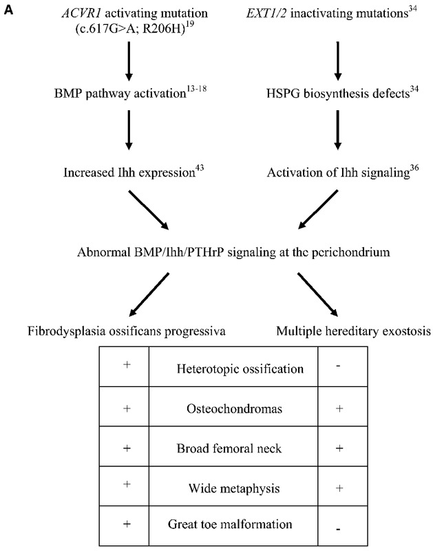
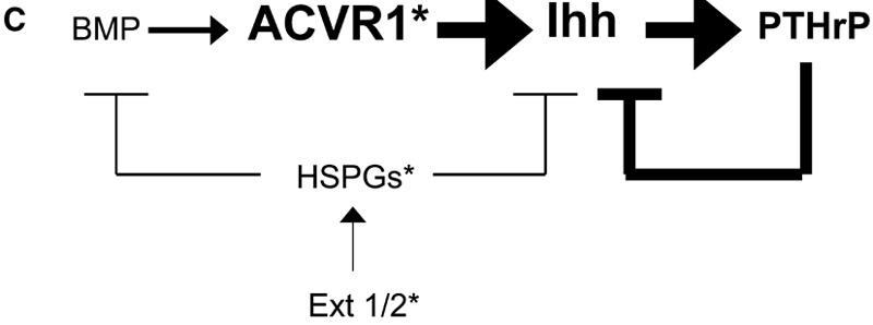
Background: Fibrodysplasia ossificans progressiva is a rare autosomal dominant disorder characterized by congenital malformation of the great toes and by progressive heterotopic ossification of skeletal muscle and soft connective tissues. The disorder is caused by a recurrent missense mutation in the glycine-serine activation domain of activin A receptor type I, a bone morphogenetic protein (BMP) type-I receptor, in all classically affected individuals. Osteochondromas of the proximal part of the tibia are benign osteochondral neoplasms or orthotopic lesions of skeletal remodeling associated with dysregulated BMP signaling and have been considered an atypical feature of fibrodysplasia ossificans progressiva, but they may be underdiagnosed because of their often asymptomatic nature. The purpose of the present study was to determine the prevalence and characteristics of proximal tibial osteochondromas in individuals who have fibrodysplasia ossificans progressiva.
Methods: Over a period of thirty months, we evaluated all patients with new and established fibrodysplasia ossificans progressiva for the presence of proximal tibial osteochondromas on the basis of medical history, physical examination, and radiographic studies. We quantified the prevalence of osteochondromas and characterized the types of osteochondromas to identify relevant trends.
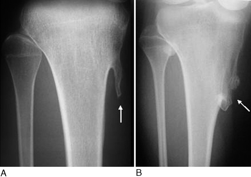
Results: Ninety-six patients (including fifty-two female patients and forty-four male patients) with fibrodysplasia ossificans progressiva were evaluated on the basis of a history and physical examination. Plain radiographs were available for sixty-seven patients. Ninety percent of all patients had osteochondroma of the proximal part of the tibia. These lesions usually were asymptomatic, most commonly were bilateral, and typically were located at the pes anserinus. Seventy-five percent of the lesions were pedunculated, and 25% were sessile.
Conclusions: Proximal tibial osteochondromas are a common phenotypic feature of fibrodysplasia ossificans progressiva, a finding that expands the recognized consequences of recurrent activating mutations in activin A receptor type I to include not only congenital skeletal malformations and heterotopic skeletogenesis but also benign osteochondral neoplasms or orthotopic lesions of skeletal modeling. The present study provides insight into the genetic basis of osteochondroma formation in patients with fibrodysplasia ossificans progressiva and possibly into that of more common conditions in which these lesions occur.
Figures


Similar articles
-
Early diagnosis of fibrodysplasia ossificans progressiva.Pediatrics. 2008 May;121(5):e1295-300. doi: 10.1542/peds.2007-1980. Pediatrics. 2008. PMID: 18450872 Free PMC article.
-
Fibrodysplasia ossificans progressiva: a current review of imaging findings.Skeletal Radiol. 2018 Aug;47(8):1043-1050. doi: 10.1007/s00256-018-2889-5. Epub 2018 Feb 14. Skeletal Radiol. 2018. PMID: 29445932 Review.
-
Pay Attention to the Osteochondromas in Fibrodysplasia Ossificans Progressiva.Orthop Surg. 2024 Mar;16(3):781-787. doi: 10.1111/os.13956. Epub 2024 Jan 7. Orthop Surg. 2024. PMID: 38185793 Free PMC article.
-
Hematopoietic stem-cell contribution to ectopic skeletogenesis.J Bone Joint Surg Am. 2007 Feb;89(2):347-57. doi: 10.2106/JBJS.F.00472. J Bone Joint Surg Am. 2007. PMID: 17272450
-
Challenges in the diagnosis of fibrodysplasia ossificans progressiva with the ACVR1 mutation (c.774G > C, p.R258S): a case report and review of literature.Orphanet J Rare Dis. 2024 Sep 30;19(1):360. doi: 10.1186/s13023-024-03363-y. Orphanet J Rare Dis. 2024. PMID: 39350127 Free PMC article. Review.
Cited by
-
Inherited human diseases of heterotopic bone formation.Nat Rev Rheumatol. 2010 Sep;6(9):518-27. doi: 10.1038/nrrheum.2010.122. Epub 2010 Aug 10. Nat Rev Rheumatol. 2010. PMID: 20703219 Free PMC article. Review.
-
Is fibrodysplasia ossificans progressiva ever seen without skeletal abnormalities?Rheumatol Int. 2012 May;32(5):1475-6; author reply 1477-8. doi: 10.1007/s00296-011-1904-0. Epub 2011 Mar 25. Rheumatol Int. 2012. PMID: 21437684 No abstract available.
-
When one skeleton is enough: approaches and strategies for the treatment of fibrodysplasia ossificans progressiva (FOP).Drug Discov Today Ther Strateg. 2008;5(4):255-262. doi: 10.1016/j.ddstr.2008.11.004. Drug Discov Today Ther Strateg. 2008. PMID: 23599718 Free PMC article.
-
Glycobiology and the growth plate: current concepts in multiple hereditary exostoses.J Pediatr Orthop. 2011 Jul-Aug;31(5):577-86. doi: 10.1097/BPO.0b013e31821c7738. J Pediatr Orthop. 2011. PMID: 21654469 Free PMC article. Review.
-
BMP signaling and skeletal development in fibrodysplasia ossificans progressiva (FOP).Dev Dyn. 2022 Jan;251(1):164-177. doi: 10.1002/dvdy.387. Epub 2021 Jun 26. Dev Dyn. 2022. PMID: 34133058 Free PMC article. Review.
References
-
- Kaplan FS, McCluskey W, Hahn G, Tabas JA, Muenke M, Zasloff MA. Genetic transmission of fibrodysplasia ossificans progressiva. Report of a family. J Bone Joint Surg Am. 1993;75:1214-20 - PubMed
-
- Kaplan FS, Tabas JA, Gannon FH, Finkel G, Hahn GV, Zasloff MA. The histopathology of fibrodysplasia ossificans progressive. An endochondral process. J Bone Joint Surg Am. 1993;75:220-30 - PubMed
-
- Schroeder HW, Jr, Zasloff M. The hand and foot malformations in fibrodysplasia ossificans progressiva. Johns Hopkins Med J. 1980;147:73-8 - PubMed
-
- Cohen RB, Hahn GV, Tabas JA, Peeper J, Levitz CL, Sando A, Sando N, Zasloff M, Kaplan FS. The natural history of heterotopic ossification in patients who have fibrodysplasia ossificans progressiva. A study of forty-four patients. J Bone Joint Surg Am. 1993;75:215-9 - PubMed
-
- Rogers JG, Geho WB. Fibrodysplasia ossificans progressiva. A survey of forty-two cases. J Bone Joint Surg Am. 1979;61:909-14 - PubMed
Publication types
MeSH terms
Grants and funding
LinkOut - more resources
Full Text Sources
Medical
