Review
. 2008 Mar;172(3):555-65.
doi: 10.2353/ajpath.2008.070442. Epub 2008 Feb 2.

Prion diseases: from protein to cell pathology

Affiliations
Review

Prion diseases: from protein to cell pathology

Gabor G Kovacs et al. Am J Pathol. 2008 Mar.

Abstract

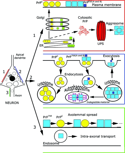
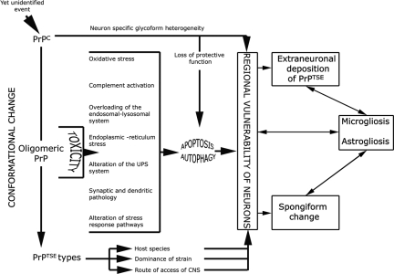
Prion diseases or transmissible spongiform encephalopathies are fatal neurodegenerative conditions in humans and animals that originate spontaneously, genetically or by infection. Conformational change of the normal (cellular) form of prion protein (PrP c) to a pathological, disease-associated form (PrP TSE) is considered central to pathogenesis and formation of the infectious agent or prion. Neuronal damage is central to clinical manifestation of prion diseases but poorly understood. In this review, we analyze the major pathogenetic pathways that lead to tissue pathology in different forms of disease. Neuropathogenesis of prion diseases evolves in complex ways on several front lines, most but not all of which exist also in other neurodegenerative as well as infectious diseases. Whereas intracellular accumulation of PrP forms might significantly impair cell function and lead to cytopathology, mere extracellular deposition of PrP TSE is questionable as a direct cytotoxic factor. Tissue damage may result from several parallel, interacting, or subsequent pathways. Future studies should clarify the trigger(s) and sequence of these processes and whether, and which, one is dominating or decisive.


Figures

Figure 1
Figure 1
Summary of cytopathology and PrP processing pathways. Pathway 1 (intracellular PrP processing, red): The PrPC polypeptide (yellow circles), including genetic mutants (green circles), is synthesized in the ER, processed in the Golgi apparatus, and then carried in its mature form to the cell surface where most of it is found in lipid rafts. Generation of PrPTSE, consisting of a mixture of dominant and subdominant types (blue and dark blue boxes, A and B), from PrPC would occur after the arrival of PrPC at the cell surface. Another hypothetical pathway would form misfolded cytosolic PrP, associated with neurotoxicity, involving the ubiquitin-proteasome system and forming aggresomes.,, The same would be promoted by mutant PrPTSE (green quadrangles). Pathway 2 (processing of external PrPC and PrPTSE, blue): PrPC from the plasma membrane is internalized and processed in lysosomes. Exogenic PrPTSE, consisting of a mixture of dominant and subdominant types (blue and dark blue boxes, A and B), leads to conformational change of PrPC before or during internalization via endosomes. Overloading of the endosomal-lysosomal system may lead to accumulation of indigestible material or exocytosis of PrPTSE that forms extracellular aggregated deposits that presumably lack direct neurotoxic effects. This process may be accompanied by an outburst of lysosomal enzymes leading to tissue damage. The detailed delineation of endosomal and lysosomal compartments is omitted for clarity. Pathway 3 (spread of PrPTSE, green): Endosomes may transport PrPTSE in the axons, in addition to domino-like spread of PrPTSE axolemmally.
Figure 2
Figure 2
Histological and immunohistochemical demonstration of pathological alterations discussed in the present review. Technical details are detailed in our previous studies.,,, a: Shrinkage of the nucleus in a pyramidal neuron of the frontal cortex accompanied by intense PAS positivity in a representative case with sporadic CJD, suggesting lysosomal overload. b: Terminal deoxynucleotidyl transferase-mediated dUTP nick end-labeling positivity, indicated by blue color, of a representative neuronal nucleus in the same case as in Figure 1a, suggesting apoptosis. c: Immunostaining for RNA-derived 8-hydroxy-guanosine (8-OHG) and DNA-derived 8-hydroxydeoxy-guanosine (8-OHdG) in CJD demonstrates predominantly cytoplasmic immunoreactivity. d: Cathepsin D (CathD) immunoreactivity in a normal brain without neurological disease. e: CathD immunopositivity in a representative case of CJD. Note accumulation of enlarged dot-like cytoplasmic immunoreactivity as compared to Figure 1d, suggesting lysosomal overload. f: Intra- and perineuronal colocalization (indicated by yellow color) of PrPTSE (indicated by green color) and CathD (indicated by red color) demonstrated by laser confocal scanning microscopy in CJD, suggesting interaction of PrPTSE with the lysosomal system. g: Immunodeposits of the neoantigen of C9 on a neuron in CJD, suggesting involvement of the terminal complement. h: Neuronal immunoreactivity for the membrane attack complex (C5b-9) in CJD, demonstrated by LCM, supporting the involvement of the terminal complement. i: Immunostaining for the dendritic marker MAP-2 in the frontal cortex in CJD demonstrates distorted and fragmented dendritic arborization. j: MAP-2 immunoreactive atrophic dendrites in the frontal cortex in CJD. Scale bars represent 10 μm for a–h, 40 μm for i, and 20 μm for j.
Figure 3
Figure 3
Diagram of pathogenetic events in prion disease. A yet unidentified event (external prions, spontaneous conversion, or awakening of silent prions50) initiates conformational change of PrPC with potentially reversible functional impairment of neurons. During this process oligomeric forms of PrP are present that may have a direct neurotoxic effect initiating a cascade of events leading to either or both apoptosis and autophagy. This process is potentially influenced by the loss of protective function of PrPC. According to the neuron-specific glycoform heterogeneity, the species of host, dominance of PrPTSE type, or route of access of external prions to the CNS, tissue pathology comprising spongiform change, astro- and microgliosis develops in regional variability. Astro- and microgliosis are initiated by the neuronal damage or toxic intermediate forms of PrP, but inversely may also contribute to neuronal death and formation of spongiform change.

References

    1. Prusiner SB. Prions. Proc Natl Acad Sci USA. 1998;95:13363–13383. - PMC - PubMed
    1. Aguzzi A, Heikenwalder M. Pathogenesis of prion diseases: current status and future outlook. Nat Rev Microbiol. 2006;4:765–775. - PubMed
    1. Caughey B, Baron GS. Prions and their partners in crime. Nature. 2006;443:803–810. - PubMed
    1. Westergard L, Christensen HM, Harris DA. The cellular prion protein (PrP(C)): its physiological function and role in disease. Biochim Biophys Acta. 2007;1772:629–644. - PMC - PubMed
    1. Ironside JW, Ritchie DL, Head MW. Phenotypic variability in human prion diseases. Neuropathol Appl Neurobiol. 2005;31:565–579. - PubMed

Publication types