. 2008 May;49(5):1015-23.
doi: 10.1194/jlr.M700535-JLR200. Epub 2008 Feb 2.

Human C-reactive protein promotes oxidized low density lipoprotein uptake and matrix metalloproteinase-9 release in Wistar rats

Affiliations

Human C-reactive protein promotes oxidized low density lipoprotein uptake and matrix metalloproteinase-9 release in Wistar rats

U Singh et al. J Lipid Res. 2008 May.

Abstract

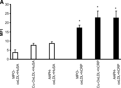
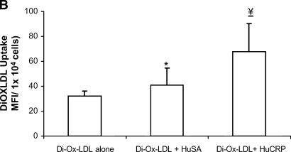
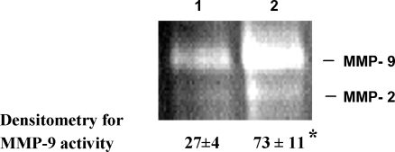
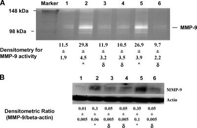
C-reactive protein (CRP) is present in the atherosclerotic plaques and appears to promote atherogenesis. Intraplaque CRP colocalizes with oxidized low density lipoprotein (OxLDL) and macrophages in human atherosclerotic lesions. Matrix metalloproteinase-9 (MMP-9) has been implicated in plaque rupture. CRP promotes OxLDL uptake and MMP induction in vitro; however, these have not been investigated in vivo. We examined the effect of CRP on OxLDL uptake and MMP-9 production in vivo in Wistar rats. CRP significantly increased OxLDL uptake in the peritoneal and sterile pouch macrophages compared with human serum albumin (huSA). CRP also significantly increased intracellular cholesteryl ester accumulation compared with huSA. The increased uptake of OxLDL by CRP was inhibited by pretreatment with antibodies to CD32, CD64, CD36, and fucoidin, suggesting uptake by both scavenger receptors and Fc-gamma receptors. Furthermore, CRP treatment increased MMP-9 activity in macrophages compared with huSA, which was abrogated by inhibitors to p38 mitogen-activated protein kinase, extracellular signal-regulated kinase (ERK), and nuclear factor (NF)-kappaB but not Jun N-terminal kinase (JNK) before human CRP treatment. Because OxLDL uptake by macrophages contributes to foam cell formation and MMP release contributes to plaque instability, this study provides novel in vivo evidence for the role of CRP in atherosclerosis.


Figures

Fig. 1.
Fig. 1.
A: Effects of human C-reactive protein (hCRP) compared with human serum albumin (huSA) on 1,1′-dioctadecyl-3,3,3′,3′ tetramethylindocarbocyanide perchlorate (DiI) oxidized low density lipoprotein (OxLDL) uptake in human monocyte-derived macrophages (HMDMs). LDL was oxidized using 2,2-azobis-2-amidino-propane dihydrochloride (AAPH), copper (Cu), and myeloperoxidase (MPO) as described in Materials and Methods. HMDMs were cultured for 7 days and treated with DiI-OxLDL alone or in the presence of huSA/hCRP. DiI uptake was measured as described in Materials and Methods. * P < 0.05 compared with DiI-OxLDL + huSA (n = 3 experiments). MFI, mean fluorescence intensity. B: Effects of intraperitoneal hCRP compared with huSA administration along with DiI-OxLDL in Wistar rats on DiI uptake. The rats were injected with hCRP/huSA for 2 days at 20 mg/kg body weight. On the 2nd day, DiI-OxLDL was injected along with hCRP/huSA. Peritoneal macrophages were isolated and plated on 24-well plates. After adherence to the surface, DiI was extracted with isopropanol and fluorescence was measured as described in Materials and Methods. DiI uptake is expressed as MFI per 104 cells. * P = 0.049 compared with Di-OxLDL alone; ¥ P = 0.03 compared with huSA + DiI-OxLDL (n = 4 rats/group). C: Effects of intraperitoneal hCRP compared with huSA administration along with DiI-OxLDL in Wistar rats on free cholesterol (FC) and intracellular cholesteryl ester (EC) accumulation. The rats were injected with hCRP/huSA for 2 days at 20 mg/kg body weight. On the 2nd day, DiI-OxLDL was injected along with hCRP/huSA. Peritoneal macrophages were isolated and plated on 12-well plates. Total and free cholesterol were quantitated, and cholesteryl ester content was determined as detailed in Materials and Methods. * P < 0.05 compared with buffer, ** P < 0.05 compared with OxLDL alone, *** P < 0.003 compared with DiI-OxLDL + huSA, ¥ P < 0.05 compared with OxLDL alone, ¥¥ P < 0.01 compared with DiI-OxLDL + huSA (n = 4 rats/group). Values shown are means ± SD.
Fig. 1.
Fig. 1.
A: Effects of human C-reactive protein (hCRP) compared with human serum albumin (huSA) on 1,1′-dioctadecyl-3,3,3′,3′ tetramethylindocarbocyanide perchlorate (DiI) oxidized low density lipoprotein (OxLDL) uptake in human monocyte-derived macrophages (HMDMs). LDL was oxidized using 2,2-azobis-2-amidino-propane dihydrochloride (AAPH), copper (Cu), and myeloperoxidase (MPO) as described in Materials and Methods. HMDMs were cultured for 7 days and treated with DiI-OxLDL alone or in the presence of huSA/hCRP. DiI uptake was measured as described in Materials and Methods. * P < 0.05 compared with DiI-OxLDL + huSA (n = 3 experiments). MFI, mean fluorescence intensity. B: Effects of intraperitoneal hCRP compared with huSA administration along with DiI-OxLDL in Wistar rats on DiI uptake. The rats were injected with hCRP/huSA for 2 days at 20 mg/kg body weight. On the 2nd day, DiI-OxLDL was injected along with hCRP/huSA. Peritoneal macrophages were isolated and plated on 24-well plates. After adherence to the surface, DiI was extracted with isopropanol and fluorescence was measured as described in Materials and Methods. DiI uptake is expressed as MFI per 104 cells. * P = 0.049 compared with Di-OxLDL alone; ¥ P = 0.03 compared with huSA + DiI-OxLDL (n = 4 rats/group). C: Effects of intraperitoneal hCRP compared with huSA administration along with DiI-OxLDL in Wistar rats on free cholesterol (FC) and intracellular cholesteryl ester (EC) accumulation. The rats were injected with hCRP/huSA for 2 days at 20 mg/kg body weight. On the 2nd day, DiI-OxLDL was injected along with hCRP/huSA. Peritoneal macrophages were isolated and plated on 12-well plates. Total and free cholesterol were quantitated, and cholesteryl ester content was determined as detailed in Materials and Methods. * P < 0.05 compared with buffer, ** P < 0.05 compared with OxLDL alone, *** P < 0.003 compared with DiI-OxLDL + huSA, ¥ P < 0.05 compared with OxLDL alone, ¥¥ P < 0.01 compared with DiI-OxLDL + huSA (n = 4 rats/group). Values shown are means ± SD.
Fig. 2.
Fig. 2.
A: Effects of hCRP/huSA intrapouch administration (25 μg/ml) on DiI-OxLDL uptake. Sterile pouches were made in rats, and hCRP/huSA was injected in the pouch cavities of Wistar rats on the 3rd day after the formation of the pouch. For CRP-mediated OxLDL uptake experiments, DiI-OxLDL (50 μg/ml protein) was injected in the pouch along with CRP. The next day, the pouch fluid was aspirated as described in Materials and Methods and the cells isolated were used for DiI uptake. * P < 0.05 compared with huSA + DiI-OxLDL (n = 5 rats/group). B: Demonstration of the involvement of various receptors involved in CRP-mediated DiI-OxLDL uptake. Rats were injected with hCRP/huSA as described above in the pouch cavities of Wistar rats on the 3rd day after the formation of the pouch. Antibodies to CD32, CD64, CD36, and control isotype as well as the combinations CD32 + CD36, CD32 + CD64, and CD36 + fucoidin were also injected at 1–2 h before hCRP treatment. DiI uptake was analyzed by flow cytometry as described in Materials and Methods. **P < 0.05 versus DiI-OxLDL + CRP (n = 3–5 rats/group). Values shown are means ± SD.
Fig. 3.
Fig. 3.
Effects of CRP on matrix metalloproteinase-9 (MMP-9) production in peritoneal macrophages of Wistar rats administered huSA or hCRP (20 mg/kg body weight/day for 2 days). Peritoneal macrophages were isolated, and lysates were run on a SDS-PAGE gelatin gel for MMP-9 zymography at 4°C. After electrophoresis, the gel was subjected to washing, development, and staining as described in Materials and Methods. Active MMP-9 was visualized as proteolytic (white) bands on a dark background. Column 1, huSA; column 2, hCRP. The gel shown is representative of samples from four different rats. * P < 0.01 compared with huSA.
Fig. 4.
Fig. 4.
A: Effects of pretreatment with various inhibitors to p38, extracellular signal-regulated kinase (ERK), Jun N-terminal kinase (JNK), and NF-κB in the rat sterile air pouch model in Wistar rats on MMP-9 activity. Column 1, huSA; column 2, hCRP; column 3, p38 MAPK inhibitor + hCRP; column 4, ERK inhibitor + hCRP; column 5, JNK inhibitor + CRP; column 6, NF-κB inhibitor + CRP. The pouch fluids were run on SDS-PAGE gelatin gels for zymography as described in Materials and Methods. The gel shown is representative from four rats per group. Densitometric values (means ± SD) are given below the gel in arbitrary units. * P < 0.001 compared with huSA, δ P > 0.05 compared with hCRP. B: MMP-9 total protein mass as described in Materials and Methods. Column 1, huSA; column 2, hCRP; column 3, p38 MAPK inhibitor + hCRP; column 4, ERK inhibitor + hCRP; column 5, JNK inhibitor + CRP; column 6, NF-κB inhibitor + CRP. The pouch cell lysates were run on SDS-PAGE gelatin gels for MMP-9 total protein mass as described in Materials and Methods. The gel shown is representative from four rats per group. Densitometric ratios (means ± SD) are given below the gel. * P < 0.05 compared with huSA; δ P > 0.05 compared with hCRP.

Similar articles

Cited by

References

    1. Croce K., and P. Libby. 2007. Intertwining of thrombosis and inflammation in atherosclerosis. Curr. Opin. Hematol. 14 55–61. - PubMed
    1. Jialal I., S. Devaraj, and S. K. Venugopal. 2004. C-reactive protein: risk marker or mediator in atherothrombosis? Hypertension. 44 6–11. - PubMed
    1. Verma S., S. Devaraj, and I. Jialal. 2006. Is C-reactive protein an innocent bystander or proatherogenic culprit? C-reactive protein promotes atherothrombosis. Circulation. 113 2135–2150. - PubMed
    1. Ishikawa T., K. Hatakeyama, T. Imamura, Y. Shibata, Y. Hikichi, Y. Asada, and T. Eto. 2003. Involvement of CRP obtained by directional coronary atherectomy in plaque instability and developing restenosis in patients with stable or unstable angina pectoris. Am. J. Cardiol. 91 287–292. - PubMed
    1. Ballou C. P., and G. Lozanski. 1992. Induction of inflammatory cytokine release from cultured human monocytes by CRP. Cytokine. 4 361–368. - PubMed

Publication types